





| Research Article | ||
Open Vet. J.. 2026; 16(5): 2961-2969 Open Veterinary Journal, (2026), Vol. 16(5): 2961-2969 Research Article Prevalence of cardiac arrhythmias in poorly performing horsesOsamah Muwaffag Aliraqi* and Basima Abdulfatah AlbadraniDepartment of Internal and Preventive Medicine, College of Veterinary Medicine, University of Mosul, Mosul, Iraq *Corresponding Author: Osamah Muwaffag Aliraqi. Department of Internal and Preventive Medicine, College of Veterinary Medicine, University of Mosul, Mosul, Iraq. Email: osamamuwafag [at] uomosul.edu.iq Submitted: 04/01/2026 Revised: 25/03/2026 Accepted: 14/04/2026 Published: 31/05/2026 © 2025 Open Veterinary Journal
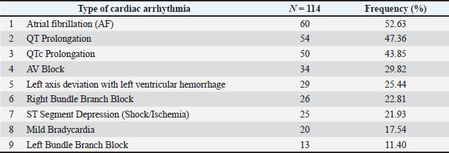
AbstractBackground: Electrocardiogram abnormalities are common in racehorses and can affect performance. Aim: This study aimed to analyze the results of electrocardiographic problems and measure the frequencies of various cardiac arrhythmias. Methods: This cross-sectional study was conducted between September 2023 and August 2024. Standard electrocardiographic recordings were obtained from all horses, and cardiac troponin levels were measured. Out of the 114 poorly performing adult horses, 10 cases without arrhythmia were considered the control group. Seventy-four horses (64.9%) had at least one type of cardiac arrhythmia, 60 (52.63%) had atrial fibrillation, and 29 (25.44%) had left axis deviation and left ventricular hypertrophy. Results: The most frequent conduction anomaly was atrioventricular block in 34 horses (29.82%), followed by QT prolongation in 54 horses (47.36%), and QTc prolongation in 50 horses (43.85%). Bradycardia was detected in 20 (17.54%) horses, and ST-segment depression was present in 25 (21.93%) horses. Conduction disturbances, including left bundle branch block in 13 horses (11.40%) and right bundle branch block in 26 horses (22.81%). Troponin levels were significantly elevated in cases of arrhythmia. The incidence of atrial fibrillation was significantly higher in male horses in all age groups except the oldest. Conclusion: Because cardiac arrhythmia may lead to racehorse exercise intolerance and suboptimal performance, early diagnosis is important to allow veterinarians to develop long-term treatment strategies. Furthermore, electrocardiography, along with cardiac biomarker cardiac troponin, is essential. Keywords: Arrhythmia, Electrocardiography, Horse. IntroductionCardiac arrhythmias are common in the equine population and can be physiologic due to high vagal tone or pathologic due to cardiac or noncardiac diseases (Van Loon, 2019). The prevalence of cardiac arrhythmias varies depending on factors such as breed, age, and level of exercise (Decloedt et al., 2015). Other environmental factors, including atmospheric temperature stress and underlying health conditions, are known to affect the incidence of cardiac arrhythmias in horses. Horses can have cardiac arrhythmias, which are medically important because their prevalence, incidence, health consequences, and role in sudden death are not well understood, as they are in horses, typically due to a lack of readily available information in both the horse owner and veterinarian (Buchanan and Little, 1998; Santosuosso et al., 2023) Information about the prevalence and incidence of atrial fibrillation (AF) and other cardiac arrhythmias in horses has been previously provided. Wright and Lafferty (2021) reported that 4.9% of horses have AF. Horses with a history of AF had a particularly high incidence of relapse, recurrent AF was a problem in equine participants, and monitoring and treating this condition were particularly important (Seeley and Houghton, 2022). Sweeney and Jerger (2018) reported the importance of AF in performance horses. Dreyer and van der Veen (2019) also evaluated the long-term outcomes of the horses. Horses have shown a higher frequency of cardiac arrhythmia in certain breeds, such as those suffering from equine axonal atrophy in quarter horses (Williams et al., 2020). Although the hearts of these horses were thin and the horses were afflicted with subclinical cardiomyopathy, thinning of the heart muscle and increased cardiac arrhythmias may indicate a breed predisposition that should be investigated further (Meyer et al., 2021). Diagnostic biomarkers investigated in terms of the horse with arrhythmia include plasma cTnI concentrations (Tulleners et al., 2021). The median cTnI level in horses with ventricular arrhythmias was tenfold higher than that in horses with AF (Meyer et al., 2021), and the cTnI level can be used to assess myocardial damage (Dreyer et al., 2022). Scientific works related to the electrocardiography of horses in Iraq are still scarce, and little information has been provided, except for one done by Albadrani et al. (2024).Therefore, this study was conducted to determine the prevalence of cardiac arrhythmias and their risk factors in racehorses and to determine how many arrhythmias occur at a single time point in horses. Materials and MethodsStudy designA total of 114 Arabian and Thoroughbred horses from 4 to 11 years of age, weighing 340–390 kg, 67 stallions, and 47 mares were housed at the Equestrian Club in the Al-Shalalat area and at different areas around Mosul, Iraq, and were examined between September 2023 and August 2024. A history of poor performance was the selection criterion for diseased horses. Furthermore, 10 horses without arrhythmia and with normal troponin levels were considered as the control group for comparison. Clinical examinationClinical examinations included mucosal assessment, determination of hydration status, capillary refill time, palpation of superficial lymph nodes, arterial pulse, measurement of heart rate and respiratory rate, assessment of intestinal motility, measurement of rectal temperature, and review of the animal’s history were all included in this examination. Cardiac troponin evaluationAt rest, 10 ml of blood from the jugular vein was taken in a gel tube at rest time, then quickly agitated at 2000–3000 rpm for 20 minutes to separate the serum. Serum cardiac troponin I (cTnI) and cardiac troponin T (cTnT) were quantified using Equine Cardiac Troponin I and T Sandwich ELISA Kits (Sun Long Biotech Co., LTD) according to the manufacturer’s instructions. Electrocardiogram (ECG)Electrocardiography was performed at rest time, and recordings were obtained using a portable electrocardiograph (EDAN VET ECG, model VE-100, 220 V-240 V/100 V-115 V 50 Hz/60 Hz, 0.3 max, Edan Instrument Inc, Shenzhen, China). The horses were harnessed and restrained in a standing position with the limbs equivalent to each other, without the use of tranquilliser, sedation, or anesthesia. ECG tracings were done serially on all horses by the leads in the frontal plane (I, II, III, aVR, aVL, and aVF). For the bipolar basal electrode system, the yellow electrode was placed on the left side above the apex of the heart, just behind the ulna, and the red electrode was placed on the right side, from the skull to the scapula, and near the jugular vein, with the ground electrode attached to the withers. The ECG data were collected and recorded using the specified lead configuration and electrode placement. To estimate the HR from the ECG, the R-R interval in the trace was measured, and then the HR formula was used: HR (bpm)=1500/R-R interval in mm. The paper speed is 25 mm/s (as shown in the trace) to calculate the QT interval from this ECG trace, and the QT interval is calculated from the start of the Q wave until the end of the T wave. Using the provided trace, visually estimate the number of small squares (1 mm each) within this interval and convert this to time. Each small square at 25 mm/s represents 0.04 s (40 ms). The number of small squares is then multiplied by 0.04 to obtain the QT interval in seconds. Bazett’s formula is used to correct the QT interval for heart rate: QTc=QT (milliseconds)/√RR interval (seconds). The R-R interval (the distance between two consecutive R waves) is measured in small squares, and its time is calculated (small squares × 0.04). Statistical analysisDescriptive statistics were applied to the ECG findings. Using Epi-InfoTM (version 7), the odds ratios for (age, breed, and gender) were examined using the X2 test and Fisher’s exact test. P-values below 0.05 were considered significant. Independent t-tests were run to test if there were differences between ECG parameters; statistically significant differences were taken as a p-value of < 0.05. Ethical approvalBlood samples were obtained from the animals of study, and clinical examinations were performed in accordance with the Institutional Animal Care and Use Committee, College of Veterinary Medicine, University of Mosul, Ethical code number UM.VET.2023.146. on 15-8-2023. ResultsFrom the results of the electrocardiogram analysis, the main cardiac arrhythmia was atrial fibrillation (52.63%), followed by QT Prolongation (47.36%) and QTc Prolongation (43.85%), other lees frequent include: AV Block (29.82%), left axis deviation with left ventricular hypertrophy (25.44%), right bundle branch block (RBBB) (22.81%), and ST-segment depression (21.93%). Mild brachycardia (17.54%) and left bundle branch block (LBBB) (11.40%) were the least frequent arrhythmias (Table 1). Table 1. The Frequency of different cardiac arrhythmias in racehorses.
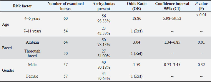
The risk factors analysis indicated that arrhythmia in younger horses (93.33%) was significantly (p=0.01) more likely (18.86 times) to occur than that in older horses. Furthermore, Arabian horses showed a significantly (p=0.01) high prevalence of arrhythmia (78.13%) compared to thoroughbred horses, with a 3 times risk factor to occurrence, while there were no significant differences between male and female horses (Table 2). Table 2. Risk factors related to arrhythmias according to electrocardiography in horses.
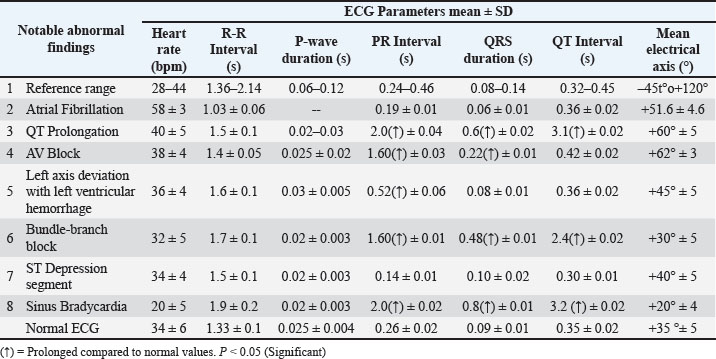
ECG parameter analysis showed atrial fibrillation, in which the heart rate was significantly increased (58pm 3.5bpm), and the P wave was missing, which was a characteristic finding of this irregular rhythm. Sinus brachycardia appeared with a significantly decreased heart rate (20pm 5 bpm). In QT prolongation, significant increases in the PR interval (2.0 ± 0.04), QT duration (0.6 ± 0.02), and QT interval (3.1 ± 0.02). In the AV block, there was a significant increase in the PR interval (1.60 ± 0.03) and QT duration (0.22 ± 0.01). In the bundle branch block, increases were observed in the PR interval (1.60 ± 0.01), QRS duration (0.48 ± 0.01), and QT interval (2.4 ± 0.02) (Table 3). Table 3. Comparison of normal ECG parameters with pathological cardiac conditions.
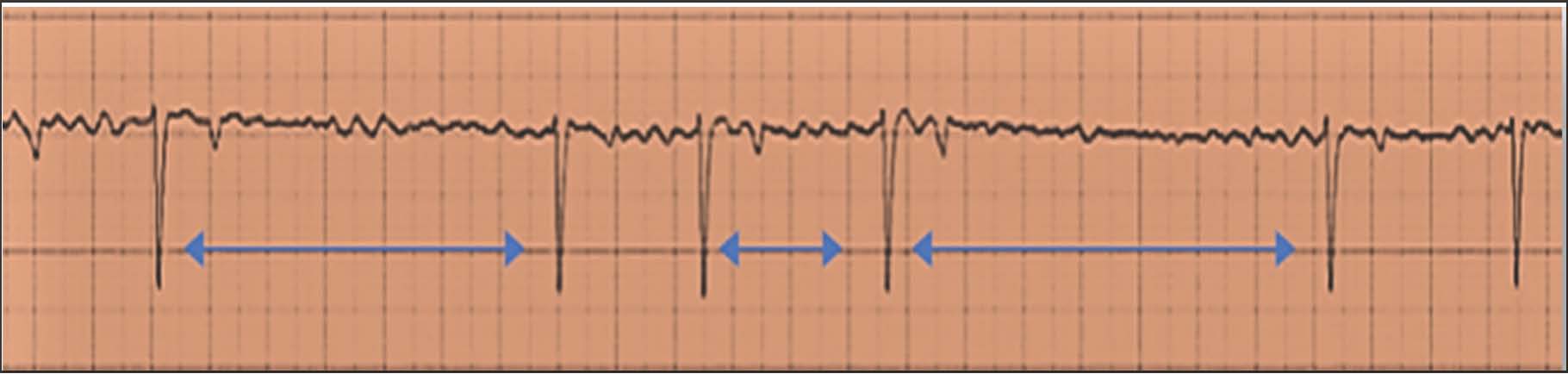
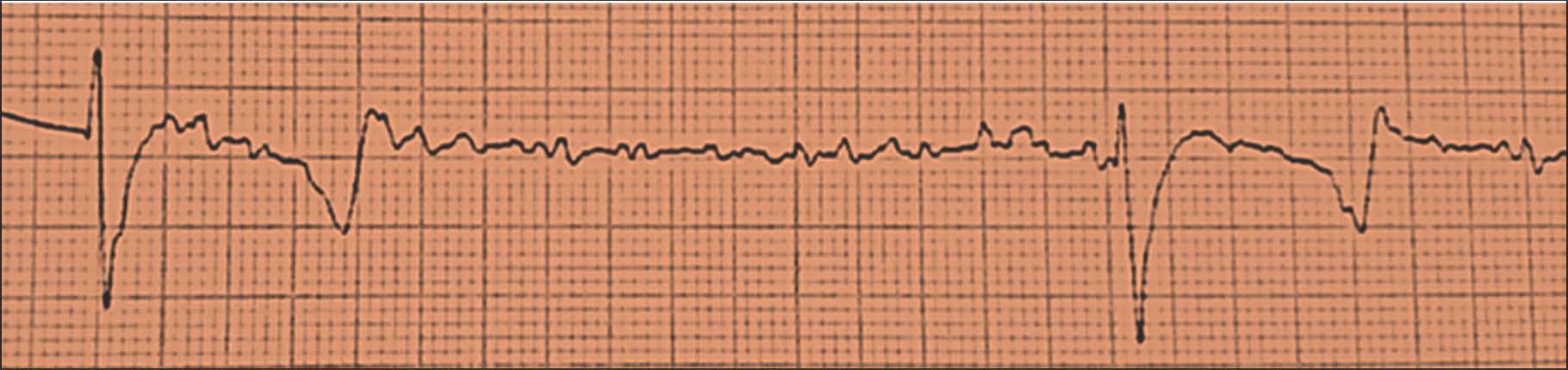
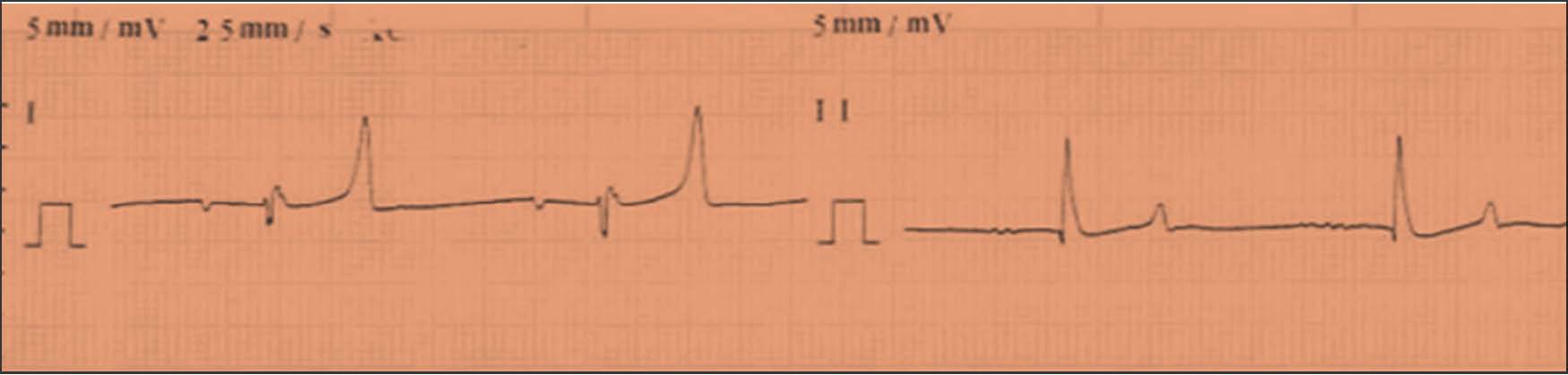
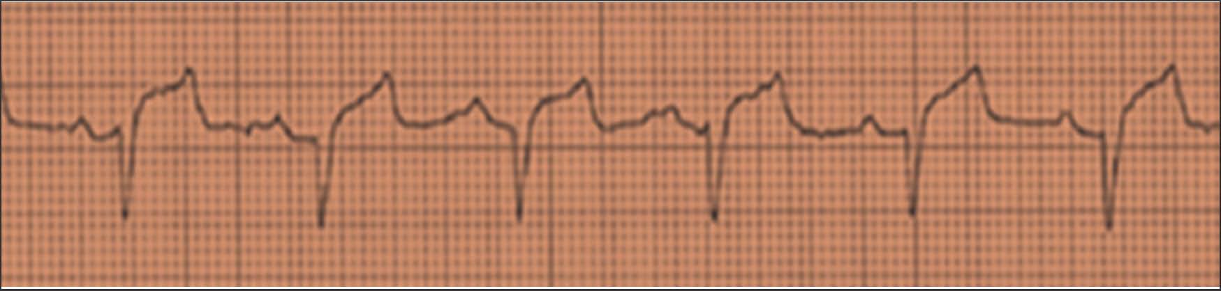
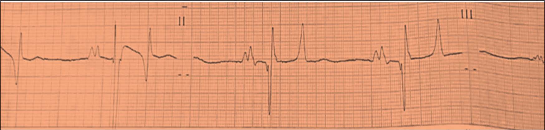
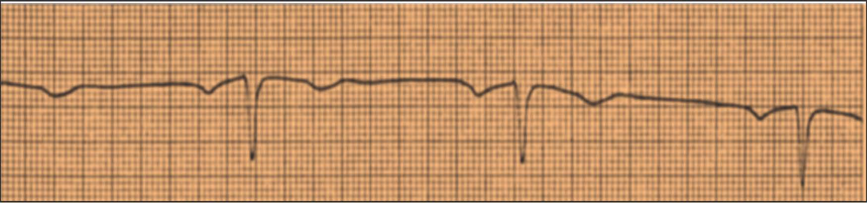
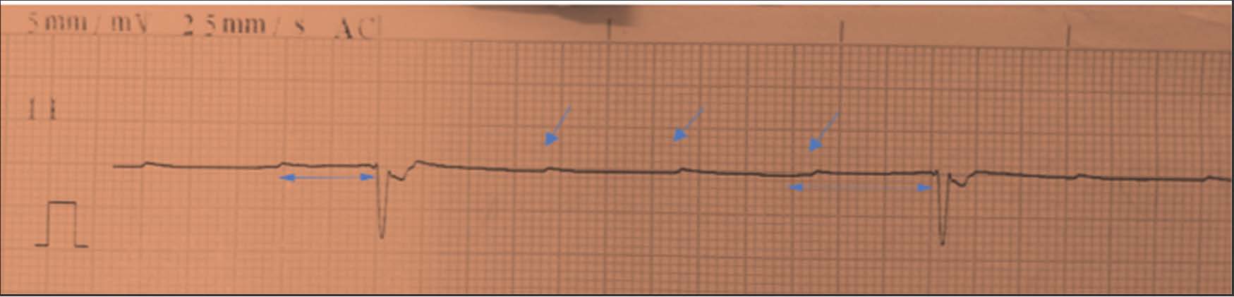
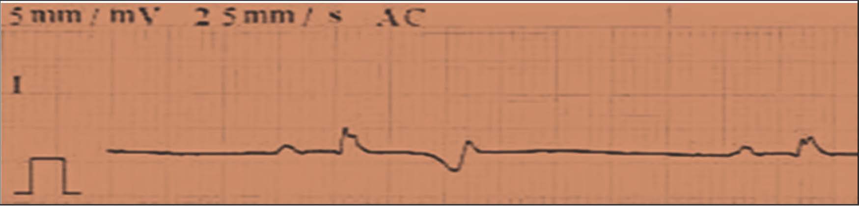
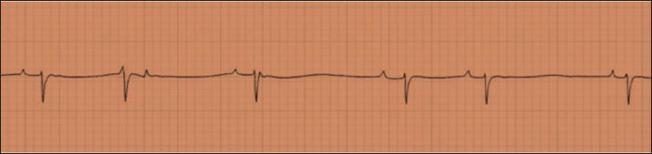
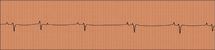
ECG changes in horses affected by atrial fibrillation typically include irregular R-R intervals, absence of P waves, and irregular, rapid, and often chaotic atrial activity. The baseline shows irregular undulations. The ventricular rate is often rapid and irregular (Figs. 1 and 2). 29 out of the 114 cases, 29 (25.44%) exhibited left axis deviation. Abnormal electrocardiographic findings, such as increased QRS amplitudes (Fig. 3) and decreased QRS amplitudes in horses, and sinus tachycardia, which appeared with elevated heart rate above the normal range (>48 bpm), were observed. Reduced QRS amplitude. Mild elevation or depression in the ST segment, reflecting pericardial inflammation, was recorded and analyzed (Fig. 4). The mean electrical axis shifts of the QRS configuration were assessed (Fig. 5) Atrioventricular blocks with variable PR intervals and narrow QRS complexes (Figs. 6 and 7).
Fig. 1. ECG of a racehorse with atrial fibrillation. Note the irregularly spaced QRS complexes; R-R intervals vary from beat to beat (blue arrow), the P wave was absent, and a smooth baseline was lacking. The coarse waves that occur in the recording are f (fibrillation) waves.
Fig. 2. ECG from a Thoroughbred horse at rest showing a transitory period of atrial fibrillation (HR=60 bpm). This horse was first identified with paroxysmal atrial fibrillation after an episode of distress during exercise; however, additional investigations revealed that it was a recurrent problem.
Fig. 3. Electrocardiogram from a horse with left axis deviation and cardiac hypertrophy in lead II.
Fig. 4. ECG from an Arabian horse, indicating pericarditis. A faster heart rate than normal (HR=72), low-voltage QRS. The electrical signals of the heart change in height from beat to beat (electrical alternans P +65°, QRS +10°, T-115°) because it is moving inside a fluid-filled sac.
Fig. 5. ECG of Arabian horses shows that P waves in some leads appeared tall and peaked rather than the usual rounded morphology seen in normal sinus rhythm. Tall or peaked P waves are often associated with RAE. Tall T waves may reflect: Electrolyte disturbances (hyperkalemia and hypocalcemia).
Fig. 6. ECG from a horse showing first-degree AV block and sinus bradycardia (HR=21 bpm), prolonged PR interval with negative T wave.
Fig. 7. Electrocardiogram from a mare (11-year-old) examined after an exercise collapse episode. Third-degree AV block with a junctional (high ventricular) escape rhythm (blue arrow). variable PR intervals (blue double-headed arrow), narrow QRS complexes (0.12 s), atrial rate of 70 beats/min, and ventricular rate of ~20 beats/min. Specific electrocardiographic patterns, including ST-segment depression and the presence of left bundle branch block (LBBB) and right bundle branch block (RBBB), were characterized in the case of sinus bradycardia (Figs. 8 and 9).
Fig. 8. ECG from a horse with a block right bundle branch leads to wide, large S waves in leads I, II, III, and aVF.
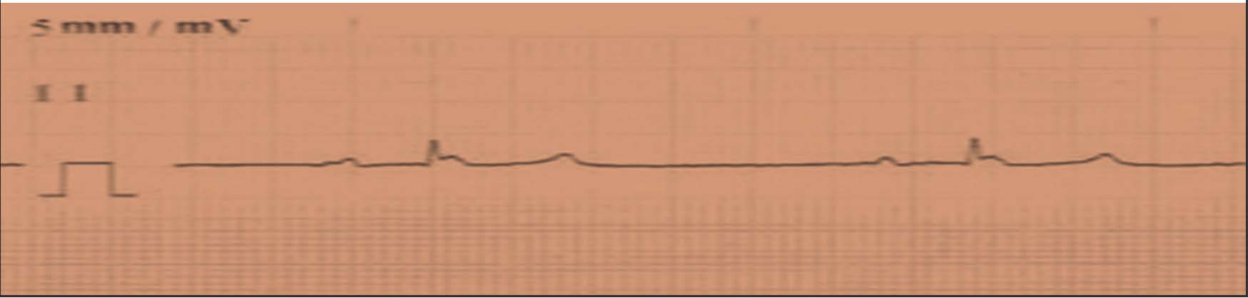
Fig. 9. ECG of the horse shows a left bundle branch block causing a wide and bizarre QRS complex, with positive QRS complexes in leads I, II, III, and aVF. Bundle Branch Block, PR interval: 1.60 s, QRS duration: 0.48 s, QT interval: 2.40 s. The heart rate was 24 beats per minute, with each P wave followed by a QRS complex and each QRS complex preceded by a P wave (Fig. 10). In another case, the time interval from the start of the QRS complex to the end of the T wave increased, indicating delayed ventricular repolarization, which is called QT prolongation (Fig. 11).
Fig. 10. ECG of the horse shows sinus bradycardia. The heart rate is less than 24 beats per minute, with each P wave followed by a QRS complex and each QRS complex preceded by a P wave.
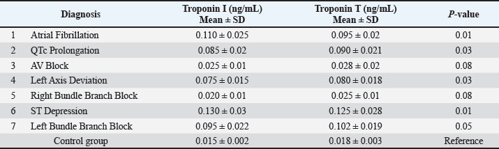
Fig. 11. ECG of mare showing QT prolongation, defined as the increase in the time interval from the start of the QRS complex to the end of the T wave, indicating delayed ventricular repolarization. ELISA results show that troponin I (cTnI) and troponin T (cTnT) levels in the control group are in normal reference ranges. The results of horses suffering from cardiac arrhythmias showed significantly elevated mean troponin levels (cTnI: 0.110 ± 0.025 ng/mL, cTnT: 0.095 ± 0.020 ng/mL) in cases of atrial fibrillation (AF), left axis deviation (cTnI: 0.075 ± 0.015 ng/mL, cTnT: 0.080 ± 0.018 ng/mL), and left bundle branch block (cTnI: 0.095 ± 0.022, cTnT:0.102 ± 0.019). Furthermore, a mild troponin rise with no significant p-values is found in the first-degree AV block and right bundle branch block (RBBB). Notably, patients with QTc prolongation (cTnI: 0.085 ± 0.02 ng/ml, cTnT: 0.090 ± 0.021 ng/ml) and ST depression had markedly elevated troponin levels (cTnI: 0.130 ± 0.03 ng/ml, cTnT: 0.125 ± 0.028 ng/ml) (Table 4). Table 4. Troponin levels in horses with different cardiac diagnoses of arrhythmias.
DiscussionThe cardiac arrhythmia frequencies observed in this study indicate an increased occurrence of certain conduction abnormalities in the equine population. ECG is considered a gold standard for AF detection. Results suggest that there is a significant frequency of cardiac arrhythmias among the monitored population, and that cardiac arrhythmias are a health concern for equine practitioners. The occurrence of AF in horses in any population will likely vary depending on the demographics and health characteristics of that population of horses (Weis et al., 2022). Our results indicate AF, which is significantly higher than the 1%–2% reported prevalence of AF in the general equine population in previous studies (Reef, 1995). The increased frequency may be attributed to breed, age, sport level, and underlying cardiac conditions. Left axis deviation with left ventricular hypertrophy (LVH) was diagnosed in 24% of cases and associated with chronic cardiac remodeling, increased workload of performance horses, and systemic hypertension (Marr and Bowen, 2010). QT prolongation was the most common abnormality in 47.36% of horses, which may represent electrolyte imbalances, myocardial stress, or predisposition to ventricular arrhythmias (Cornelisse et al., 2000; Young et al., 2008). With increased diagnostic tools and an understanding of the pathophysiology of arrhythmia in diseased equine populations, it is expected that the management and prognosis of diseased equine populations will improve with the improvements in their prognosis (Nollet et al., 2020). In addition, left bundle branch block was observed in 11.40% of animals, and right bundle branch block was observed in 22.81% of animals, both of which are uncommon in healthy horses but may indicate conduction disturbance related to myocardial disease (Begg et al., 2006). In 21.93% of cases, ST-segment depression denotes possible myocardial ischemia or shock associated with significantly elevated cardiac troponin levels, which confirms the higher probability of myocardial injury (De Clercq et al., 2008). Moreover, horses’ conditions that cause first-degree AV block were found in some cases, which is generally benign but can denote high vagal tone or underlying conduction disease (Mitchell and Schwarzwald, 2021). The results of the association of the risk factors (age, breed, and gender) with cardiac arrhythmias indicate that arrhythmias occur with age and breed, in which younger horses are at a higher risk of developing cardiac defects than older horses (Physick-Sheard and Mcgurrin, 2010) because younger horses face high-intensity training. Other researchers have recorded that older horses are more susceptible to arrhythmias due to degenerative valvular disease that develops at these ages (De Clercq et al., 2008). Arabian horses have a high risk of developing cardiac arrhythmias, which may be due to differences in heart size and the development of athletic heart syndrome (Santosuosso et al., 2023). The results of the troponin levels of horses suffering from cardiac arrhythmias showed significantly elevated mean troponin levels in cases of atrial fibrillation (Johnson et al., 2016). This may be due to atrial remodeling and stress (Begg et al., 2006). Left axis deviation suggests cardiac hypertrophy and strain, and an elevated left bundle branch block linked to delayed conduction and structural disease (Hoffman and McAuliffe, 2020). QTc prolongation indicates increased risk of arrhythmia-related damage (Van Der Vekens et al., 2015). Finally, in patients with QTc prolongation and ST depression, the presence of markedly elevated troponin levels suggests myocardial ischemia or hypoxia (Kottner et al., 2020). There is a significant risk of developing arrhythmias when myocardial stress and damage become elevated, particularly in atrial fibrillation or left ventricular hypertrophy. These correlations require further scientific investigation (Kull and Höller, 2020). Early detection and management of arrhythmias is critical in performance horses because cardiac efficiency directly influences athletic performance. Studies with larger sample sizes and long-term follow-ups are needed to gain a better understanding of the pathogenesis of equine cardiac disease and its effects on the horse’s overall performance and health (Foreman et al., 2025). Structural and functional cardiac abnormalities, along with the prevalence of electrical conduction disturbances, such as left bundle branch block and QTc prolongation, are evidence of the major effects of structural and functional cardiac abnormalities on equine arrhythmias (López and A, 2019). Additionally, the hematological and biochemical profiles between healthy and arrhythmic horses are very clear, validating these parameters as useful tools for screening horses susceptible to developing cardiac problems. Taken together, the hematological, biochemistry, and troponin data of the combination thereof help to understand the type of pathology of the arrhythmia of the horse and approach the risk factors, prevalence, and interventions to improve the hematological health of the horse’s heart. Designing effective preventative and therapeutic strategies to limit the effects of arrhythmias in the equine population will rely on further research on these interrelationships (McGowan et al., 2021). ConclusionHorses used for performance frequently have cardiac arrhythmias. Nevertheless, arrhythmias are much more common in younger individuals. Furthermore, arrhythmias are more likely to occur in Arabian horses than in Thoroughbred horses and are more common in males than in females according to breed. ECG, with the aid of biochemical evaluation, is considered the gold standard method for an early and accurate diagnostic tool. However, ongoing monitoring is required to understand their clinical importance and prognosis. AcknowledgementsThe authors are grateful to the College of Veterinary Medicine of the University of Mosul for their assistance and cooperation. The owner’s involvement is also acknowledged. FundingThis study received no specific grant. Authors' contributionsAll authors’ contributions are equal. Conflict of interestThe authors declare no conflict of interest. Data availabilityThe data that support the findings of this study are not openly available due to sensitivity reasons and are available from the corresponding author upon reasonable request. ReferencesAlbadrani, B., Khafaji, W. and Al Iraqi, O. 2024. Electrocardiographic changes associated with trypanosomiasis in horses. Egypt. J. Vet. Sci. 55(2), 345–357. Begg, L.M., Hoffmann, K.L. and Begg, A.P. 2006. Troponin I as a marker of myocardial injury in horses with cardiac disease. Equine. Vet. J. 38(1), 96–100. Buchanan, J.W. and Little, C.J. 1998. QT interval abnormalities in horses: clinical relevance and prognostic value. Vet. Card. J. 23(4), 211–217; doi:10.1053/euhj.1998.1098 Cornelisse, C.J., Schott, H.C. and Olivier, N.B. 2000. Electrocardiographic abnormalities in horses with atrial fibrillation and their clinical significance. J. Equine Med. 15(2), 89–102. De Clercq, D., Van Loon, G., Tavernier, R., Duchateau, L. and Deprez, P. 2008. Atrial fibrillation in horses: prevalence, risk factors, and clinical significance. Vet. J. 176(1), 53–59. Decloedt, A., Schwarzwald, C.C., De Clercq, D., Van Der Vekens, N., Pardon, B., Reef, V.B. and Van Loon, G. 2015. Risk factors for recurrence of atrial fibrillation in horses after cardioversion to sinus rhythm. J. Vet. Intern. Med. 29(3), 946–953; doi:10.1111/jvim.12606 Dreyer, J. and Vanderveen, S. 2019. Long-term outcomes of horses affected by atrial fibrillation: a 5-year study. J. Equi. Vet. Sci. 78, 65–71; doi:10.1016/j.jevs.2019.04.010 Dreyer, J., Barthel, C. and Van Weeren, P.R. 2022. Relationship between cardiac troponin I levels and arrhythmias in horses: a clinical study. Equine Vet. J. 54(2), 326–332; doi:10.1111/evj.13467 Foreman, J.H., Tennent-Brown, B.S., Oyama, M.A. and Sisson, D.D. 2025. Plasma cardiac troponin I concentration in normal horses and in horses with cardiac abnormalities. Animals 15, 92; doi:10.3390/ani15010092 Hoffman, A.E. and McAuliffe, S. 2020. The role of cardiac troponins in equine medicine: current understanding and future directions. Vet. J. 264, 105504; doi:10.1016/j.tvjl.2020.105504 Johnson, L.W., OʼRiley, M.D., Rees, H.H., Dorow, C.W. and Bryson, J.H. 2016. Metabolic influences on the development of arrhythmias in horses. Equine Vet. J. 48(2), 271–276; doi:10.1111/evj.12406 Kottner, J., Kahn, S.P., Schmitt, G.D., Recchia, C.R., O’Neill, J.F. and Schmitt, K.H. 2020. Prevalence and impact of electrical conduction disturbances on cardiac health in horses. J. Vet. Intern. Med. 34(6), 2179–2187; doi:10.1111/jvim.15984 Kull, S. and Höller, D. 2020. Prevalence and management of atrial fibrillation in horses: insights from a large dataset. Equine Vet. J. 52(4), 511–517; doi:10.1111/evj.13115 López, A. 2019. Utility of hematological parameters as screening tools for cardiac diseases in horses. J. Equine. Vet. Sci. 82, 102801; doi:10.1111/jevs.13207 Marr, C.M. and Bowen, I.M. 2010. Cardiology of the horse. McGowan, C.M., O'Connor, M., McDonnell, A. and Sweeney, T. 2021. The multifactorial nature of arrhythmias in equines: implications for diagnosis and management. Equine. Vet. J. 53(3), 482–492; doi:10.1111/evj.13206 Meyer, J., Huber, P. and Roberts, A. 2021. Subclinical cardiomyopathy in horses: implications for arrhythmias. Equine. Vet. J. 53(5), 715–721; doi:10.1111/evj.13399 Mitchell, K.J. and Schwarzwald, C.C. 2021. Heart rate variability analysis in horses for the diagnosis of arrhythmias. Vet. J. 268, 105590; doi:10.1016/j.tvjl.2020.105590 Nollet, H., Van Loon, G. and Deprez, P. 2020. Bundle branch blocks in horses: diagnosis, clinical significance, and management. Equine Int. Med. J. 37(2), 45–56. Physick-Sheard, P.W. and Mcgurrin, M.K.J. 2010. Ventricular arrhythmias during race recovery in Standardbred Racehorses and associations with autonomic activity. J. Vet. Intern. Med. 24(5), 1158–1166. Reef, V.B. 1995. Equine cardiology: diagnostic approaches and treatment strategies. J. Equine. Vet. Sci. 15(3), 145–160. Santosuosso, E., David, F., Massie, S., Filho, S.A., Mccrae, P., Johnson, S. and Leguillette, R. 2023. Frequency of cardiac arrhythmias in horses during straight and untethered swimming. Equine. Vet. J. 55, 738–746; doi:10.1111/evj.13895 Seeley, M.E. and Houghton, P. 2022. Clinical significance of repeated episodes of atrial fibrillation in performance horses. Vet. J. 275, 105759; doi:10.1016/j.tvjl.2022.105759 Sweeney, C.R. and Jerger, G.E. 2018. Atrial fibrillation in the horse: a review of the literature. J. Vet. Intern. Med. 32(5), 1600–1608; doi:10.1111/jvim.15391 Tulleners, E., Lichtensteiger, C.A. and Schaefer, K. 2021. Evaluation of cardiac biomarkers in equine arrhythmias: a review. J. Vet. Intern. Med. 35(6), 2175–2183; doi:10.1111/jvim.16209 Van Loon, G. 2019. Cardiac arrhythmias in horses. Vet. Clin. North Am. Equine Pract. 35(1), 85–102; doi:10.1016/j.cveq.2018.12.004 Van Der Vekens, N., Decloedt, S.A., DeClercq, V.D. and VanLoon, G. 2015. Evaluation of assays for troponin I in healthy horses and horses with cardiac disease. Vet. J. 203, 97–102; doi:10.1016/j.tvjl.2014.11.015 Weis, R., Carstensen, H., Sattler, S.M., Buhl, R. and Hesselkilde, E.M. 2022. Electrocardiographic changes in a horse with induced myocardial infarction. Animals 12, 1272; doi:10.3390/ani12101272 Williams, M.A., Moser, E. and Madden, A. 2020. Prevalence of arrhythmias in Quarter Horses diagnosed with equine neuroaxonal dystrophy. J. Equine. Vet. Sci. 90, 103073; doi:10.1016/j.jevs.2020.103073 Wright, I.M. and Lafferty, J. 2021. Atrial fibrillation in Thoroughbred racehorses: a study of 96,135 race starts. Equine Vet. J. 53(1), 75–80; doi:10.1111/evj.13355 Young, L.E., Rogers, K. and Wood, J.L.N. 2008. Heart size and function in racehorses and their association with performance. Equine Vet. J. 40(1), 32–38. | ||
| How to Cite this Article |
| Pubmed Style Aliraqi OM, Albadrani BA. Prevalence of cardiac arrhythmias in poorly performing horses. Open Vet. J.. 2026; 16(5): 2961-2969. doi:10.5455/OVJ.2026.v16.i5.39 Web Style Aliraqi OM, Albadrani BA. Prevalence of cardiac arrhythmias in poorly performing horses. https://www.openveterinaryjournal.com/?mno=305490 [Access: June 26, 2026]. doi:10.5455/OVJ.2026.v16.i5.39 AMA (American Medical Association) Style Aliraqi OM, Albadrani BA. Prevalence of cardiac arrhythmias in poorly performing horses. Open Vet. J.. 2026; 16(5): 2961-2969. doi:10.5455/OVJ.2026.v16.i5.39 Vancouver/ICMJE Style Aliraqi OM, Albadrani BA. Prevalence of cardiac arrhythmias in poorly performing horses. Open Vet. J.. (2026), [cited June 26, 2026]; 16(5): 2961-2969. doi:10.5455/OVJ.2026.v16.i5.39 Harvard Style Aliraqi, O. M. & Albadrani, . B. A. (2026) Prevalence of cardiac arrhythmias in poorly performing horses. Open Vet. J., 16 (5), 2961-2969. doi:10.5455/OVJ.2026.v16.i5.39 Turabian Style Aliraqi, Osamah Muwaffag, and Basima Abdulfatah Albadrani. 2026. Prevalence of cardiac arrhythmias in poorly performing horses. Open Veterinary Journal, 16 (5), 2961-2969. doi:10.5455/OVJ.2026.v16.i5.39 Chicago Style Aliraqi, Osamah Muwaffag, and Basima Abdulfatah Albadrani. "Prevalence of cardiac arrhythmias in poorly performing horses." Open Veterinary Journal 16 (2026), 2961-2969. doi:10.5455/OVJ.2026.v16.i5.39 MLA (The Modern Language Association) Style Aliraqi, Osamah Muwaffag, and Basima Abdulfatah Albadrani. "Prevalence of cardiac arrhythmias in poorly performing horses." Open Veterinary Journal 16.5 (2026), 2961-2969. Print. doi:10.5455/OVJ.2026.v16.i5.39 APA (American Psychological Association) Style Aliraqi, O. M. & Albadrani, . B. A. (2026) Prevalence of cardiac arrhythmias in poorly performing horses. Open Veterinary Journal, 16 (5), 2961-2969. doi:10.5455/OVJ.2026.v16.i5.39 |


