





| Research Article | ||
Open Vet. J.. 2026; 16(5): 2757-2765 Open Veterinary Journal, (2026), Vol. 16(5): 2757-2765 Research Article Epidemiological investigation of Mycobacterium bovis infection by PCR in dairy cattle milk in Pangalengan, Bandung Regency, West Java, IndonesiaRoostita L. Balia1*, Kuswandewi Mutyara2, Ardini Saptaningsih Raksanagara2, Tyagita Hartady1, Sarasati Windria1, Shafia Khairani1, Suseno Amien3, Nilla Krisna Sari3 and Muhammad Farid Rizal41Veterinary Study Program, Faculty of Medicine, Padjadjaran University, Bandung, Indonesia 2Medicine Study Program, Faculty of Medicine, Padjadjaran University, Bandung, Indonesia 3Department of Biotechnology, Graduate School, Padjadjaran University, Bandung, Indonesia 4Urban Animal Pet Care Clinic, Bandung, Indonesia *Corresponding Author: Roostita L Balia. Veterinary Study Program, Faculty of Medicine, Padjadjaran University, Bandung, Indonesia. Email: roostita [at] gmail.com Submitted: 16/12/2025 Revised: 11/04/2026 Accepted: 23/04/2026 Published: 31/05/2026 © 2025 Open Veterinary Journal
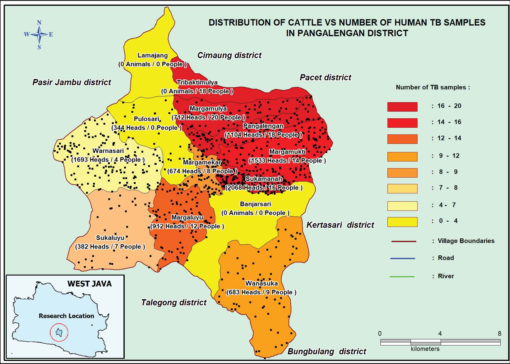
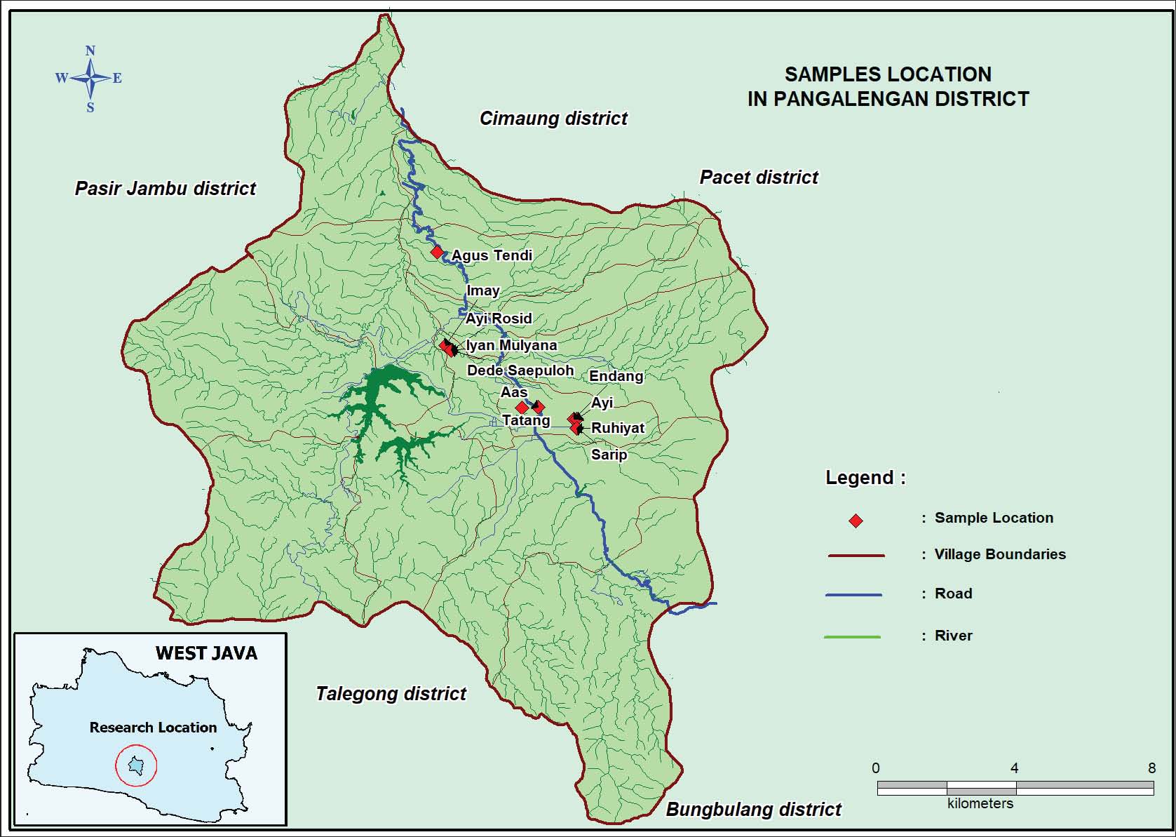
AbstractBackground: Bovine tuberculosis (bTB), caused by Mycobacterium bovis, is a zoonotic disease that primarily affects cattle and may be transmitted to humans through aerosol exposure or consumption of unpasteurized milk. Although Indonesia has no officially reported clinical cases of bTB, molecular surveillance remains essential in dairy production centers. Aim: This study aimed to detect M. bovis in dairy cattle milk using polymerase chain reaction (PCR) and to assess the potential risk of zoonotic transmission in Pangalengan, West Java. Methods: A total of 60 milk samples were collected from lactating dairy cows in three villages (Pangalengan, Sukamanah, and Margamulya). Deoxyribonucleic acid extraction was performed using a commercial kit, followed by conventional PCR targeting CSB1 and CSB2 gene regions specific for M. bovis. PCR products were analysed by agarose gel electrophoresis. Results: All 60 milk samples (100%) tested negative for M. bovis. No specific 168 bp amplicon was detected in any sample; the positive control showed clear amplification. Conclusion: No evidence of M. bovis infection was detected in dairy milk samples from Pangalengan. These findings suggest a minimal current risk of zoonotic tuberculosis transmission via milk in this region; however, continued surveillance is recommended. Keywords: Bovine tuberculosis, Dairy cattle, Milk, PCR, Zoonosis. IntroductionBovine tuberculosis (bTB) is a chronic zoonotic disease caused by Mycobacterium bovis, a member of the Mycobacterium tuberculosis complex (MTBC). The bacterium primarily infects cattle but may also affect goats, buffalo, pigs, companion animals, and wildlife. Transmission to humans occurs mainly through inhalation of infectious aerosols or consumption of contaminated and unpasteurized dairy products (Taye et al., 2021). Mycobacterium bovis is the causative agent of bTB (Blanco et al., 2025). Rod-shaped bacteria are slender, straight or curved, sometimes filamentous or branched in the shape of the letters X, Y, or V (Tkachenko et al., 2020). Mycobacterium bovis bacteria have metachromatic granules called much granules, do not form spores, are immobile, and have waxy cell walls (Palmer et al., 2022). The characteristics of M. bovis colonies are flat, smooth, white, colourless, moist, friable/brittle, and slow-growing (visible after 4 or 5 weeks) (Tkachenko et al., 2021). Tuberculosis (TB) remains a fatal disease that is primarily transmitted through airborne droplets and milk consumption, with children being the most vulnerable group. Furthermore, the live-attenuated strain M. bovis Bacillus vaccine developed by Albert Calmette and Camille Guérin (BCG) comes from M. bovis isolates and is widely used throughout the world, significantly reducing child mortality rates in areas severely affected by TB (Starshinova et al., 2025). Mycobacterium bovis is declared as the pathogenic agent causing bTB, which causes significant economic losses to the cattle industry. Worldwide, M. bovis frequently kills wildlife & cattle. Although M. bovis can spread in several ways, the respiratory & gastrointestinal systems are the primary entry routes. Up to 10% of human TB cases in some countries are believed to be caused by bTB (Mah Noor, 2025). Improved pasteurisation, standardised diagnostics using molecular methods such as polymerase chain reaction (PCR), and enhanced surveillance are key to reducing risk, along with global food safety standards and public health campaigns to protect vulnerable groups (Sabuz et al., 2025). Infected animals have a high potential to infect humans (zoonotic TB). Therefore, M. bovis poses a potential health hazard to both animals and humans (Putra et al., 2023a). In Indonesia, dairy farming plays a significant economic role, particularly in West Java Province. Pangalengan Subdistrict is one of the largest dairy production centres in the region. Close contact between farmers and cattle, along with routine milk handling, may increase the potential risk of zoonotic transmission. The most common vaccine for M. bovis is BCG, which uses live, attenuated M. bovis bacteria to prevent TB in humans. In livestock, modified live M. bovis vaccines (such as Protivity Zoetis) are used to stimulate cellular immunity in calves. Vaccinations BCG have never been carried out in that study area. Mitigating zoonotic diseases necessitates targeted measures to control and eradicate M. bovis infections in cattle populations. Detection of TB lesions at slaughterhouses should prompt epidemiological tracing of the cattle's origin to identify additional cases. Numerous antemortem and postmortem approaches exist for early bTB identification, each with distinct diagnostic strengths and limitations (Borham et al., 2022). Antemortem techniques, including PCR, facilitate definitive bTB diagnosis, complemented by emerging sequencing and bioinformatics approaches. Postmortem methods encompass macroscopic lesion inspection at abattoirs and acid-fast microscopic staining to confirm M. bovis presence (Sari et al., 2025), culture (Ferrari et al., 2024), and PCR (Elsayed and Amer, 2019), blood tests using Enzyme-Linked Immunosorbent Assay (Van Der Heijden et al., 2020), and histological preparation (Cadavid et al., 2024). Data from the Office International des Epizooties in 2025 states that in Indonesia, including Pangalengan, West Java Province, there have never been any clinically reported cases of bTB includes in 2023, in Central and Eastern Java Island, Indonesia too. The potential transmission of TB cases between dairy cattle has a score of 0.44 (moderate), as does the potential transmission to humans (0.40). The study concluded that the risk of bTB transmission between dairy cows and people is moderate (Putra et al., 2023b). In 2024, studies in Lekok Sub-District (Pasuruan Regency) and Surabaya revealed discrepancies between diagnostics: Ziehl-Neelsen staining detected no Mycobacterium spp., yet PCR identified M. tuberculosis in 60% (30/50) and M. bovis in 4% (2/50) of Lekok samples, and M. bovis in 6.8% (3) plus M. tuberculosis in 70.45% (31) of Surabaya samples (Vera-Salmoral et al., 2023; Desire et al., 2024). Although Indonesia is officially considered free of clinically reported bTB, several molecular-based studies conducted in Java have emphasized the importance of surveillance using sensitive diagnostic methods. Conventional techniques such as Ziehl–Neelsen staining and culture may have limited sensitivity, especially in milk samples with low bacterial load. Polymerase chain reaction offers higher sensitivity and specificity for detecting MTBC members in clinical and environmental samples. To date, no molecular screening study has specifically evaluated M. bovis in dairy milk from Pangalengan. Therefore, this study aimed to detect the presence of M. bovis in dairy cattle milk using PCR and to assess the potential risk of zoonotic transmission in this important dairy production area. Materials and MethodsAnimals and the study areaThe sampled cows were predominantly Friesian Holstein crossbreeds aged 3–7 years. This study was conducted between July and December 2025 in Pangalengan Subdistrict, Bandung Regency, West Java, Indonesia. Laboratory analysis was performed at the Biotechnology Laboratory, Veterinary Centre Subang. Sample collectionA total of 60 milk samples were collected from lactating dairy cows using purposive and proportional sampling from three villages: Pangalengan, Sukamanah, and Margamulya (20 samples per village) (Fig. 1). The selected villages represent areas with high dairy cattle populations and reported human TB cases (Fig. 2).
Fig. 1. Distribution of Cattle VersuS Number of Human TB Patients, cattle density is depicted in dots, each dot representing 10 head of cattle.
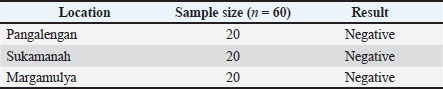
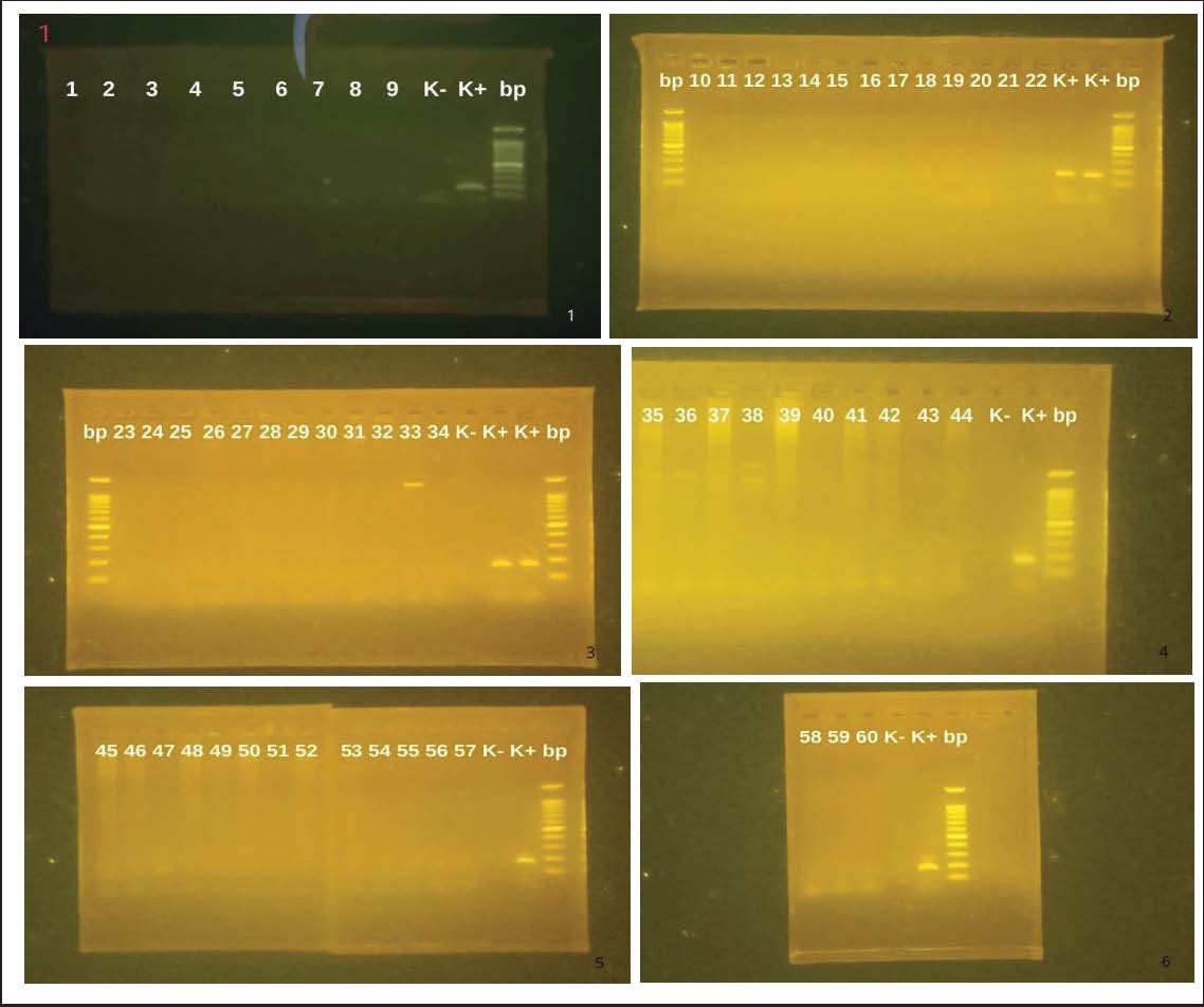
Fig. 2. The location of the farm of each sample was taken. Milking was conducted manually in most farms, with some using semi-mechanical systems. All farms applied basic biosecurity practices Nor et al. (2025). Milk samples were collected during morning milking, labelled appropriately, and transported under cold chain conditions to the laboratory within 48 hours. Deoxyribonucleic acid (DNA) extraction and PCR amplificationTotal DNA was extracted from milk samples using the QIAamp® DNA Mini Kit (Germany) following the manufacturer's instructions. Conventional PCR was performed targeting CSB1 (5'-TTCCGAATCCCTTGTGA-3') and CSB2 (5'-GGAGAGCGCCGTTGTA-3') primers specific for M. bovis. Each 25 µl reaction mixture consisted of 12.5 µl master mix, 1 µl of each primer, 6.5 µl nuclease-free water, and 3 µl DNA template. PCR amplification conditions included initial denaturation at 95°C for 1 minute, 35 cycles of denaturation at 95°C for 15 seconds, annealing at 50°C for 15 seconds, extension at 72°C for 30 seconds, and final extension at 72°C for 5 minutes. PCR products were separated by 1.5% agarose gel electrophoresis and visualised under UV illumination. The expected amplicon size was 168 bp. Ethical approvalThis study received ethical approval from the Research Ethics Committee of Universitas Padjadjaran (No. 119/UN6.KEP/EC/2025) 05 February 2025. Milk sampling was non-invasive and conducted in accordance with animal welfare standards. ResultsPCR analysis of all 60 milk samples showed negative results for M. bovis. No 168 bp amplification band was observed in any sample. The positive control demonstrated a clear band at the expected size, confirming proper PCR performance (Fig. 3). The distribution of results by village is presented in Table 1. Table 1. PCR test results for milk samples from dairy farms in each village in the Pangalengan area of West Java.
Fig. 3. (a–f). Detection of M. bovis in milk samples in the Pangalengan region of West Java. DNA marker (bp). Representative electrophoregram of the target gene. (K+) M. bovis positive control, (K–) M. bovis negative control, as well as negative sample results (1–60). DiscussionThis study did not detect M. bovis in any of the dairy milk samples collected from Pangalengan. These findings are consistent with previous studies conducted in Central and Eastern Java, which also reported negative PCR results in milk samples. PCR has been demonstrated to be more sensitive than conventional microscopic methods for detecting MTBC members, particularly in samples with low bacterial load. While culture remains the gold standard for confirmation, it requires viable organisms and prolonged incubation time. PCR provides rapid and specific detection of bacterial DNA, making it suitable for surveillance purposes. The absence of M. bovis detection in this study may reflect effective farm management practices and biosecurity implementation. However, negative PCR results do not entirely rule out infection, especially if bacterial DNA concentration is below the detection threshold. Given that Pangalengan is a major dairy production area with reported human TB cases, continued surveillance using molecular methods remains essential to ensure food safety and public health protection. Tuberculosis is transmitted from humans to cattle (Szacawa et al., 2025b). Among cows, the highest incidence was found in cows kept together, as in dairy cows (Weldegebriel et al., 2025). Beef cattle can also have high incidence rates when animals are crowded together due to limited feed and water supplies (Phiri et al., 2025). This occurs mainly because dairy cows that have never undergone tuberculin testing are brought into a farm (Ngo et al., 2025). Infected milk is a source of disease transmission for calves and humans Desu et al. (2025) and Umer et al. (2025). About 5% of cows suffering from infection show signs of TB mastitis (tuberculous mastitis) (Tomanić et al., 2025). Reach the mucous membranes through the respiratory tract, the digestive tract, or by contact (Mtetwa et al., 2022). TB may show complete recovery (Ramanujam et al., 2024). The number of M. bovis germs that cause infection, often known as the infecting dose, is important in the development of disease (Matthews et al., 2025; Parchinski et al., 2025). Once infection has taken hold, the disease can spread in various ways, including direct spread from TB lesions, natural channels in the body, lymphatic system, bloodstream, and serous surfaces (Khairullah et al., 2024), either through coughing or aspiration (Silva-Angulo, 2021). Sputum may be coughed up and then swallowed again, causing it to spread to the gastrointestinal tract (Khairullah et al., 2024). The germ enters the body through the airways and causes primary lesions in the lungs in the form of caseating tubercles (Ullah et al., 2025). Mycobacterium bovis can cause TB in humans, which can affect the lungs, lymph nodes, and other parts of the body (Commandeur et al., 2025). Humans are most often infected with M. bovis through contaminated food or drink, unpasteurized dairy products, direct contact with wounds, or by inhaling bacteria in the air exhaled by animals infected with M. bovis (Devi et al., 2021). Mycobacterium bovis infection spreads to livestock mainly through aerosols from coughing or sneezing of animals with active TB or from infected dust particles (Lu et al., 2025). The spread of M. bovis through the consumption of infectious materials has also been reported, namely through drinking infected milk (Kayukova et al., 2025) or eating grass or through contaminated feed (Szacawa et al. 2025a). In cattle, there are no clinical signs in the early stages of infection (Bueno et al., 2025). Nests of bTB in cattle are found in the lungs and pleura, liver, spleen, peritoneum, lymph glands, and sometimes in the skin and bones (Kriek et al., 2019). TB of the udder is more common in cows than in other species (Willgert et al., 2025). Diagnosis of bTB is based on clinical symptoms, isolation and identification of bacteria (culture and acid-fast staining), tuberculin test, blood tests [Gamma Interferon assay (IFNɣ assay) and lymphocyte proliferation assay], and molecular testing, PCR Elbarbary et al. (2024). Considerations regarding the slaughter of animals and the utilisation of meat bTB in cattle including in areas where bTB is being eradicated, at the end of an eradication program, and at the start of eradication in infected areas (Dibaba and Kriek, 2019). Microscopic examination of M. bovis is performed by staining acid-fast bacilli using the heating method (Ziehl-Neelsen) or the non-heating method (Gabbett's or Kinyoun's). A minimum of 100 fields of view must be examined before declaring the test result negative (Sari et al., 2025). AFB examination results recommended by RNTCP: (3+) if more than 10 AFBs are found per field of view; (2+) if 1-10 AFBs are found per field of view; and (1+) if 10-99 AFBs are found per 100 fields of view (Mitra et al., 2024). The culture techniques used in veterinary laboratories differ from those used in medical laboratories (Chatterjee et al., 2025 Franklin-Guild et al., 2025). Mycobacterium bovis strains grow poorly or not at all on Lowenstein Jensen medium (egg-based medium) containing glycerol, which is commonly used as a medium for the growth of M. tuberculosis, but the growth of M. bovis can be stimulated with sodium pyruvate as a substitute for glycerol (Pal et al., 2025). Mycobacterium bovis cultures can also use Stonebrink medium (pyruvate-based medium) (Turgenbayev et al., 2025). PCR was first discovered by Kary B. Mullis in 1985 (Strinden, 2022). At the end of each cycle, DNA fragments are duplicated using the appropriate primers. In this study, it took 1 minute to ensure that the targeted DNA molecules to be replicated had completely denatured into single-stranded DNA. For subsequent denaturation, the time required is only 15 seconds. Incomplete denaturation causes the DNA to renature (form double-stranded DNA again) quickly, resulting in PCR failure (Huang et al., 2025; Hussain et al., 2025). In this study, the temperature was used for the primary annealing. The longer the primer, the higher the temperature during the primary extension phase. The speed of nucleotide synthesis by the enzyme in this study was 72°C, estimated to be between 35 and 100 nucleotides per second, depending on the buffer, pH, salt concentration, and target DNA molecule (Assal, 2021; Kaur, 2021). Thus, for PCR products 2,000 base pairs long, 1 minute is more than enough time for this primer extension stage (Sathyanarayana and Wainman, 2024). No evidence of M. bovis infection was detected in dairy cattle milk samples from Pangalengan using PCR. These findings suggest that the current risk of zoonotic transmission through milk in this region is minimal. Continued monitoring and surveillance are recommended to maintain bTB-free status. AcknowledgmentsThis research was supported by an Academic Leadership Grant (ALG) research assignment to Professor Drh. Roostita L Balia, M.App.Sc., PhD, to increase the number of publications and citations at Padjadjaran University Bandung. The researcher is expressing gratitude to BVET Subang and KPBS Pangalengan. Drh. Asep Khoirudin, M.Pt. as the Head of KPBS Pangalengan, for his support and assistance during this study. This research is funded by an ALG from Universitas Padjadjaran under the Department of Research and Community Services (DRPMH). We express our sincere gratitude to the Dean of the Faculty of Medicine, Prof. Dr. Yudi Mulyana Hidayat, Dr. SpOG(K), the Director of DRPMH, Prof. Dr. Nur Atik, M.Kes., PhD, for the grants. Conflict of interestThe authors have not declared any conflict of interest. FundingThis study received financial support from an Academic Leadership Grant (ALG) research assignment from Universitas Padjadjaran with contract number: 5317/UN6.C/PT.02/2025. Authors’ contributionRoostita L. Balia and Suseno Amien were responsible for the overall design and execution of the research. Nilla Krisna Sari and Ardini Saptaningsih Raksanagara were responsible for data collection and analysis. Writing the manuscript and ensuring all necessary revisions were made by all authors. Sarasati Windria and Shafia Khairani assisted in conceptualizing the study and contributed to the experimental design. Additionally, Tyagita Hartadi and Kuswandewi Mutyara helped with data analysis and interpretation. Roostita L. Balia and Suseno Amien are responsible as the supervisor. Muhammad Farid Rizal contributed to the literature review, interpretation of results, and final editing of the manuscript. All authors have read and approved the final version of the manuscript before publication in the present journal. Data availabilityAll data are available in the manuscript. ReferencesAssal, N. 2021. High throughput discovery of novel diagnostic antigens for Mycobacterium bovis using a whole genome approach. Thesis, University of Ottawa. Blanco, F.C., Marini, M.R., Klepp, L.I., Vázquez, C.L., García, E.A., Bigi, M.M., Canal, A. and Bigi, F. 2025. Long-term evaluation in BALBc mice of a triple mutant of Mycobacterium bovis and the Bacillus Calmette-Guérin as potential vaccines against bovine tuberculosis. Vet. Microbiol. 302, 110371; doi:10.1016/j.vetmic.2025.110371 Borham, M., Oreiby, A., El-Gedawy, A., Hegazy, Y., Khalifa, H.O., Al-Gaabary, M. and Matsumoto, T. 2022. Review on Bovine Tuberculosis: an Emerging Disease Associated with Multidrug-Resistant Mycobacterium Species. Pathogens 11(7), 715; doi:10.3390/pathogens11070715 Bueno, N.M.M., Reck, C., Nogueira, L.L., Pinto, A.R. and Menin, A. 2025. Development and evaluation of an ELISA based on recombinant Mb1454 antigen for the detection of specific anti-Mycobacterium bovis antibodies in cattle. Vet. Immunol. Immunopathology. 290, 111025; doi:10.1016/j.vetimm.2025.111025 Cadavid, P.P., Balvin, D.I., Julio, R.V., Ramos, E.M., Gutierrez, J.B., Buitrago, J.D.R. and Garcia, R.R. 2024. Bovine tuberculosis testing in Colombia: comparative histopathological, microbiological, and molecular biology findings. J. Buffalo Sci. 13, 53–63; doi:10.6000/1927-520X.2024.13.06 Chatterjee, B., Banerjee, A., Chatterjee, D., Chakraborty, O., Dey, S. and Mitra, A.K. 2025. Biofilm Threats in Livestock: a Growing Concern. In Biofilm Associated Livestock Diseases and their Management. Eds., Lahiri, D., Nag, M., Bhattacharya, D. and Ray, R.R. Singapore: Gateway East, pp: 367–92. https://doi.org/10.1007/978-981-96-1885-9_17 Commandeur, S., Van Der Most, M., Koomen, J., Van Keulen, L., Dinkla, A., Luinenburg, X., Escher, M., Jacobs, P., Keur, I., Grinwis, G.C.M., Weerts, E., Broens, E.M., Anthony, R., Agterveld, M.K.V., Rebel, K., Huisman, E., Heijne, M. and Koets, A. 2025. Mycobacterium bovis infected domestic cats in an officially bovine tuberculosis free country resulting in human infection. One. Health. 20, 101048; doi:10.1016/j.onehlt.2025.101048 Desire, I.A., Luqman, M., Puspitasari, Y., Tyasningsih, W., Wardhana, D.K., Meles, D.K., Dhamayanti, Y., Permatasari, D.A., Witaningrum, A.M., Perwitasari, A.D.S., Raharjo, H.M., Ayuti, S.R., Kurniawan, S.C., Kamaruzaman, I.N.A. and Silaen, O.S.M. 2024. First detection of bovine tuberculosis by Ziehl–Neelsen staining and polymerase chain reaction at dairy farms in the Lekok Sub-District, Pasuruan Regency, and Surabaya region, Indonesia. Vet. World. 17, 577–584; doi:10.14202/vetworld.2024.577-584 Desu, M., Fasil, N. and Abebe, R. 2025. Apparent prevalence, lesion distribution and risk factors of bovine tuberculosis in cattle slaughtered at the Shashemene and Arsi Negelle municipal abattoirs, Ethiopia. One. Health. 21, 101200; doi:10.1016/j.onehlt.2025.101200 Devi, K.R., Lee, L.J., Yan, L.T., Syafinaz, A., N., Rosnah, I. and Chin, V.K. 2021. Occupational exposure and challenges in tackling M. bovis at human–animal interfac A narrative review. Int. Arch. Occupational. Environ. Health. 94(6), 1147–1171; doi:10.1007/s00420-021-01677-z Dibaba, A.B. and Kriek, N.P. Thoen, C.O. 2019. The Control of Bovine Tuberculosis in Africa. In Tuberculosis in Animals: an African Perspective. Switzerland: Springer International Publishing, pp: 237–70; doi: 10.1007/978-3-030-18690-6_10 Elbarbary, N.K., Darwish, W.S., Bekhit, M.M., Salem, M.M., Abdelhaseib, M., Madkour, B.S., El-Malah, S.S., El-Hawary, S.F., Salama, M. and Dandrawy, M.K. 2024. Unseen Threats to Meat Safety: exposing the Hidden Epidemic to Bovine Tuberculosis in Slaughterhouses. Food Sci. Anim. Resour. 45(5), 1309–1325; doi:10.5851/kosfa.2024.e95 Elsayed, M.S.A.E. and Amer, A. 2019. The rapid detection and differentiation of Mycobacterium tuberculosis complex members from cattle and water buffaloes in the delta area of Egypt, using a combination of real-time and conventional PCR. Mol. Biol. Rep. 46(4), 3909–3919; doi:10.1007/s11033-019-04834-3 Ferrari, S., Zanoni, M., Mangeli, A., Pigoli, C., D'Incau, M., Alborali, G.L., Pacciarini, M.L. and Boniotti, M.B. 2024. Bacteriological culture and direct PCR for detecting the Mycobacterium tuberculosis complex in the Italian eradication campaign: a decade of experience at the National Reference Laboratory. J. Appl. Microbiol. 135(3), 64; doi:10.1093/jambio/lxae064 Franklin-Guild, R., Pinn-Woodcock, T. and Guarino, C. 2025. Enhanced guidance for veterinary microbiological culture specimen handling will improve quality of results: a survey of best practices. J. Am. Vet. Med. Assoc. 263(S1), S17–S23; doi:10.2460/javma.24.11.0754 Huang, F., Shi, M., Chen, L., Li, X., Wang, Z., Jiang, D., Chen, J., Zhang, H. and He, Z.G. 2025. ACA kills Mycobacterium tuberculosis and M. bovis by targeting cell wall core assembling protein CpsA. Commun. Biol. 8(1), 1706; doi:10.1038/s42003-025-09107-3 Hussain, S.M., Sharif, A., Bashir, F., Ali, S., Javid, A., Hussain, A.I., Ghafoor, A., Alshehri, M.A., Naeem, A., Naeem, E. and Amjad, M. 2025. Polymerase chain reaction: a toolbox for molecular discovery. Mol. Biotechnol. 68(2), 409–421;doi:10.1007/s12033-025-01390-z Kaur, H. 2021. Veterinary microbiology (Minor Subject: Animal Biotechnology). Kayukova, L., Bismilda, V., Turgenbayev, K., Uzakova, A., Baitursynova, G., Jussipbekov, U., Mukanova, M., Chingissova, L., Dyussembayeva, G., Borsynbayeva, A., Yerlanuly, A. and Auyezov, A. 2025. Β-Aminopropioamidoximes derivatives as potential antitubercular agents against anthropozoonotic infections caused by Mycobacterium tuberculosis and Mycobacterium bovis. Vet. World 18(3), 731–745; doi:10.14202/vetworld.2025.731-745 Khairullah, A., Moses, I., Kusala, M., Tyasningsih, W., Ayuti, S., Rantam, F., Fauziah, I., Silaen, O., Puspitasari, Y., Aryaloka, S., Raharjo, H., Hasib, A., Yanestria, S. and Nurhidayah, N. 2024. Unveiling insights into bovine tuberculosis: a comprehensive review. Open. Vet. J. 14(6), 1330; doi:10.5455/OVJ.2024.v14.i6.2 Kriek, N.P.J., Areda, D.B. and Dibaba, A.B. 2019. The Diagnosis of Bovine Tuberculosis. In Tuberculosis in Animals: an African Perspective. Springer International Publishing, pp: 171–235; doi: 10.1007/978-3-030-18690-6_9 Lu, H., Xie, Y., Chen, L., Song, Y., Zhang, L., Li, R., Nie, X., Liu, Y., Zhu, G., Ding, X. and Wang, L. 2025. Microbial Aerosols in Livestock Farming Environment: a Threat That Cannot Be Ignored. Vet. Sci. 12(12), 1147; doi:10.3390/vetsci12121147 Mah Noor, I.T. 2025. Inquisition of Mycobacterium bovis in Cattles and its Critiques (Version v2). Zenodo; doi:10.5281/ZENODO.17072033 Matthews, M.C., Toorians, M.E.M., Davies, T.J., Stewart, R.D., Goosen, W.J. and Miller, M.A. 2025. A microbial safari: finding evidence of Mycobacterium bovis DNA in soil from the Kruger National Park, South Africa. Microbiol. Spectr. 14(1), 1658; doi:10.1128/spectrum.01658-25 Mitra, S., Saikia, P.J., Dutta, A., Das, R., Das, G., Baishya, T., Das, C., Sarma, T., Das, S., Pathak, L. and Das, B. 2024. A novel comprehensive Tuberculosis (TB) control programme methodology based on the nexus of participatory action research inspired public health and precision treatment approach. Public Global Health; doi:10.1101/2024.01.02.23300347 Mtetwa, H.N., Amoah, I.D., Kumari, S., Bux, F. and Reddy, P. 2022. The source and fate of Mycobacterium tuberculosis complex in wastewater and possible routes of transmission. BMC. Public. Health. 22(1), 145; doi:10.1186/s12889-022-12527-z Ngo, V.M., Kallu, V., Walkowiak, B., Barrett, D., Clarke, P., Roantree, M. and McCarren, A. 2025. Predicting herd-level bovine tuberculosis breakdowns in Ireland based on a Graph Database and AI Techniques. Comp. Sci. Mathematics; doi: 10.20944/preprints202512.0096.v1 Nor, A.K.C.M., Citartan, M., Pin, L.L., Manickam, R., Zainuddin, Z.F. and Tang, T.H. 2025. Development of thermostabilized “ready-to-use” multiplex PCR assay for the rapid detection and distinction between Mycobacterium tuberculosis complex members and non-tuberculous mycobacteria. Diagnostic. Microbiol. Infect. Dis. 113(3), 117010; doi:10.1016/j.diagmicrobio.2025.117010 Pal, M., Regassa, M., Gizachewu, M., Shifara, K., Zende, R., Nair, A. and Sgroi, G. 2025. Battling bovine tuberculosis: modern approaches to diagnosis and control. Am. J. Infect. Dis. Microbiol. 13(1), 5–14; doi:10.12691/ajidm-13-1-2 Palmer, M.V., Kanipe, C. and Boggiatto, P.M. 2022. The Bovine Tuberculoid Granuloma. Pathogens 11(1), 61; doi:10.3390/pathogens11010061 Parchinski, K., Pascopella, L. and Barry, P. 2025. Central nervous system tuberculosis: characteristics, risks, and outcomes in California adults, 2010–2022. J. Clin. Tuberculosis. Other. Mycobacterial. Dis. 42, 100574; doi:10.1016/j.jctube.2025.100574 Phiri, A., Likulunga, E., Chauwa, A., Zulu, M., Kankhuni, B., Monde, N. and Malama, S. 2025. Knowledge and awareness of bovine tuberculosis associated with raw milk and under-cooked meat contamination among cattle farmers in selected parts of zambia. PLos Neglected. Trop. Dis. 19(4), 12870; doi:10.1371/journal.pntd.0012870 Putra, A.E., Basri, C. and Sudarnika, E. 2023b. Potential of Bovine Tuberculosis Transmission in Dairy Cattle and Humans in the Central and Eastern Regions of Java Island, Indonesia. Acta VETERINARIA Indonesiana 11(2), 139–147; doi:10.29244/avi.11.2.139-147 Putra, A.E., Basri, C., Sudarnika, E. and Lestari, S. 2023a. Milk Screening Test for Mycobacterium bovis from Dairy Farms in Central and Eastern Java Island, Indonesia. Jurnal. Sain. Veteriner. 41(3), 306; doi:10.22146/jsv.82787 Ramanujam, H., Refaya, A.K., Thiruvengadam, K., Pazhanivel, N., Kandasamy, D., Shanmugavel, A., Radhakrishnan, A., Radhika, G., Ravi, R., Ravi, N., Palanisamy, M., Shanmugam, S., Stuber, T.P., Kapur, V. and Palaniyandi, K. 2024. Recovery of Mycobacterium tuberculosis Complex Isolates Including Pre–Extensively Drug-Resistant Strains From Cattle at a Slaughterhouse in Chennai, India. Open. Forum. Infect. Dis. 12(1), 733; doi:10.1093/ofid/ofae733 Sabuz, S.H., Jahan, I., Debnath, B.K., Islam, M.M. and Islam, M.S. 2025. Zoonotic Tuberculosis and Dairy Products: comprehensive Meta‐Analysis of Prevalence and Public Health Implications. Vet. Med. Sci. 11(5), e70556; doi:10.1002/vms3.70556 Sari, N.K., Amien, S. and Balia, R.L. 2025. Rapid Identification Prevalence Zoonotic Tuberculosis (Mycobacterium bovis) in West Bandung and Pangalengan. Sathyanarayana, S.H. and Wainman, L.M. 2024. Laboratory approaches in molecular pathology: the polymerase chain reaction. In Diagnostic molecular pathology. Elsevier, pp: 13–25; doi: 10.1016/B978-0-12-822824-1.00041-9 Silva-Angulo, F. 2021. From bcg vaccination routes to lung and gut microbiota: avenues to tackle Mycobacterium tuberculosis infection. Starshinova, A., Kudryavtsev, I., Rubinstein, A., Dovgalyuk, I., Kulpina, A., Churilov, L.P. and Kudlay, D. 2025. BCG vaccination: historical role, modern applications, and future perspectives in tuberculosis and beyond. Front. Pediatrics 13, 1603732; doi:10.3389/fped.2025.1603732 Strinden, M.J. 2022. Development and application of nanopore sequencing based methods for rapid, culture-free diagnosis of tuberculosis. Szacawa, E., Kozieł, N., Brzezińska, S., Augustynowicz-Kopeć, E., Weiner, M., Szulowski, K. and Krajewska-Wędzina, M. 2025a. Laboratory Diagnosis of Animal Tuberculosis in Tracing Interspecies Transmission of Mycobacterium bovis. Pathogens 14(5), 459; doi:10.3390/pathogens14050459 Szacawa, E., Radulski, Ł., Weiner, M., Szulowski, K. and Krajewska-Wędzina, M. 2025b. Mycobacterium tuberculosis complex infections in animals: a comprehensive review of species distribution and laboratory diagnostic methods. Pathogens 14(10), 1004; doi:10.3390/pathogens14101004 Taye, H., Alemu, K., Mihret, A., Wood, J.L.N., Shkedy, Z., Berg, S. and Aseffa, A. 2021. Global prevalence of Mycobacterium bovis infections among human tuberculosis cases: systematic review and meta‐analysis. Zoonoses. Public. Health. 68(7), 704–718; doi:10.1111/zph.12868 Tkachenko, O., Bilan, M., Hlebeniuk, V., Kozak, N., Nedosekov, V. and Galatiuk, O. 2020. Dissociation of Mycobacterium bovis: morphology, Biological Properties and Lipids. Adv. Anim. Vet. Sci. 8(3), 317–326; doi:10.17582/journal.aavs/2020/8.3.317.326 Tkachenko, O., Kozak, N., Bilan, M., Hlebeniuk, V., Alekseeva, N., Kovaleva, L., Nedosekov, V. and Galatiuk, O. 2021. The Effect of Long-Term Storage on Mycobacterium bovis. Polish J. Microbiol. 70(3), 327–337; doi:10.33073/pjm-2021-031 Tomanić, D., Kladar, N. and Kovačević, Z. 2025. Antibiotic Residues in Milk as a Consequence of Mastitis Treatment: balancing Animal Welfare and One Health Risks. Vet. Sci. 12(12), 1159; doi:10.3390/vetsci12121159 Turgenbayev, K., Borsynbayeva, A., Ozatbekuly, A., Dyusenov, S., Tlepov, A. and Turgenbayev, R. 2025. Development and evaluation of a formaldehyde-stabilised tuberculin as a safe and potent alternative to phenol-based purified protein derivative for the diagnosis of animal tuberculosis. Vet. World 3268, 3268–3287; doi:10.14202/vetworld.2025.3268-3287 Ullah, A., Hafeez, F., Taj, R., Gul, S., Khan, I., Faheem, B., Ahmad, M., Ullah, R., Ahmad, A., Hanif, M., Khan, A.U., Khan, M.O., Basit, A., Khan, M.I., Khan, S. and Islam, M. 2025. Comparative Study on Detection of Mycobacterium bovis Infection in Bovine Tuberculous Lesions. Sarhad J. Agriculture 41(1), 110–125; doi:10.17582/journal.sja/2025/41.1.110.125 Umer, A.A., Moti, T.B. and Garoma, A. 2025. Prevalence and factors associated of bovine tuberculosis in north shawa oromia and Addis Ababa dairy farms, Ethiopia. J. Tuberculosis 8(1), 1039. Van Der Heijden, E.M.D.L., Cooper, D.V., Rutten, V.P.M.G. and Michel, A.L. 2020. Mycobacterium bovis prevalence affects the performance of a commercial serological assay for bovine tuberculosis in African buffaloes. Comparative Immunol. Microbiol. Infect. Dis. 70, 1–101369; doi:10.1016/j.cimid.2019.101369 Vera-Salmoral, E., Gómez-Laguna, J., Galán-Relaño, A., Ruedas-Torres, I., Carrasco, L., Luque, I., Huerta, B. and Sánchez-Carvajal, J.M. 2023. Optimization of real-time PCR protocols from lymph node bovine tissue for direct detection of Mycobacterium tuberculosis complex. Microbiol. Spectr. 11(5), e00348–e00-23; doi:10.1128/spectrum.00348-23 Weldegebriel, M., Hailu, K., Seid, K., Negash, L., Weldu, Y., Fantay, H., Mekonnen, B. and Abebe, N. 2025. Prevalence of Mycobacterium Bovis infection and associated risk factors among dairy farm cattle in Mekelle and Wukro towns, Northern Ethiopia. BMC. Microbiol. 25(1), 539; doi:10.1186/s12866-025-04267-y Willgert, K., Cliff, M., Meinke, S., Messina, D., Broom, D.M., Wood, J. and Conlan, A.J.K. 2025. Burden of Bovine Tuberculosis on Animal Health, Welfare and Production: a Systematic Review. Transboundary Emerg. Dis. 1(1), 6541298; doi:10.1155/tbed/6541298 | ||
| How to Cite this Article |
| Pubmed Style Balia RL, Mutyara K, Raksanagara AS, Hartady T, Windria S, Khairani S, Amien S, Sari NK, Rizal MF. Epidemiological investigation of Mycobacterium bovis infection by PCR in dairy cattle milk in Pangalengan, Bandung Regency, West Java, Indonesia. Open Vet. J.. 2026; 16(5): 2757-2765. doi:10.5455/OVJ.2026.v16.i5.18 Web Style Balia RL, Mutyara K, Raksanagara AS, Hartady T, Windria S, Khairani S, Amien S, Sari NK, Rizal MF. Epidemiological investigation of Mycobacterium bovis infection by PCR in dairy cattle milk in Pangalengan, Bandung Regency, West Java, Indonesia. https://www.openveterinaryjournal.com/?mno=303395 [Access: June 26, 2026]. doi:10.5455/OVJ.2026.v16.i5.18 AMA (American Medical Association) Style Balia RL, Mutyara K, Raksanagara AS, Hartady T, Windria S, Khairani S, Amien S, Sari NK, Rizal MF. Epidemiological investigation of Mycobacterium bovis infection by PCR in dairy cattle milk in Pangalengan, Bandung Regency, West Java, Indonesia. Open Vet. J.. 2026; 16(5): 2757-2765. doi:10.5455/OVJ.2026.v16.i5.18 Vancouver/ICMJE Style Balia RL, Mutyara K, Raksanagara AS, Hartady T, Windria S, Khairani S, Amien S, Sari NK, Rizal MF. Epidemiological investigation of Mycobacterium bovis infection by PCR in dairy cattle milk in Pangalengan, Bandung Regency, West Java, Indonesia. Open Vet. J.. (2026), [cited June 26, 2026]; 16(5): 2757-2765. doi:10.5455/OVJ.2026.v16.i5.18 Harvard Style Balia, R. L., Mutyara, . K., Raksanagara, . A. S., Hartady, . T., Windria, . S., Khairani, . S., Amien, . S., Sari, . N. K. & Rizal, . M. F. (2026) Epidemiological investigation of Mycobacterium bovis infection by PCR in dairy cattle milk in Pangalengan, Bandung Regency, West Java, Indonesia. Open Vet. J., 16 (5), 2757-2765. doi:10.5455/OVJ.2026.v16.i5.18 Turabian Style Balia, Roostita L., Kuswandewi Mutyara, Ardini Saptaningsih Raksanagara, Tyagita Hartady, Sarasati Windria, Shafia Khairani, Suseno Amien, Nilla Krisna Sari, and Muhammad Farid Rizal. 2026. Epidemiological investigation of Mycobacterium bovis infection by PCR in dairy cattle milk in Pangalengan, Bandung Regency, West Java, Indonesia. Open Veterinary Journal, 16 (5), 2757-2765. doi:10.5455/OVJ.2026.v16.i5.18 Chicago Style Balia, Roostita L., Kuswandewi Mutyara, Ardini Saptaningsih Raksanagara, Tyagita Hartady, Sarasati Windria, Shafia Khairani, Suseno Amien, Nilla Krisna Sari, and Muhammad Farid Rizal. "Epidemiological investigation of Mycobacterium bovis infection by PCR in dairy cattle milk in Pangalengan, Bandung Regency, West Java, Indonesia." Open Veterinary Journal 16 (2026), 2757-2765. doi:10.5455/OVJ.2026.v16.i5.18 MLA (The Modern Language Association) Style Balia, Roostita L., Kuswandewi Mutyara, Ardini Saptaningsih Raksanagara, Tyagita Hartady, Sarasati Windria, Shafia Khairani, Suseno Amien, Nilla Krisna Sari, and Muhammad Farid Rizal. "Epidemiological investigation of Mycobacterium bovis infection by PCR in dairy cattle milk in Pangalengan, Bandung Regency, West Java, Indonesia." Open Veterinary Journal 16.5 (2026), 2757-2765. Print. doi:10.5455/OVJ.2026.v16.i5.18 APA (American Psychological Association) Style Balia, R. L., Mutyara, . K., Raksanagara, . A. S., Hartady, . T., Windria, . S., Khairani, . S., Amien, . S., Sari, . N. K. & Rizal, . M. F. (2026) Epidemiological investigation of Mycobacterium bovis infection by PCR in dairy cattle milk in Pangalengan, Bandung Regency, West Java, Indonesia. Open Veterinary Journal, 16 (5), 2757-2765. doi:10.5455/OVJ.2026.v16.i5.18 |


