





| Research Article | ||
Open Vet. J.. 2026; 16(5): 3094-3103 Open Veterinary Journal, (2026), Vol. 16(5): 3094-3103 Research Article Evaluation of seroprevalence and risk factors for Q fever (Coxiella burnetii) infection in sheep from Tiaret province, northwestern AlgeriaImen Safer1,2, Fatima Mahouz1,2, Mohamed Amine Ayad2,3, Houari Hemida2, Saad Aissat2, Amine Abdelli4, George Cosmin Nadăş5* and Sofiane Derrar2,31Laboratory of Improvement and Valorization of Local Animal Production, University of Tiaret, Tiaret, Algeria 2Institute of Veterinary Sciences, University of Tiaret, Tiaret, Algeria 3Laboratory of Animal Hygiene and Pathologies, University of Tiaret, Tiaret, Algeria 4Faculty of Science of Nature and Life, Akli Mohand Oulhadj, University of Bouira, Bouira, Algeria 5Department of Microbiology, Immunology and Epidemiology, Faculty of Veterinary Medicine, University of Agricultural Sciences and Veterinary Medicine, Cluj-Napoca, Romania *Corresponding Author: George Cosmin Nadăş. Department of Microbiology, Immunology and Epidemiology, Faculty of Veterinary Medicine, University of Agricultural Sciences and Veterinary Medicine, Cluj-Napoca, Romania. Submitted: 20/11/2025 Revised: 14/04/2026 Accepted: 27/04/2026 Published: 31/05/2026 © 2025 Open Veterinary Journal
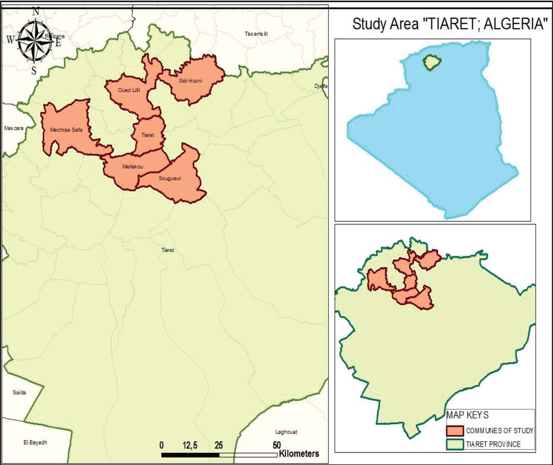
AbstractBackground: Coxiella burnetii causes Q fever and represents a globally important zoonotic disease affecting both humans and animals. In ruminants, particularly sheep, the infection is often subclinical but can lead to reproductive losses, including abortion, stillbirth, weak lambs, and reduced fertility, with significant economic impact. In humans, C. burnetii causes acute febrile illness, pneumonia, or hepatitis and may progress to endocarditis. Despite its public and veterinary health relevance, information on the epidemiology and distribution of Q fever in Algeria remains limited, especially in major sheep-producing areas such as Tiaret Province. Aim: This study aimed to determine the seroprevalence of C. burnetii infection in sheep from Tiaret Province, northwestern Algeria, and to identify key animal-level factors associated with seropositivity. Methods: A cross-sectional study was conducted between December 2023 and July 2024. A total of 184 serum samples collected from sheep of different ages, genders, herd sizes, and management systems were tested for anti-C. burnetii antibodies using a commercial indirect enzyme-linked immunosorbent assay (IDVet®, France). Doubtful samples were excluded from the statistical analysis. Univariate and multivariate logistic regression analyses were performed to assess the relationship between seropositivity and selected explanatory variables. Results: The obtained results reveal a seroprevalence of 37.50% at the animal level. Univariate analysis indicated that seropositivity was significantly associated with age, herd size, herd composition, and farm type. Multivariable analysis confirmed that sheep aged ≥4 years (odds ratio (OR)=4.16; p=0.02) and herds containing >250 animals (OR=37.47; p < 0.001) were at significantly higher risk of infection. Conclusion: Coxiella burnetii infection is endemic among sheep in Tiaret Province. The identification of age and herd size as major risk factors underscores the need for improved reproductive management, enhanced biosecurity practices, and targeted surveillance measures. Integrating these practices within a One Health framework is essential for reducing disease transmission, protecting public health, and mitigating economic losses in Algerian sheep production systems. Keywords: Algeria, Coxiella burnetii, Q fever, Risk factors, Seroprevalence. IntroductionCoxiella burnetii is the etiological agent of Q fever (in humans) or coxiellosis (in animals), a worldwide occurrence zoonotic disease. It is an obligate intracellular Gram-negative bacterium with remarkable environmental resilience (surviving in spore-like forms in dust or birth products) and has been detected in a wide range of vertebrate and invertebrate hosts (including livestock, wildlife, and arthropods) (Eldin et al., 2017, Ahaduzzaman and Reza, 2024). Domestic ruminants, especially sheep, goats, and cattle, are considered the primary reservoirs for human infection, primarily via inhalation of contaminated aerosols (placenta, birth fluids, feces, urine, and milk) and environmental spread over distances (Cho et al., 2023, Celina and Cerný, 2022). In livestock, particularly small ruminants, the infection is often subclinical but can lead to significant reproductive disorders (abortion, stillbirth, weak offspring, and infertility) as well as prolonged shedding of bacteria into the environment, thus presenting both economic and public-health concerns (Gisbert et al., 2024; Rahravi et al., 2022). For example, in goat herds, persistent infections have caused abortion rates up to 80%–90% over successive kidding seasons (Zendoia et al., 2024). In humans, acute Q fever may manifest as febrile illness, pneumonia, or hepatitis, whereas a smaller proportion of infections progress to chronic forms (e.g., endocarditis and vascular infection) if untreated or undiagnosed (Meles et al., 2024, Cho et al., 2023). Despite its significance, Q fever remains under-recognized and under-reported in many regions, particularly in low- and middle-income countries, due to non-specific clinical signs, under-diagnosis, lack of routine surveillance in livestock and humans, and limited awareness of its zoonotic potential (Guerrero-Freire et al., 2024, Ahaduzzaman and Reza, 2024). In the context of small ruminants globally, a recent comprehensive meta-analysis reported variable seroprevalence estimates and highlighted substantial regional heterogeneity, underlying differences in husbandry systems, animal movement, biosecurity levels, climate, and surveillance effort (Ahaduzzaman and Reza, 2024). Several studies on small ruminants in the North African region (including Algeria) have reported that the seroprevalence of C. burnetii varies widely (for example, from modest to very high levels depending on the province, species, and sampling methodology), but major gaps remain. Data from many production zones are limited, and risk-factor analyses (at animal and flock level) are scarce (Devaux et al., 2020, Haif et al., 2021). Given that sheep production plays a major role in the agricultural economy of many Algerian provinces and that environmental, husbandry, and trade conditions may favor the persistence and dissemination of C. burnetii, targeted epidemiological studies are required (Mezienne et al., 2024). Tiaret Province (northwestern Algeria) is an important sheep-producing region. However, to date, no comprehensive serological survey and risk-factor analysis focused on sheep in this province has been published (or is very limited). Therefore, understanding the local seroprevalence and associated risk factors is essential to inform livestock health management, reduce economic losses, and assess zoonotic risks at the human–animal interface. Therefore, the present study aimed to (i) determine the seroprevalence of C. burnetii infection in sheep in Tiaret Province, Algeria, and (ii) identify the potential animal-level and risk factors associated with infection. The findings of this study will provide new baseline data for the region, support veterinary public health measures, and contribute to the broader understanding of the epidemiology of C. burnetii in small ruminants. Materials and MethodsStudy areaTiaret Province (35° 22' 15.128'' N and 1° 19' 18.427 E) is characterized by two main periods during the year: a harsh winter, often accompanied by snowfall, with an average temperature of 7.2°C, and a hot, dry summer with an average temperature of 40°C. During normal periods, the province of Tiaret receives 300–400 mm of rain per year, with seasonal fluctuations in rainfall ranging from 157 mm in winter to 31 mm in summer. This province has an estimated sheep population of 2.300.756 and 194.876 goats. The sheep flock size varied from 20 to 800, and all flocks were destined for meat production. Study designA cross-sectional study was conducted in various geographical areas of this province during December 2023 and July 2024 from different communes in Tiaret (Fig. 1) to establish the individual prevalence of Q fever in sheep and identify potential associated risk factors. Moreover, the sampling covered a significant number of randomly selected animals per flock. The sample size was calculated using the following formula for estimating prevalence in a finite population (Thursfield, 2018):
Fig. 1. Map of Algeria showing the geographical locations of the Tiaret province and the communes included in the study. Figure 1 Map of the study area showing the distribution of sampling sites across Tiaret, Algeria. The base map was generated using the ArcGIS software. The scale bar and north arrow are included for spatial reference.
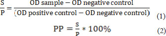
where n=required sample size, Z=Z-value for 95% confidence interval (CI)=1.96, Pexp=expected prevalence of 20% (0.20) as a conservative estimate, d=desired precision (margin of error) [7% (0.07)], and N=total sheep population in Tiaret Province (2,300,756 sheep). Accordingly, the minimum required sample size was approximately 125 sheep. However, to increase the precision of the study, a total of 184 sheep [males (41) and females (143)] belonging to flocks localized in Tiaret were enrolled, and the collected sera were examined. Data collection and blood samplingOn the day of sampling, face-to-face interviews were conducted with the flock owners to complete the questionnaire. The questionnaire was administered to herd owners, and the investigator explained the questions to them individually. We designed a structured questionnaire that begins with a short paragraph that clarifies the objective of our study and highlights the farm owners’ answers’ relevance. This study includes four sections. In the first section, closed-ended questions (Yes/No and multiple choice questions) are used to collect information about herd sanitary status, such as the farms’ locations, breed, number of sheep, herd type, age, and gender. The second section is devoted to the owners’ knowledge of the disease, including its zoonotic nature. The third section focused on the hygiene practices within the herd, and the fourth section asked questions about the general management-related aspects. Very limited data are available about the geographic distribution of the disease in Algeria. Blood samples were collected from the jugular vein using a 4 ml vacuum tube (CDHEALTHR, SARL SIREM MED) without anticoagulants. The samples were then stored in a refrigerated bag and transferred to the laboratory. Sera were removed by centrifugation at 3,500 ×g for 10 minutes and stored at −20°C until further testing. Furthermore, no vaccination against Q fever has ever been administered in Algeria. Serological testsOvine sera were tested for the presence of anti-C. burnetii antibodies using a commercial enzyme-linked immunosorbent assay (ELISA) test (ID SCREEN® Q fever indirect multi-species, IDVet, France), according to the manufacturer’s instructions. The optical density (OD) values were read at 450 nm using an ELx808 absorbance microplate reader (BIO-TEK INSTRUMENTS®, Inc., Vermont, USA). The manufacturer had previously validated the ELISA kit, estimating sensitivity at 100% (95% CI: 89.28%–100%) and specificity at 100% (95% CI: 97.89%–100%). The results were expressed as the sample/positive (S/P) ratio and percentage positive (PP). They were calculated using the following formula:
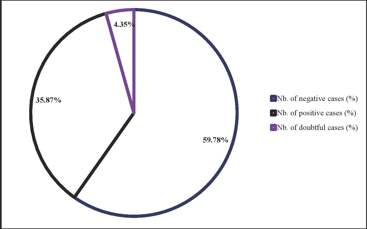
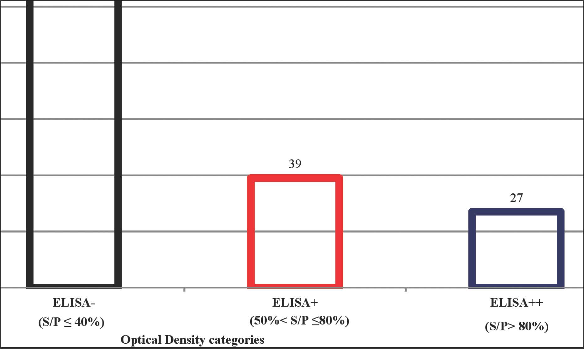
The results were interpreted as follows: PP ≤ 40% was defined as negative (−), 50% < PP ≤ 80% was defined as positive (+), PP > 80% was defined as strong positive (++), and 40%< PP ≤ 50% was defined as doubtful. Samples with a PP between 40% and 50% were classified as “doubtful” according to the manufacturer’s instructions.” These doubtful samples were excluded from all prevalence calculations and regression analyses to avoid potential misclassification bias, as doubtful results cannot be reliably interpreted as either positive or negative without confirmatory testing (e.g., polymerase chain reaction (PCR) or re-testing with a different ELISA kit). Excluding these samples ensured that only clearly defined serological outcomes were used for prevalence estimation and risk factor analysis, thereby increasing the statistical results’ accuracy and robustness. Statistical analysisThe data were coded using Microsoft Excel 2007, imported, and analyzed in R (version 3.5.1; R Foundation for Statistical Computing, Vienna, Austria) via RStudio (version 1.1.383, RStudio Inc., Boston, MA). To assess potential risk factors, odds ratios (ORs) and corresponding p-values with 95% CI were calculated for all examined variables. A p-value < 0.05 was considered statistically significant. The explanatory variables included age (≤1, 2, 3, and ≥4 years), sex (male and female), herd size (≤150, 150–250, and ≥250 animals), farm type (private or state-owned), and herd composition (mixed flocks or sheep-only flocks). Associations between the individual serological status of an animal (X) and the independent variables were assessed using univariate logistic regression. The dependent variable for serological status was binomial, with animals classified as positive or negative. Multicollinearity among predictor variables was assessed using the variance inflation factor. Multilevel generalized mixed effects models based on restricted maximum likelihood were constructed for the risk factor analysis. The lme4 software glmer function was used to execute the model. Multivariate models were constructed using a stepwise approach of manual backward elimination, and Akaike’s information criteria was used to select the final multivariate model. Non-significant variables (p > 0.05) were then excluded. The adapted ORs and corresponding CIs for each category were compared with the reference category. Prevalence is the proportion of individuals in a population that tests positive for Q fever disease at a specific time point. In this study, the observed prevalence was calculated as the number of sheep testing positive by ELISA divided by the total number of sheep tested, excluding doubtful results. A farm is considered seropositive if at least one animal has tested positive for the relevant antibodies. Ethical approvalThe Scientific Council of the Institute of Veterinary Sciences, University of Tiaret, 14000 Tiaret, Algeria approved the study protocol (Report of Scientific Council Ref: 463/VRPG/dated March 30, 2022). Verbal consent was obtained from the livestock farmers before the collection of samples and data for the study. The samples were collected in accordance with international ethical guidelines. ResultsA total of 184 sheep were tested for antibodies to C. burnetii; 35, 87% (66/184) were positive, 4.35% (8/184) were doubtful, and 59.78% (110/184) were negative by ELISA (Fig. 2). Doubtful animals were excluded from the study. Therefore, 176 sheep were retained for statistical analysis. Among the 66 positive cases, 27 had a %S/P value higher than 80% (ELISA++) and were considered as strong positive, and 39 were positive (ELISA+) with a %S/P value between 50% and 80% (Fig. 3).
Fig. 2. Percentage of animals diagnosed with C. burnetii (ELISA test). Figure 2 illustrates the distribution of the ELISA test results. The data are divided into three categories: positive cases with a percentage of 35.87%, negative cases with a percentage of 59.78%, and doubtful cases with a percentage of 4.35%.
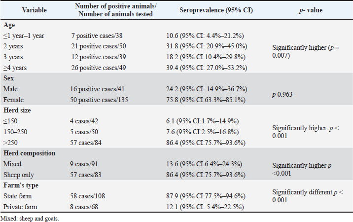
Fig. 3. Serological distribution of animals according to ELISA OD Values for antibodies to C. burnetii. Figure 3 represents a bar graph illustrating the distribution of animals across three different S/P ratio categories based on ELISA test results; ELISA- (S/P 40%) indicates animals that were tested negative, ELISA+ (S/P between 50% and 80%) indicates animals that were positive, and ELISA++ (S/P > 80%) indicates animals with strong positive results. The samples tested positive for C. burnetii antibodies (66/176), with an observed seroprevalence of 37.50% (95% CI: 31.5%–46.7%). Risk factorsThe flock management factors associated with sheep seropositivity in a univariate analysis with a p < 0.05 are presented in Table 1. Table 1. Univariate analysis of the association of flock management risk factors with Q fever seropositivity among sheep sampled from flocks in Tiaret province, Algeria.
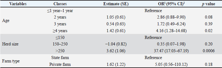
The univariable analysis at the animal level in this study indicated that four factors were significantly (p < 0.05) associated with C. burnetii seropositivity: age, herd size, herd composition, and farm type. Seroprevalence was significantly higher in older animals (≥4 years: 39.4%, 95% CI: 27.0%–53.2%; p=0.007), larger herd size (≥ 250 animals 86.4, 95% CI: 75.7%–93.6%; p < 0.001), sheep-only flocks (86.4%, 95% CI: 75.7%–93.6%; p < 0.001), and state farms (87.9, 95% CI: 77.5%–94.6%; p < 0.001). Although the sero-prevalence was higher in female sheep (75.8%, 95%CI: 63.3%–85.1%) than in male sheep (24.2%, 95%CI: 14.9%–36.7%). However, this difference was not statistically significant (p=0.963). The sero-prevalence was higher in females than in older sheep, and this difference was found to be statistically significant (p=0.007). Table 2 presents flock management factors associated with sheep seropositivity in a multivariate analysis at p < 0.05. Age and herd size were the only two factors that were significantly associated with Q fever seropositivity in the final multivariate logistic regression model. However, the farm type was no longer significant in the final multivariate logistic regression model. Table 2. Multivariable logistic regression model analysis for risk factors presumed of flock. management (p < 0.05) with Q fever seropositivity among sheep sampled from flocks in Tiaret province, Algeria.
Results from the multivariable model showed that animals aged >4 years were three times more likely to suffer from the disease than younger animals (OR=4.16; p=0.02). Herds with a size of more than 250 animals are 36.47% more likely to experience Q fever than herds with fewer animals (p=0.0006). However, no significant effect of farm type on the odds of being seropositive was observed (p=0.18). DiscussionSheep, goats, and dairy cattle are the main livestock species kept in Tiaret Province, Algeria. In Algeria, C. burnetii infection in livestock is still poorly investigated. During abortion outbreaks and in veterinary diagnostic laboratories, veterinarians frequently fail to suspect the infection. Q fever tests are not a part of the routine differential diagnosis for abortion cases, which induced significant economic losses, and they could also cause zoonotic epidemic infections in humans. Various techniques have been used to diagnose coxiellosis in domestic animals. The choice of the diagnostic test largely depends on the type and number of samples to be analyzed and the availability of the test. In our study, ELISA was chosen for mass screening because it is a sensitive, specific, and rapid diagnostic test (Lyo et al., 2017). This study was conducted to investigate the seroprevalence status of C. burnetii in sheep and its associated risk factors. The findings of this study established the widespread spread of Q fever in sheep in Tiaret (northwest of Algeria). The obtained results in this study demonstrate a seroprevalence of 37.50% (95% CI: 31.5%–46.7%) at the animal level. These values are significantly higher than those reported in Constantine (northeast of Algeria) (12.4%) (HIRECHE et al., 2020), in eight departments of Algeria (the seroprevalence rate was 14.1%) (Khaled et al., 2016), M’Sila Governorate (27.2%) (Zemmouri et al., 2020), northwest of Algeria (28%) (Karim et al., 2017), in Tunisia (7.31% (12/164)) (Guesmi et al., 2023), in Nandi County Kenya [seroprevalence at the animal-level 1.413% (4/283) CI:95% (1.0%–7.78%)] (Kiptanui et al., 2022), in Yobe State, Nigeria [(49/420) (11.7%)] (Amumu et al., 2019), in northern Cote d’Ivoire [9.4% (95% CI 5.7–15] (Kanouté et al., 2017) and in Ghana (16.57%). However, our results were lower than the findings in Assiut, Egypt (60%) (Abbass et al., 2020). The differences in the seroprevalence of Q fever in sheep across various studies can be attributed to several factors, including geographical location, seasonal variations, farming practices, and sampling methodologies. These elements significantly influence the detection rates of C. burnetii antibodies in sheep populations. The infection can occur at any age, whereas in our study, a significantly higher serological prevalence of the seroprevalence of C. burnetii was recorded in adult animals (p=0.02) than in young animals. This result is consistent with the findings of most studies that have examined the correlation between age and seroprevalence in C. burnetii. The higher seroprevalence observed in older animals likely reflects cumulative exposure over time rather than increased susceptibility. Coxiella burnetii infection typically occurs through the inhalation of contaminated aerosols or contact with infected birth materials, which accumulate in the environment and within herds as the animals age. Consequently, older sheep have more opportunities for repeated exposure and antibody development. In addition, persistent environmental contamination, particularly during lambing seasons, may maintain continuous transmission cycles within large flocks. This age-related pattern is consistent with C. burnetii’s chronic environmental persistence and the bacterium’s ability to remain viable in dust and soil for extended periods, thereby contributing to its endemic maintenance in endemic zones. Previous studies conducted in Algeria (Karim et al., 2017), Kenya (Larson et al., 2019), Iran (Ezatkhah et al., 2015), Saudi Arabia (Jamelnab et al., 2018), Jordan (Lafi et al., 2020), and Egypt (Klemmer et al., 2018) reported similar results. The seroprevalence was higher in female sheep than in male sheep. However, this difference was not statistically significant (p > 0.05). This result showed that gender is not a risk factor for cancer. The same idea was supported by Emaeili et al. (2014) in northwestern Iran, who found no relationship between gender and the seroprevalence rate. In addition, Wolff et al. (2020) discovered that gender did not have a significant influence on the detection of C. burnetii infection using ELISA and quantitative Polymerase Chain Reaction. This study demonstrated that larger herds are highly infected by C. burnetii compared with small and medium herds. This finding is consistent with that of Villari et al. (2018), who found that farms housing more than 50 animals have a risk almost four times higher (p < 0.001) of contracting C. burnetii than those with fewer than 50 animals. The authors suggested that this increased risk could be attributed to two potential factors. First, there was a possibility that more random contact between uninfected and infected animals occurred in overcrowded livestock buildings. Second, a decreased chance of clearing the infection in the presence of more infected animals and lambing. In the present study, the seroprevalence observed among ewes kept in sheep flocks (86.4%, 95% CI: 75.7%–93.6%) was higher than that among ewes kept in mixed flocks (with goats) (13.6%, 95% CI: 6.4%–24.3%), although without significance. Our results were similar to those reported in Algeria (Khaled et al., 2016), whereas the seroprevalence of C. burnetii in mixed flocks was lower (46.4%) than that in flocks with single species (71.4%). In northwestern Italy, the highest flock true seroprevalence was recorded in mixed flocks (48.5%), indicating that this flock type, characterized by larger size and co-presence of the two species, more likely represents a risk factor for infection spread (Rizzo et al., 2016). In central Portugal (Anastacio et al., 2013), the prevalence of C. burnetii was higher in mixed herds (38.4%) than in sheep herds (37.5%). In Algeria (Belhouari et al., 2022), the sero-prevalence among ewes kept in mixed flocks (with cattle or goats) was higher (28.07%) than that of ewes kept in sheep flocks (18.64%). In Germany (Wolf et al., 2020), researchers observed that goats shed C. burnetii more than sheep, which can elevate the risk of infection in sheep within the same flock. This study has several limitations that should be considered when interpreting the results. First, the cross-sectional design captures exposure and serological status at a single point in time; therefore, causal inferences and temporal relationships between potential risk factors and C. burnetii seropositivity cannot be established. In addition, most explanatory variables evaluated in this study (including age category, herd size, herd composition, and farm management practices) are dynamic and may change over time, except for fixed characteristics such as sex. Consequently, the measured exposures may not accurately reflect the conditions present at the time of infection or seroconversion, and the identified associations should be interpreted as correlation indicators rather than causation indicators. Furthermore, seropositivity reflects past exposure to C. burnetii and does not allow differentiation between recent and historical infections, limiting insight into current infection dynamics and active shedding within flocks. Although the ELISA kit used in this study was reported by the manufacturer to have 100% sensitivity and specificity, these estimates were obtained under controlled validation conditions and have not been independently verified under local field conditions in Algeria. Diagnostic performance may vary according to the tested population, epidemiological context, sample handling, and laboratory conditions. Moreover, no reference (gold standard) method, such as PCR or bacterial culture, was used to confirm the ELISA results. Therefore, the true diagnostic accuracy of the assay in this specific setting cannot be fully assessed, and serological status misclassification cannot be excluded. Finally, the relatively limited number of farms included in the study may restrict the generalizability of the findings to the wider sheep population of Tiaret Province. Future longitudinal studies incorporating repeated sampling, molecular diagnostics, and environmental assessments are needed to better elucidate the transmission dynamics and causal pathways of C. burnetii infection in sheep. ConclusionThis study provides the first detailed assessment of C. burnetii exposure among sheep in Tiaret Province, Algeria, revealing a high individual seroprevalence of 37.50% (95% CI: 31.5%–46.7%). The detection of antibodies suggests the circulation of C. burnetii among the sampled flocks. Multivariate analysis identified animal age (≥4 years) and herd size (>250 sheep) as significant risk factors, reflecting cumulative exposure over time and the role of animal density in disease maintenance and transmission. In contrast, gender and farm type did not significantly influence the infection risk. These results underscore the urgent need to integrate C. burnetii into national livestock surveillance systems and strengthen biosecurity and reproductive management practices. Preventive actions should include improved hygiene during lambing, control of animal movements, and awareness campaigns among farmers and veterinarians. In line with the One Health approach, collaboration between veterinary and human health authorities is crucial for preventing spillover to humans, protecting animal productivity, and reducing the economic and public health burden of Q fever in Algeria. Future work should combine serological, molecular, and spatial analyses to better understand transmission dynamics and identify reservoirs contributing to environmental contamination. AcknowledgmentsWe would like to thank the farm owners who allowed us to collect sera for the study and answered the research survey. We also express our appreciation and recognition to Dr. Assia Boumezrag and Mr. Redouane Doucene, who have helped in this work, in addition to the DGRSDT-MERRS-ALGERIA. Conflict of interestThe authors declare no conflict of interest related to this article. FundingThis research is a contribution to the PRFU D01N01UN140120220003 project, funded by the Directorate-General for Scientific Research and Technological Development DGRSDT-MESRS-ALGERIA. Authors' contributionsImen Safer: Fatima Mahouz and Saad Aissat: Fatima Mahouz and Saad Aissat: Mohamed Amine Ayad: Providing the materials and the samples of the study; Houari Hemida: Conducting the test of the study; Amine Abdelli and Sofiane Derrar: Amine Abdelli and Sofiane Derrar: Software and formal analysis; George Cosmin Nadăș Reviewing and editing the manuscript. Data availabilityAll data supporting this study’s findings are available within the manuscript. ReferencesAbbass, H., Selim, S.A.K., Sobhy, M.M., El-Mokhtar, M.A., Elhariri, M. and Abd-Elhafeez, H.H. 2020. High prevalence of Coxiella burnetii infection in humans and livestock in Assiut, Egypt: a serological and molecular survey. Vet. World 13(12), 2578–2586. Ahaduzzaman, M. and Reza, M.M.B. 2024. Global and regional seroprevalence of coxiellosis in small ruminants: a systematic review and meta-analysis. Vet. Med. Sci. 10, e1441. Amumu , S., Tijani, A., Amumu, N., Atsanda, N., Dava, J. and Lawan, F. 2019. Epidemiology of Q fever in sheep flocks in Yobe State, Nigeria. J. Vet. Biomed. Sci. 2, 70–76. Anastacio, S., Tapares, N., Carlino, N., Sidi-Boumedin, K. and Silva, G. 2013. Serological evidence of Coxiella burnetii exposure in sheep and goats in central Portugal. Vet. Microbiol. 167, 500–505. Belhouari, A., Souames, S., Berrama, Z. and Ouchene, N. 2022. Seroprevalence of Q fever among ewes and associated risk factors in the Ain Defla region of north-central Algeria. Comparative Immunol. Microbiol. Infect. Dis. 87, 101853; doi:10.1016/j.cihd.2011.01.013 Celina, S.S. and Cerný, J. 2022. Coxiella burnetii in ticks, livestock, pets, and wildlife: a mini-review. Front. Vet. Sci. 9, 1068129. Cho, Y.S., Park, J., -H.., Kim, J.W., Lee, J., -J.., Youn, S.Y., Byeon, H.S., Jeong, H.W., Kim, D.-M., Yu, S.N. and Yoon, J.W. 2023. Current status of Q fever and the challenge of outbreak preparedness in Korea: one health approach to zoonoses Remark 1. J. Korean. Med. Sci. 38, 24. Devaux, C.A., Osman, I.O., Million, M. and Raoult, D. 2020. Coxiella burnetii in dromedary camels (Camelus dromedarius): a possible threat for humans and livestock in North Africa and the near and Middle East?. Front. Vet. Sci. 7, 558481. Eldin, C., Mélenotte, C., Mediannikov, O., Ghigo, E., Million, M., Edouard, S., Mege, J.L., Maurin, M. and Raoult, D. 2017. From Q fever to Coxiella burnetii infection: a paradigm change. Clin. Microbiol. Rev. 30, 115–190. Emaeili, S., Bagheri Amiri, F. and Mastavi, E. 2014. Seroprevalence of Q fever among sheep in northwestern Iran. Vector-Borne Zoonotic Dis. 14, 189–192. Ezatkhah, M., Alimolaei, M., Khalili, M. and Sharifi, H. 2015. Seroepidemiological study of Q fever in small ruminants from Southeast Iran. J. Infect. Public. Health 8, 170–176. Gisbert, P., Garcia-Ispierto, I., Quintela, L.A. and Guatteo, R. 2024. Coxiella burnetii and reproductive disorders in cattle: a systematic review. Animals 14, 1313. Guerrero-Freire, M.S., Ledesma, Y., Echeverría, G., Blanco, F.C. and De Waard, J.H. 2024. Light on risk: seroprevalence of Q fever among farm animals and workers in Ecuador. One Health 19, 100939. Guesmi, K., Kalthoum, S., Mamluk, A., Baccar, M.N., Belhajmohamed, B., Hajlaoui, H., Toumi, A., Cheroni, J., Seghaier, C. and Messadi, L. 2023. Seroprevalence of zoonotic abortive diseases and associated risk factors in Tunisian sheep. BMC. Vet. Res. 19, 50. Haif, A., Khelifi-Ouchene, N.A., Khelifi, M., Ouchetati, I., Zeroual, F. and Ouchene, N. 2021. Abortive diseases and associated risk factors in small ruminants in Algeria: a systematic review. Trop. Anim. Health Prod. 53, 520. Hireche, S., Agabou, A. and Bouaziz. 2020. Seroprevalence of Coxiella burnetii among ewes and associated risk factors in Constantine, Algeria. J. Hellenic Vet. Med. Soc. 71, 2383–2390. Jamelnab, A.A., Al-Shaikh, M.A., Bakhiet, A.O., Omer, S.A., Aljumah, R.S., Hamriss, G.D., Mohammed, O.B. and Hussein, M.F. 2018. Seroprevalence of Q fever in farm animals. Biomed. Res. 29, 895–900. Kanouté, Y.B., Grangnon, B.G., Schindler, C., Bofoh, B. and Schelling, E. 2017. Epidemiology of brucellosis, Q fever, and Rift Valley fever at the human–vegetable interface in northern Côte d’Ivoire. Acta. Tropica. 165, 66–75. Karim, A., Ait, O.K. and Khelef, D. 2017. Seroprevalence of chlamydial abortion and Q fever in ewes aborted in the North-West of Algeria. J. Vet. Med. Anim. Health. 9, 246–249; doi:10.1016/j.jvmah.2017.09.016 Khaled, H., Sidi-Boumedine, K., Merdja, S., Duffour, P., Dahamani, A., Thiéry, R., Rousset, E. and Bouyoucef, A. 2016. Serological and molecular evidence of Q fever in small ruminant flocks in Algeria. Comparative. Immunol. Microbiol. Infect. Dis. 47, 19–25. Kiptanui, J., Gathura, P.B., Kitala, P.M. and Bett, B. 2022. Seroprevalence estimates of Q fever and infection predictors in cattle, sheep, and goats in Nandi County, Kenya. Vet. Med. Int. 2022, 3741285. Klemmer, J., Njeru, J., Emam, A., El-Sayed, A., Moawad, A.A., Henning, K., Elbeskawy, M.A., Sauter-Louis, C., Straubinger, R.K., Neubauer, H. and El-Diasty, M.M. 2018. Q fever in Egypt: epidemiological survey of Coxiella burnetii-specific antibodies in cattle, buffaloes, sheep, goats, and camels. PLos One 13, 192188. Lafi, S.Q., Tarafh, A.Q., Abou-Dalibouh, M.A., Hailat, R.S. and Khalifeh, M.S. 2020. Seroprevalence and associated risk factors of Coxiella burnetii (Q fever) infection in goats and sheep in northern Jordan. Trop. Anim. Health. Prod. 52, 1553–1559. Larson, P.S., Espira, L., Grabow, C., Wang, C.A., Muloi, D., Browne, A.S., Deem, S.L., Fèvre, E.M., Foufopoulos, J., Hardin, R. and Eisenberg, J.N.S. 2019. The sero‐epidemiology of Coxiella burnetii (Q fever) across livestock species and herding contexts in Laikipia County, Kenya. Zoonoses Public Health 66, 316–324. Lyo, K.-S., Kim, D., Jang, H.G., Lee, S.-J., Park, M.Y. and Hahn, T.-W. 2017. Prevalence of antibodies against Coxiella burnetii in South Korean Native Cattle, dairy cattle, and dogs. Vector-Borne. Zoonotic. Dis. 17, 213–216. Meles, D.K., Khairullah, A.R., Mustofa, I., Wurlina, W., Akintunde, A.O., Suwasanti, N., Mustofa, R.I., Putra, S.W., Moses, I.B. and Kusala, M.K.J. 2024. Q fever: current perspectives and challenges in preparedness for an outbreak. Open. Vet. J. 14, 2509. Mezienne, R., Mouss, A.K., Hammouches, D., Boughiris, M. and Boughiris, F. 2024. Practical management of sheep farming in Eastern Algeria: situation, constraints, and perspectives. World 14, 389–399. Rahravi, M., Moravedji, M., Mastavi, E., Mouhammadi, M., Seyfi, H., Bastri, N., Mozoun, M.M., Latifani, M. and Emaeili, S. 2022. Epidemiological survey of Coxiella burnetii in small ruminants and ticks in western Iran. BMC Vet. Res. 18, 292. Rizzo, F., Vitale, N., Ballardini, M., Borromeo, V., Luzzago, C., Chiavacci, L. and Mandola, M.L. 2016. Seroprevalence of Q fever and risk factors in sheep and goats in northwest Italy. Prev. Vet. Med. 130, 10–17. Thrusfield, M. 2018. Veterinary Epidemiology, 4th ed. Chichester, UK: John Wiley & Sons. Villari, S., Galluzzo, P., Arnone, M., Alfano, M., Geraci, F. and Chiarenza, G. 2018. Seroprevalence of Coxiella burnetii infection (Q fever) in sheep farms in Sicily (Southern Italy) and related risk factors. Small. Ruminant. Res. 164, 82–86. Wolff, A., Prüfer, T., Schoneberg, C., Campe, A., Runge, M., Ganter, M. and Büuer, B. 2020. Risk factors for Coxiella burnetii infection in German sheep flocks. Epidemiol. Infect. 148, 260. Zemmouri, L., Besbaci, M., Mammeri, A. and Lafri, M. 2020. Seroepidemiological investigation of major abortive bacterial agents in Ewes of the M'Sila Governorate, Algeria. Bull. Univ. Agricult. Sci. Vet. Med. 77. Zendoia, I.I., Barandika, J.F., Cavianes, A., Hurtado, A. and García-Pérez, A.L. 2024. Coxiella burnetii infection persistence in a goat herd during seven kidding seasons after an abortion outbreak: the effect of vaccination. Appl. Environ. Microbiol. 90, e02201–e02223. | ||

| How to Cite this Article |
| Pubmed Style Safer I, Mahouz F, Ayad MA, Hemida H, Aissat S, Abdelli A, Nadăş GC, Derrar S. Evaluation of seroprevalence and risk factors for Q fever (Coxiella burnetii) infection in sheep from Tiaret province, northwestern Algeria. Open Vet. J.. 2026; 16(5): 3094-3103. doi:10.5455/OVJ.2026.v16.i5.51 Web Style Safer I, Mahouz F, Ayad MA, Hemida H, Aissat S, Abdelli A, Nadăş GC, Derrar S. Evaluation of seroprevalence and risk factors for Q fever (Coxiella burnetii) infection in sheep from Tiaret province, northwestern Algeria. https://www.openveterinaryjournal.com/?mno=298269 [Access: June 26, 2026]. doi:10.5455/OVJ.2026.v16.i5.51 AMA (American Medical Association) Style Safer I, Mahouz F, Ayad MA, Hemida H, Aissat S, Abdelli A, Nadăş GC, Derrar S. Evaluation of seroprevalence and risk factors for Q fever (Coxiella burnetii) infection in sheep from Tiaret province, northwestern Algeria. Open Vet. J.. 2026; 16(5): 3094-3103. doi:10.5455/OVJ.2026.v16.i5.51 Vancouver/ICMJE Style Safer I, Mahouz F, Ayad MA, Hemida H, Aissat S, Abdelli A, Nadăş GC, Derrar S. Evaluation of seroprevalence and risk factors for Q fever (Coxiella burnetii) infection in sheep from Tiaret province, northwestern Algeria. Open Vet. J.. (2026), [cited June 26, 2026]; 16(5): 3094-3103. doi:10.5455/OVJ.2026.v16.i5.51 Harvard Style Safer, I., Mahouz, . F., Ayad, . M. A., Hemida, . H., Aissat, . S., Abdelli, . A., Nadăş, . G. C. & Derrar, . S. (2026) Evaluation of seroprevalence and risk factors for Q fever (Coxiella burnetii) infection in sheep from Tiaret province, northwestern Algeria. Open Vet. J., 16 (5), 3094-3103. doi:10.5455/OVJ.2026.v16.i5.51 Turabian Style Safer, Imen, Fatima Mahouz, Mohamed Amine Ayad, Houari Hemida, Saad Aissat, Amine Abdelli, George Cosmin Nadăş, and Sofiane Derrar. 2026. Evaluation of seroprevalence and risk factors for Q fever (Coxiella burnetii) infection in sheep from Tiaret province, northwestern Algeria. Open Veterinary Journal, 16 (5), 3094-3103. doi:10.5455/OVJ.2026.v16.i5.51 Chicago Style Safer, Imen, Fatima Mahouz, Mohamed Amine Ayad, Houari Hemida, Saad Aissat, Amine Abdelli, George Cosmin Nadăş, and Sofiane Derrar. "Evaluation of seroprevalence and risk factors for Q fever (Coxiella burnetii) infection in sheep from Tiaret province, northwestern Algeria." Open Veterinary Journal 16 (2026), 3094-3103. doi:10.5455/OVJ.2026.v16.i5.51 MLA (The Modern Language Association) Style Safer, Imen, Fatima Mahouz, Mohamed Amine Ayad, Houari Hemida, Saad Aissat, Amine Abdelli, George Cosmin Nadăş, and Sofiane Derrar. "Evaluation of seroprevalence and risk factors for Q fever (Coxiella burnetii) infection in sheep from Tiaret province, northwestern Algeria." Open Veterinary Journal 16.5 (2026), 3094-3103. Print. doi:10.5455/OVJ.2026.v16.i5.51 APA (American Psychological Association) Style Safer, I., Mahouz, . F., Ayad, . M. A., Hemida, . H., Aissat, . S., Abdelli, . A., Nadăş, . G. C. & Derrar, . S. (2026) Evaluation of seroprevalence and risk factors for Q fever (Coxiella burnetii) infection in sheep from Tiaret province, northwestern Algeria. Open Veterinary Journal, 16 (5), 3094-3103. doi:10.5455/OVJ.2026.v16.i5.51 |


