





| Research Article | ||
Open Vet. J.. 2026; 16(5): 2869-2882 Open Veterinary Journal, (2026), Vol. 16(5): 2869-2882 Research Article Laboratory efficacy of green-synthesized silver and copper nanoparticles against malaria vector, Anopheles sergentii (Theobald, 1907) (Diptera: Culicidae)Ahmed Z.I. Shehata1, Magdy A. A. Ayad2*, Abdulnassar A. Alteab3, Ahmed N. G. Abdel-Aziz4, Mohamed A. M. Shahat1 and Raafat M. Shaapan51Department of Zoology, Faculty of Science (Boys), Al-Azhar University, Cairo, Egypt 2Department of Microbiology and Parasitology, Faculty of Veterinary Medicine, Tripoli University, Tripoli, Libya 3Department of Internal Medicine, Faculty of Veterinary Medicine, University of Tripoli, Tripoli, Libya 4Consultant Medical Entomologist at the Public Health Department, Jazan, Saudi Arabia 5Department of Zoonotic Diseases, Veterinary Research Institute, National Research Centre, Giza, Egypt *Corresponding Author: Magdy A. A. Ayad. Department of Microbiology and Parasitology, Faculty of Veterinary Medicine, Tripoli University, Tripoli, Libya. Email: magdyaa73 [at] yahoo.com Submitted: 10/11/2025 Revised: 28/03/2026 Accepted: 07/04/2026 Published: 31/05/2026 © 2025 Open Veterinary Journal
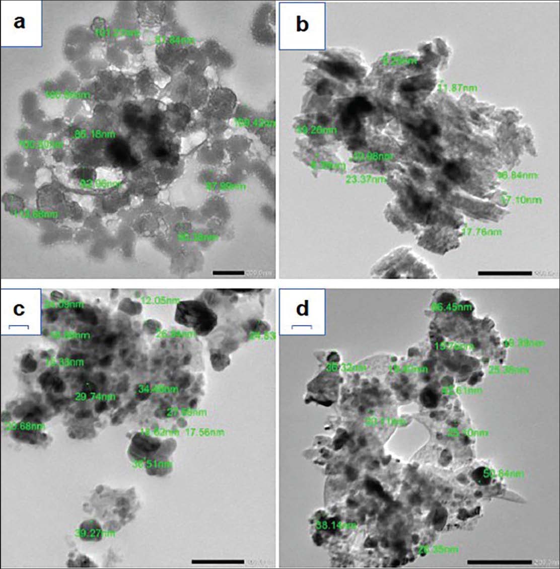
AbstractBackground: Anopheles sergentii is regarded as a significant vector-borne insect such as malaria. Aim: This work sought to investigate the efficacy of silver and copper nanoparticles (AgNPs and CuNPs) produced from aqueous extracts of Cestrum nocturnum and Salvia malticaulis against various immature stages of An. sergentii. Methods: The transmission electron microscopy (TEM) pictures of generated AgNPs and CuNPs indicated that the diameters of the nanoparticles varied from 8.39 to 113.68 nm for AgNPs and from 12.05 to 66.45 nm for CuNPs, respectively. The efficacy of synthesized AgNPs and CuNPs against various immature stages of An. sergentii was estimated after 24 and 48 hours. Results: The UV–vis Spectrophotometric analysis findings for both AgNPs and CuNPs exhibited a single absorption peak within the 300–400 nm region, signifying the existence of spherical AgNPs and CuNPs. The efficacy of the produced AgNPs and CuNPs against the immature stages of An. sergentii escalated with higher concentrations of the tested nanoparticles. Calculated LC50 values indicate that AgNPs and CuNPs produced from the aqueous extract of Cestrum nocturnum leaves were more effective against various stages of An. sergentii than those against Sa. malticaulis. In addition, the tested AgNPs were more effective against An. sergentii different immature stages than CuNPs. The LC50 values of Ce. nocturnum- AgNPs against immature stages of An. sergentii recorded 13.93 and 13.18 mg/l against first instar larvae, 15.0 and 14.22 mg/l against second instar larvae, 17.06 and 14.31 mg/l against third instar larvae, 17.24 and 14.32 mg/l against fourth instar larvae, 17.56 and 14.82 mg/l against pupae after 24- and 48-hours post-treatment, respectively. In addition, a depression in Acetylcholinesterase level in An. sergentii different immature stages were recorded. Conclusion: The Ce. nocturnum and Sa. malticaulis - synthesized AgNPs and CuNPs possess a significant activity against An. sergentii immature stages. Keywords: Anopheles sergentii, Cestrum nocturnum, Larvae, Salvia malticaulis. IntroductionVector-borne diseases in The Kingdom of Saudi Arabia (KSA) and other countries of Eastern Mediterranean region of the World Health Organization (WHO) represent more than 10% of vector-borne diseases in the whole world (WHO, 2004). Anopheles species members are malarial vectors to more than 10 million people worldwide, causing about 69,000 deaths between 2019 and 2020 (WHO 2021); however, the transmission of malaria in KSA recorded low and unstable levels as compared with other countries in South-East Asia and Africa (Hay et al., 2009; Khater et al., 2013). Within the epidemiological context of the Middle East, Anopheles sergentii is the main vector of Plasmodium falciparum malaria (Sarih et al., 2019). Despite the high efficacy of pesticide treatments against target species, vector control is jeopardized by the emergence of resistance to chemical insecticides, leading to a resurgence in vectorial capacity (Liu et al., 2006). Moreover, they pose significant risks to other non-target creatures and the environment through biomagnification (El-Mehdawy et al., 2021; Shehata et al., 2023). For many years, chemical insecticides have primarily targeted aquatic larvae of various Anopheles species; however, the development of novel control agents that are safer, more effective, and environmentally friendly is seen to be a suitable and essential substitute to avoid the risks associated with chemical insecticides (Shehata et al., 2020; El-Tabakh et al., 2023). Biosynthesized AgNPs and CuNPs are often safer and more specialized (El-Waseif et al., 2023). Biosynthesized nanoparticles possess distinctive and remarkable properties that facilitate their utilization across various disparate domains, including nanomedicine, antimicrobials, and control agents for multiple mosquito species (Agalya Priyadarshini et al., 2012; Sirelkhatim et al., 2015; Ahmed et al., 2016; Bobo et al., 2016; Chen et al., 2016). Synthesized nanoparticles are environmentally benign due to the absence of hazardous substances in their production (Bhosale et al., 2014; Abbas et al., 2022). Consequently, the pursuit of synthesized nanoparticles as novel agents for mosquito control remains essential, so this work examined the efficacy of AgNPs and CuNPs produced from aqueous extracts of Cestrum nocturnum and Salvia malticaulis against several immature stages of the malaria vector, An. sergentii. Materials and MethodsAnopheles sergentii' cultureLarvae of An. sergentii were obtained from breeding sites in the Al-Aydabi region, Jazan Governorate, Kingdom of Saudi Arabia (17°14'15.1"N, 42°56'18.6"E, altitude 1693 m) utilizing a rounded net dipper (25 cm in diameter) with a stainless-steel handle (120 cm in length) (Shehata et al., 2022). The gathered larvae were classified based on the prior keys of Kirkpatrick (1925), Harbach (1985), and Morsy et al. (1990). The larvae were cultivated for five generations using a standardized protocol in a controlled laboratory environment (27ºC ± 3ºC, 70%–80% relative humidity, and a 12:12 (dark/light) photoperiod (El-Tabakh et al., 2023). Synthesis of silver and copper nanoparticles (AgNPs and CuNPs)Preparation of aqueous extractsThe leaves of Ce. nocturnum and Sa. malticaulis, sourced from a private garden in the Al-Aydabi district of Jazan Governorate, Kingdom of Saudi Arabia, were washed and air-dried in the shade for ten days at ambient temperature. Four grams of powdered leaves from each plant were heated with 100 ml of distilled water in a water bath for 3 minutes. The solution was filtered and stored in the refrigerator at 4ºC until utilized (Zaki et al., 2024). Biosynthesis of silver nanoparticlesThe AgNO3 stock solution was prepared by dissolving 0.17 g of AgNO3 in 100 ml of distilled water. Aqueous extracts of Ce. nocturnum and Sa. malticaulis were individually combined with AgNO3 solution in a 1:9 ratio at ambient temperature for 72 hours, resulting in a reddish-brown coloration indicative of AgNPs production (Shehata and Mahmoud, 2019). Biosynthesis of CuNPsAbout 50 ml (5 mM) of copper sulfate solution was mixed with 5 ml of Ce. nocturnum and Sa. malticaulis aqueous extracts separately. The pH value 7.0 adjusted for the mixture by the addition of NaOH (1 N) solution. The green color mixture was obtained. The mixture was centrifuged, pellets collected, and dried overnight in a hot air oven at 60ºC. A dark green color powder obtained was stored at room temperature for further use (Mali et al., 2020). Characterization of AgNPs and CuNPsTransmission electron microscopyThe AgNPs and CuNPs suspensions were subjected to sonication for 10 minutes and subsequently diluted to produce somewhat turbid suspensions. The AgNPs and CuNPs suspensions were analyzed using a JEOL JEM-2100 high-resolution TEM at an accelerating voltage of 200 kV (Abdel-Shafy et al., 2019). Ultraviolet-Visible spectroscopy (UV/VIS)The AgNPs and CuNPs suspensions were diluted from 1 to 10 times using distilled water derived from colloidal solutions produced during the production process. The UV/VIS spectroscopy of the suspension was conducted utilizing a UV-Vis spectrophotometer (Model: Evolution™ 300, Serial Number: EVon 10600z, from Thermo Scientific). The bioassayActivity of AgNPs and CuNPsThe efficacy of the evaluated AgNPs and CuNPs against various immature stages of An. sergentii was conducted in accordance with the established protocol of Hassanain et al. (2019). Various quantities of AgNPs and CuNPs were formulated in 250 ml of distilled water within 500 ml beakers. Twenty-five An. sergentii larvae (I-IV) and pupae were promptly allocated into beakers containing varying amounts of AgNPs and CuNPs. All beakers were incubated under regulated conditions of the An. sergentii colony. Mortality was documented 24- and 48-hours following therapy. Typically, three replicates were employed. Acetylcholinesterase (AChE) activityThe activity of AChE in various immature stages of An. sergentii was assessed 24- and 48-hours post-treatment with half-lethal concentrations (LC50) of AgNPs and CuNPs. Approximately 10 ml of 0.1 M phosphate buffer solutions at pH 7.5 (KH2PO4-NaOH), incorporating 1% Triton X-100, 1% ethanol, and 1% Triton X-100, were utilized to homogenize three distinct batches of larvae (I-IV) and pupae individually, derived from each evaluated LC50. The Hereaeus Labofuge 400 R, manufactured by Kendro Laboratory Products GmbH, Germany, was utilized to centrifuge the homogenates for 60 minutes at 4ºC and 15,000 × g. Ten microliter aliquots of supernatant were utilized without further purification for the in vitro inhibition test of AChE (U/l) (Ellman et al., 1961). StatisticsData was encoded and input with the statistical software SPSS Version 22. The data were evaluated for compliance with the assumptions of parametric tests, and continuous variables underwent the Shapiro-Wilk and Kolmogorov-Smirnov tests for normality. Probability and percentile data were normalized for normalcy by Arcsine Square Root transformation. Data was given as mean and standard deviation. The Analysis of Variance (ANOVA) analyses were conducted for experimental groups with three replicates per group; post-hoc analysis was performed using Tukey pairwise comparison; p-values were deemed significant at < 0.05. Analysis conducted with MiniTab Version 14. Data were displayed where feasible, utilizing R Studio Version 2022.02.4. Ethical approvalThe authors confirm that the conducted research was in accordance with the ethical guidelines and international regulations. ResultsTransmission Electron MicroscopyThe appearance and size of silver and copper nanoparticles (AgNPs and CuNPs) produced from aqueous extracts of Ce. nocturnum and Sa. malticaulis leaves were analyzed using TEM. The TEM pictures indicated that the dimensions of the produced nanoparticles varied from 8.39 to 113.68 nm for AgNPs and from 12.05 to 66.45 nm for CuNPs, respectively (Fig. 1).
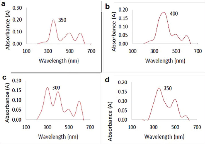
Fig. 1. The TEM images of synthesized nanoparticles. (a): Cestrum nocturnum- AgNPS; (b): Salvia malticaulis- AgNPs; (c): Cestrum nocturnum- CuNPS, and (d): Salvia malticaulis- CuNPs. Ultraviolet-Visible spectroscopyLocalized Surface Plasmon Resonance phenomenon of AgNPs and CuNPs synthesized using Ce. nocturnum and Sa. malticaulis leaves aqueous extracts were tested. The results exhibited a singular absorption peak within the region of 300–400 nm, signifying the existence of spherical-shaped AgNPs and CuNPs (Fig. 2). The reported absorption wavelength indicated an excitonic nature at ambient temperature.
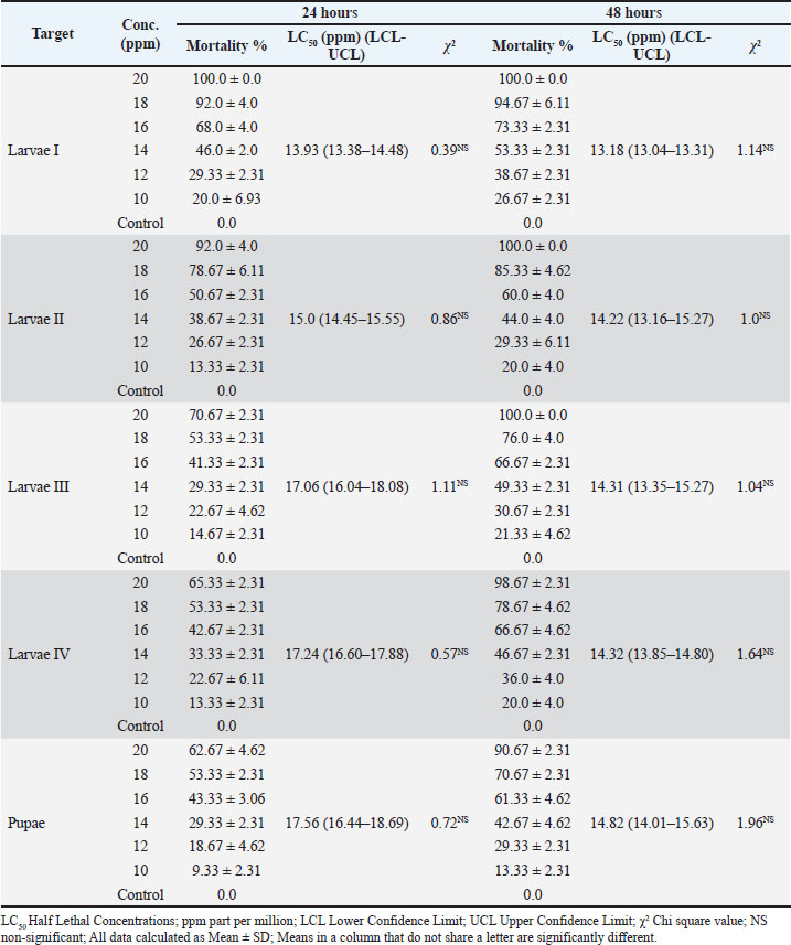
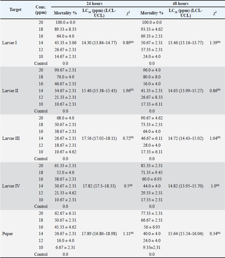
Fig. 2. Ultra-Violet (UV)—Visible curve of synthesized nanoparticles. (a): Cestrum nocturnum- AgNPS; (b): Salvia malticaulis- AgNPs; (c): Cestrum nocturnum- CuNPS, and (d): Salvia malticaulis- CuNPs. Activity of AgNPs and CuNPsAt the highest concentrations of Ce. nocturnum- AgNPs (20 and 18 mg/l), the mortality recorded 100.0%, 92.0% in An. sergentii first instar larvae (Larvae I); 92.0%, 78.67% in Larvae II, and 62.67%, 53.33% in pupae, respectively. The LC50 values of Ce. nocturnum- AgNPs against immature stages of An. sergentii recorded 13.93 and 13.18 mg/l against first instar larvae, 15.0 and 14.22 mg/l against second instar larvae, 17.06 and 14.31 mg/l against third instar larvae, 17.24 and 14.32 mg/l against fourth instar larvae, 17.56 and 14.82 mg/l against pupae after 24- and 48-hours post-treatment, respectively (Table 1 and Fig. 3). Table 1. Activity of Cestrum nocturnum-synthesized silver nanoparticles against Anopheles sergentii immature stages.
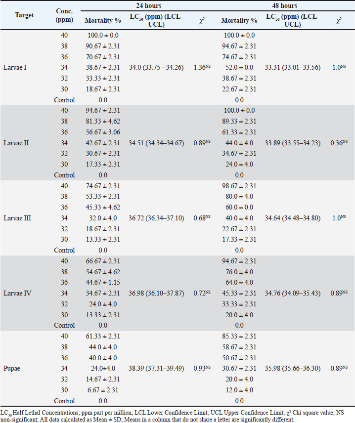
Fig. 3. Column chart represents the activity of tested silver and copper nanoparticles against Anopheles sergentii immature stages. Also, Sa. malticaulis—AgNPs recorded 100.0%, 96.0%, 90.67%, 85.33%, and 77.33% mortality in An. sergentii Larvae I, Larvae II, Larvae III, Larvae IV, and pupae at the highest concentration (20 mg/l) after 48 hours, respectively (Table 2 and Fig. 3). Table 2. Activity of Salvia malticaulis -synthesized silver nanoparticles against Anopheles sergentii immature stages.
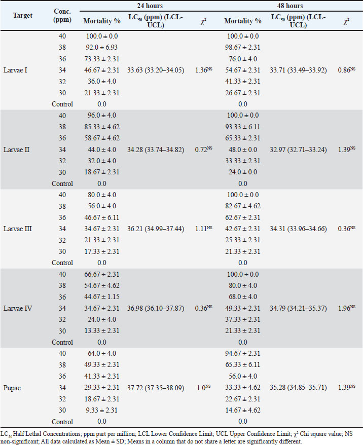
Regarding Ce. nocturnum and Sa. malticaulis synthesized CuNPs, the LC50 values recorded 33.63 & 34.0, 34.28 & 34.51, 36.21 & 36.72, 36.98 & 36.98, and 37.72 & 38.39 mg/l against Larvae I, Larvae II, Larvae III, Larvae IV, and pupae after 24 hours, respectively. Meanwhile, the LC50 values recorded 33.71 & 33.31, 32.97 & 33.89, 34.31 & 34.64, 34.79 & 34.76, and 35.28 & 35.98 mg/l against Larvae I, Larvae II, Larvae III, Larvae IV, and pupae after 48 hours, respectively (Tables 3, 4 and Fig. 3). Table 3. Activity of Cestrum nocturnum-synthesized copper nanoparticles against Anopheles sergentii immature stages.
Table 4. Activity of Salvia malticaulis -synthesized copper nanoparticles against Anopheles sergentii immature stages.
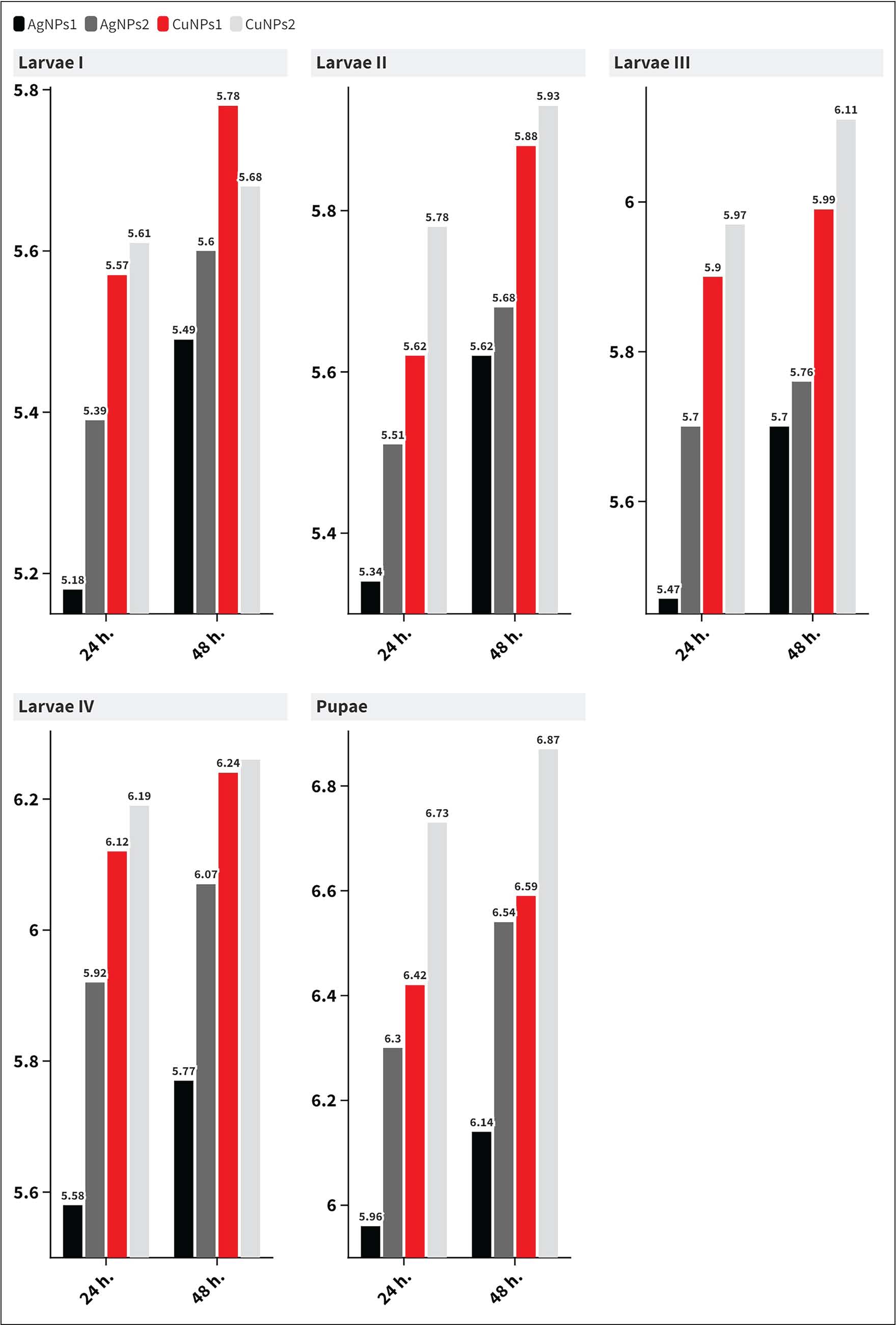
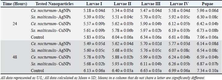
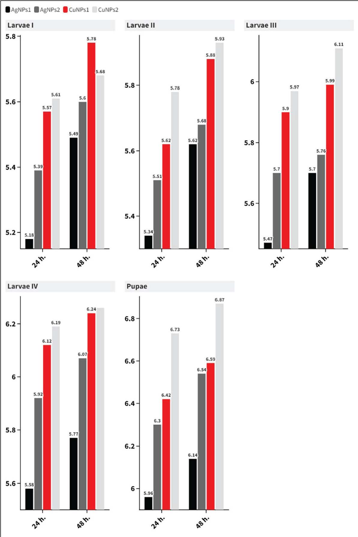
Acetylcholinesterase activityThe results of the ANOVA and Tukey's Pairwise Comparisons reveal significant differences in AChE activity between the control group and the various nanoparticle treatments in An. sergentii immature stages. At 24 hours, the control group exhibited the highest AChE activity (5.83 ± 0.05 U/l in Larvae I, 6.04 ± 0.06 U/l in Larvae II, 6.34 ± 0.06 U/l in Larvae III, 6.61 ± 0.08 U/l in Larvae IV, and 7.06 ± 0.06 U/l in Pupae), significantly higher than all treatments (p < 0.05). Tukey's pairwise comparison highlighted that CuNPs generally led to a slightly higher AChE activity compared to AgNPs, with Sa. malticaulis- CuNPs exhibiting the highest AChE activity among the treatments (6.73 ± 0.08 U/l in pupae). At 48 hours, the control group again showed the highest activity, while copper nanoparticles, particularly Sa. malticaulis- CuNPs, demonstrated prolonged inhibition of AChE, with the pupae stage showing 6.87 ± 0.07 U/l, respectively. Tukey's comparisons confirmed that treatments were significantly different (p < 0.05), suggesting that both AgNPs and CuNPs effectively reduced AChE activity in An. sergentii, with copper nanoparticles showing more persistent effects over time (Table 5 and Fig. 4). Table 5. The effect of green-synthesized silver and copper nanoparticles om Acetylcholinesterase (AChE) activity of Anopheles sergentii immature stages.
Fig. 4. Column chart represents the effect of green-synthesized silver and copper nanoparticles om Acetylcholinesterase (AChE) activity of An. sergentii immature stages. DiscussionThe TEM images of AgNPs and CuNPs synthesized using aqueous extracts of Ce. nocturnum and Sa. malticaulis leaves revealed that the sizes of the synthesized nanoparticles ranged from 8.39 to 113.68 nm for AgNPs and from 12.05 to 66.45 nm for CuNPs, respectively. The dimensions of synthesized nanoparticles corroborate the findings previously reported by Agalya Priyadarshini et al. (2012) regarding AgNPs prepared with Euphorbia hirta, Kumar et al. (2015) utilizing Morinda tinctoria leaf extract for AgNPs, Hassanain et al. (2019) employing petroleum ether extract from Lantana camara leaves for CuNPs, Shehata and Mahmoud (2019) using aqueous extract from Lagenaria siceraria leaves for AgNPs, and Zaki et al. (2024) applying aqueous extracts from Rosa arabica and Eucalyptus citriodora leaves for both AgNPs and CuNPs preparation. Furthermore, the UV–vis Spectrophotometric analysis results for both AgNPs and CuNPs exhibited a singular absorption peak within the 300–400 nm range, affirming the presence of spherical-shaped AgNPs and CuNPs, corroborating findings previously documented by El-Mehdawy et al. (2022); El-Waseif et al. (2023); and Mani et al. (2023). The efficacy of AgNPs and CuNPs, produced from aqueous extracts of Ce. nocturnum and Sa. malticaulis leaves, against An. sergentii augmented with higher concentrations of the tested nanoparticles. Calculated LC50 values indicate that AgNPs and CuNPs produced from the aqueous extract of Ce. nocturnum leaves were more efficacious against various stages of An. sergentii compared to those of Sa. malticaulis. The studied AgNPs showed greater efficacy against various immature stages of An. sergentii compared to CuNPs. The significant efficacy of green-synthesized nanoparticles against An. sergentii and various mosquito immature stages is due to their capacity to permeate the exoskeleton and infiltrate insect cells, where they impede macromolecules such as proteins and deoxyribonucleic acid, altering their structure and consequently their function (Subramaniam et al., 2015). The results obtained are consistent with those reported by Morejón et al. (2018) for AgNPs synthesized from Ambrosia arborescens extract against Aedes aegypti, Shehata and Mahmoud (2019) for AgNPs synthesized from aqueous extract of L. siceraria against Culex pipiens and An. pharoensis, Hassanain et al. (2019) for CuNPs synthesized from petroleum ether extract of La. camara leaves against An. multicolor, and Zaki et al. (2024) for AgNPs and CuNPs synthesized from aqueous extract of R. arabica and E. citriodora against Cu. antennatus larvae. A decrease in AChE levels in various immature stages of An. sergentii was observed. AChE activity assessments have become standard as a biomarker for exposure to specific types of contaminants (Grue et al., 1997). Nanoparticles may attach to and impede the function of AChE, which destroys acetylcholine, a crucial neurotransmitter in the central nervous system of insects (Nasir et al., 2022). The impact of Ce. nocturnum and Sa. malticaulis-synthesized AgNPs and CuNPs on AChE activity in various immature stages of An. sergentii corroborates the findings of Abdel-Gawad (2018), who noted a significant decrease in AChE activity in Musca domestica larvae after exposure to LC50 concentrations of Moringa oleifera-AgNPs and Moringa oleifera-ZnONPs treated diets compared to the control. Additionally, Parthiban et al. (2019) observed a notable reduction in the crucial esterase enzyme AChE in Ae. aegypti larval body homogenates following exposure to AgNPs relative to the control group. ConclusionIn conclusion, the Ce. nocturnum and Sa. malticaulis—synthesized AgNPs and CuNPs scattered uniformly in water and possess a significant activity against An. sergentii immature stages, reflecting the importance of relying on plant-synthesized nanoparticles in An. sergentii control for reducing the spread of diseases. Furthermore, there is an urgent need to develop more studies concerning the activity of green-synthesized AgNPs and CuNPs against several other mosquito species. AcknowledgmentsThe authors would like to thank colleagues in the Department of Zoology, Faculty of Science (Boys), Al-Azhar University, Egypt, Medical Entomologist at the Public Health Department, Jazan Municipality, Jazan, Saudi Arabia for their support. Conflict of interestThe authors declare no conflict of interest. FundingThis research received no fund. Authors' contributionAll authors contributed to the experimental design, hands on work, discussions, and commented on the manuscript. The final version of the manuscript read, reviewed, and approved by all authors. Data availabilityAny datasets used and/or analyzed during the current study are available from the corresponding author upon reasonable request. ReferencesA. El-Mehdawy et al.., A. 2021. The photosensitizing activity of different photosensitizers irradiated with sunlight against aquatic larvae of Culex pipiens L. (Diptera: culicidae). Egypt. J. Aquat. Biol. Fish. 25(5), 661–670; doi:10.21608/ejabf.2021.205672 Abbas, W.T., Abbas H.H., Abdel-Shafy, S., Shaapan, R.M., Ahmed, S.S. and Abdel-Kader, M.H. 2022. Evaluation of using of some novel natural nano-pesticides on fish health and water physico-chemical parameters. Egypt. J. Aquat. Biol. Fish. 26(2), 31–44; doi:10.21608/ ejabf.2022.223352 Abdel-Gawad, R. 2018. Insecticidal activity of Moringa oleifera synthesized silver and zinc nanoparticles against the house fly, Musca domestica L. Egypt. Acad. J. Biol. Sci. A. Entomol. 11(4), 19–30; doi:10.21608/eajbsa.2018.17729 Abdel-Shafy, S., Ghazy, A.A. and Shaapan, R.M. 2019. Applications of electron microscopy in ticks: description, detection of pathogens, and control. Comp. Clin. Pathol. 28(3), 585–592; doi:10.1007/s00580-018-2786-2 Agalya Priyadarshini, K., Murugan, K., Panneerselvam, C., Ponarulselvam, S., Hwang, J.S. and Nicoletti, M. 2012. Biolarvicidal and pupicidal potential of silver nanoparticles synthesized using Euphorbia hirta against Anopheles stephensi Liston (Diptera: culicidae). Parasitol. Res. 111(3), 997–1006; doi:10.1007/s00436-012-2924-8 Ahmed, S., Ahmad, M., Swami, B.L. and Ikram, S. 2016. A review on plants extract mediated synthesis of silver nanoparticles for antimicrobial applications: a green expertise. J. Ad. Res. 7(1), 17–28; doi:10.1016/j.jare.2015.02.007 Bhosale, R.R., Kulkarni, A.S., Gilda, S.S., Aloorkar, N.H., Osmani, R.A. and Harkare, B.R. 2014. Innovative eco-friendly approaches for green synthesis of silver. Int. J. Pharm. Sci. Nanotechnol. 7(1), 1; doi:10.37285/ijpsn.2014.7.1.3 Bobo, D., Robinson, K.J., Islam, J., Thurecht, K.J. and Corrie, S.R. 2016. Nanoparticle-based medicines: a review of FDA-approved materials and clinical trials to date. Pharm. Res. 33(10), 2373–2387; doi:10.1007/s11095-016-1958-5 Chen, G., Roy, I., Yang, C. and Prasad, P.N. 2016. Nanochemistry and nanomedicine for nanoparticle-based diagnostics and therapy. Chem. Rev. 116, 2826–2885; doi:10.1021/acs.chemrev.5b00148 Ellman, G.L., Courtney, K.D., Andres, V. and Featherstone, R.M. 1961. A new and rapid colorimetric determination of acetylcholinesterase activity. Biochem. Pharm. 7, 88–95; doi:10.1016/0006-2952(61)90145-9 El-Mehdawy, A.A., Koriem, M., Amin, R.M., Shehata, A.Z.I. and El-Naggar, H.A. 2022. Green synthesis of silver nanoparticles using chitosan extracted from Penaeus indicus and its potential activity as aquatic larvicidal agent of Culex pipiens. Egypt. J. Aquat. Biol. Fish. 26(1), 425–442; doi:10.21608/ejabf.2022.219887 El-Tabakh, M.A.M., Elhawary, E.A., Hwihy, H.M., Darweesh, K.F., Shaapan, R.M., Ghazala, E.A., Mokhtar, M.M., Waheeb, H.O., Emam, D.E.M., Bakr, N.A. and Shehata, A.Z.I. 2023. UPLC/ESI/MS profiling of red algae Galaxaura rugosa extracts and its activity against malaria mosquito vector, Anopheles pharoensis, with reference to Danio rerio and Daphnia magna as bioindicators. Malar. J. 22(1), 368; doi:10.1186/s12936-023-04795-w El-Waseif, A.A., Shehata, A.Z.I., Waheeb, H.O. and El-Ghwas, D.E. 2023. Correlation of probiotic synthesis nanoparticles against Rift Valley Fever Vector, Culex antennatus Becker (Diptera: culicidae). Res. J. Pharm. Technol. 16(6), 2969–2974; doi:10.52711/0974-360X.2023.00490 Grue, C.E., Gilbert, P.L. and Seeley, M.E. 1997. Neuropsychological and behavioral changes in non-target wildlife exposed to organophosphate and carbamate pesticide: thermoregulation, food consumption and reproduction. Am. Zool. 37, 369–388; https://www.jstor.org/stable/3884019 Harbach, R.E. 1985. Pictorial keys to the genera of mosquitoes, subgenera of Culex and the species of Culex (Culex) occurring in southwestern Asia and Egypt, with a note on the subgeneric placement of Culex deserticola (Diptera: culicidae). Mosq. Syst. 17, 83–107. Hassanain, N.A., Shehata, A.Z.I., Mokhtar, M.M., Shaapan, R.M. Hassanain, M.A. and Zaky, S. 2019. Comparison between insecticidal activity of Lantana camara extract and it’s synthesized nanoparticles against Anopheline mosquitoes. Pak. J. Biol. Sci. 22(7), 327–334; doi: 10.3923/PJBS.2019.327.334 Hay, S.I., Guerra, C.A., Gething, P.W., Patil, A.P., Tatem, A.J., Noor, A.M., Kabaria, C.W., Manh, B.H., Elyazar, I.R.F., Brooker, S., Smith, D.L., Moyeed, R.A. and Snow, R.W. 2009. A world malaria map: Plasmodium falciparum endemicity in 2007. PLos Med. 6(3), e1000048; doi:10.1371/journal.pmed.1000048 Khater, E.I., Sowilem, M.M., Sallam, M.F. and Alahmed, A.M. 2013. Ecology and habitat characterization of mosquitoes in Saudi Arabia. Trop. Biomed. 30(3), 409–427; https://pubmed.ncbi.nlm.nih.gov/24189671/ Kirkpatrick, T.W. 1925. The mosquitoes of Egypt. Cairo, Egypt: Egyptian Government, antimalaria commission, Government Press. pp: 224. Kumar, K.R., Nattuthurai, N., Gopinath, P. and Mariappan, T. 2015. Synthesis of eco-friendly silver nanoparticles from Morinda tinctoria leaf extract and its larvicidal activity against Culex quinquefasciatus. Parasitol. Res. 114, 411–417; doi:10.1007/s00436-014-4198-9 Liu, N., Xu, Q., Zhu, F. and Zhang, L. 2006. Pyrethroid resistance in mosquitoes. Insec. Sci. 13(3), 159–166; doi:10.1111/j.1744-7917.2006.00078.x Mali, S.C., Dhaka, A., Githala, C.K. and Trivedi, R. 2020. Green synthesis of copper nanoparticles using Celastrus paniculatus Willd. leaf extract and their photocatalytic and antifungal properties. Biotechnol. Rep. 27, e00518; doi: 10.1016/j.btre.2020 Mani, M., Sundararaj, A.S., Al-Ghanim, K.A., John, S.P., Elumalai, K., Nicoletti, M. and Govindarajan, M. 2023. Rapid synthesis of copper nanoparticles using nepeta cataria leaves: an eco-friendly management of disease-causing vectors and bacterial pathogens. Green Process. Synth. 12(1), 20230022; doi:10.1515/gps-2023-0022 Morejon, B., Pilaquinga, F., Domenech, F., Ganchala, D., Debut, A. and Neira, M. 2018. Larvicidal activity of silver nanoparticles synthesized using extracts of Ambrosia arborescens (Asteraceae) to control Aedes aegypti L. (Diptera: culicidae). J. Nanotechnol. 2018(1), 6917938; doi:10.1155/2018/6917938 Morsy, T.A., El Okbi, L.M., Kamal, A.M., Ahmed, M.M. and Boshara, E.F. 1990. Mosquitoes of the genus Culex in the Suez Canal Governorates. J. Egypt. Soc. Parasitol. 20(1), 265–268. Available via https://pubmed.ncbi.nlm.nih.gov/2332654/ Nasir, S., Walters, K.F.A., Pereira, R.M., Waris, M., Chatha, A.A., Hayat, M. and Batool, M. 2022. Larvicidal activity of acetone extract and green synthesized silver nanoparticles from Allium sativum L. (Amaryllidaceae) against the dengue vector Aedes aegypti L. (Diptera: culicidae). J. Asia Pac. Entomol. 25(3), 101937; doi:10.1016/j.aspen.2022.101937 Parthiban, E., Ramachandran, M., Jayakumar, M. and Ramanibai, R. 2019. Biocompatible green synthesized silver nanoparticles impact on insecticides resistant developing enzymes of dengue transmitted mosquito vector. SN. Appl. Sci. 1, 1282; doi:10.1007/s42452-019-1311-9 Sarih, M., Filali, O., Faraj, C., Kabine, M. and Debboun, M. 2019. Entomological status of Anopheles sergentii and the first molecular investigation of its insecticide-resistant genes, kdr and ace-1 in Morocco. J. Vector. Borne. Dis. 56(4), 308–314; doi: 10.4103/0972-9062.302033 Shehata, A.Z.I., El-Tabakh, M.A.M., Waheeb, H.O., Emam, D.E.M. and Mokhtar, M.M. 2022. Seasonal abundance and molecular identification of aquatic larvae of Culex pipiens L. and Culex antennatus Becker in Fayoum Governorate, Egypt. Egypt. J. Aquat. Biol. Fish. 26(6), 751–764; doi:10.21608/ejabf.2022.275615 Shehata, A.Z.I., Abd-Elkhalek, H.F., Hwihy, H.M., Bakr, N.A., Shahat, M.A.M., Ward, W.M.A., Elnaggar, A.M.A., Zidan, M.M.M. and El-Tabakh, M.A.M. 2023. DNA barcoding and sensitivity of Dengue Vector Aedes aegypti (Linnaeus, 1762) (Diptera: culicidae) aquatic stages to different insecticides with reference to non-target organisms, Danio rerio and Daphnia magna. Egypt. J. Aquat. Biol. Fish. 27(6), 169–186; doi:10.21608/ejabf.2023.328036 Shehata, A.Z. and Mahmoud, A.M. 2019. Efficacy of leaves aqueous extract and synthesized silver nanoparticles using Lagenaria siceraria against Culex pipiens Liston and Anopheles pharoensis Theobald. J. Egypt. Soc. Parasitol. 49(2), 381–387; doi:10.21608/jesp.2019.68148 Shehata, A.Z.I., EL-Sheikh, T.M., Shaapan, R.M., Abd El-Shafy, S. and Alanazi, A.D. 2020. Ovicidal and latent effect of Pulicaria jaubertii (Asteraceae) leaf extracts on Aedes Aegypti. J. Am. Mosq. Control Assoc. 36(3), 161–166; doi:10.2987/20-6952.1 Sirelkhatim, A., Mahmud, S., Seeni, A., Kaus, N.H.M., Ann, L.C., Bakhori, S.K.M., Hasan, H. and Mohamad, D. 2015. Review on zinc oxide nanoparticles: antibacterial activity and toxicity mechanism. Nanomicro Lett. 7(3), 219–242; doi:10.1007/s40820-015-0040-x Subramaniam, J., Murugan, K., Panneerselvam, C., Kovendan, K., Madhiyazhagan, P., Kumar, P.M., Dinesh, D., Chandramohan, B., Suresh, U., Nicoletti, M., Higuchi, A., Hwang, J.S., Kumar, S., Alarfaj, A.A., Munusamy, M.A., Messing, R.H. and Benelli, G. 2015. Ecofriendly control of malaria and arbovirus vectors using the mosquitofish Gambusia affinis and ultra-low dosages of Mimusops elengi-synthesized silver nanoparticles: towards an integrative approach?. Environ. Sci. Pollut. Res. Int. 22, 20067–20083; doi:10.1007/s11356-015-5253-5 WHO. 2004. Integrated vector management: strategic framework for the Eastern Mediterranean Region 2004–2010. Available via https://iris.who.int/handle/10665/119705 WHO. 2021. World malaria report. Geneva: World Health Organization. Available via https://www.who.int/publications/i/item/9789240040496 Zaki, M.S.M., Bream, A.S. and Shehata, A.Z.I. 2024. Biosynthesis of silver and copper nanoparticles using Rosa arabica (Rosaceae) and Eucalyptus citriodora (Myrtaceae) extracts and its biological activity against Culex antennatus becker (Diptera: culicidae). Egypt. J. Vet. Sci. 55(6), 1685–1696; doi:10.21608/ejvs.2024.265721.1806 | ||
| How to Cite this Article |
| Pubmed Style Shehata AZ, Ayad MAA, Alteab AA, Abdel-aziz ANG, Shahat MAM, Shaapan RM. Laboratory efficacy of green-synthesized silver and copper nanoparticles against malaria vector, Anopheles sergentii (Theobald, 1907) (Diptera: Culicidae). Open Vet. J.. 2026; 16(5): 2869-2882. doi:10.5455/OVJ.2026.v16.i5.29 Web Style Shehata AZ, Ayad MAA, Alteab AA, Abdel-aziz ANG, Shahat MAM, Shaapan RM. Laboratory efficacy of green-synthesized silver and copper nanoparticles against malaria vector, Anopheles sergentii (Theobald, 1907) (Diptera: Culicidae). https://www.openveterinaryjournal.com/?mno=295809 [Access: June 26, 2026]. doi:10.5455/OVJ.2026.v16.i5.29 AMA (American Medical Association) Style Shehata AZ, Ayad MAA, Alteab AA, Abdel-aziz ANG, Shahat MAM, Shaapan RM. Laboratory efficacy of green-synthesized silver and copper nanoparticles against malaria vector, Anopheles sergentii (Theobald, 1907) (Diptera: Culicidae). Open Vet. J.. 2026; 16(5): 2869-2882. doi:10.5455/OVJ.2026.v16.i5.29 Vancouver/ICMJE Style Shehata AZ, Ayad MAA, Alteab AA, Abdel-aziz ANG, Shahat MAM, Shaapan RM. Laboratory efficacy of green-synthesized silver and copper nanoparticles against malaria vector, Anopheles sergentii (Theobald, 1907) (Diptera: Culicidae). Open Vet. J.. (2026), [cited June 26, 2026]; 16(5): 2869-2882. doi:10.5455/OVJ.2026.v16.i5.29 Harvard Style Shehata, A. Z., Ayad, . M. A. A., Alteab, . A. A., Abdel-aziz, . A. N. G., Shahat, . M. A. M. & Shaapan, . R. M. (2026) Laboratory efficacy of green-synthesized silver and copper nanoparticles against malaria vector, Anopheles sergentii (Theobald, 1907) (Diptera: Culicidae). Open Vet. J., 16 (5), 2869-2882. doi:10.5455/OVJ.2026.v16.i5.29 Turabian Style Shehata, Ahmed Z.i., Magdy A. A. Ayad, Abdulnassar A. Alteab, Ahmed N. G. Abdel-aziz, Mohamed A. M. Shahat, and Raafat M. Shaapan. 2026. Laboratory efficacy of green-synthesized silver and copper nanoparticles against malaria vector, Anopheles sergentii (Theobald, 1907) (Diptera: Culicidae). Open Veterinary Journal, 16 (5), 2869-2882. doi:10.5455/OVJ.2026.v16.i5.29 Chicago Style Shehata, Ahmed Z.i., Magdy A. A. Ayad, Abdulnassar A. Alteab, Ahmed N. G. Abdel-aziz, Mohamed A. M. Shahat, and Raafat M. Shaapan. "Laboratory efficacy of green-synthesized silver and copper nanoparticles against malaria vector, Anopheles sergentii (Theobald, 1907) (Diptera: Culicidae)." Open Veterinary Journal 16 (2026), 2869-2882. doi:10.5455/OVJ.2026.v16.i5.29 MLA (The Modern Language Association) Style Shehata, Ahmed Z.i., Magdy A. A. Ayad, Abdulnassar A. Alteab, Ahmed N. G. Abdel-aziz, Mohamed A. M. Shahat, and Raafat M. Shaapan. "Laboratory efficacy of green-synthesized silver and copper nanoparticles against malaria vector, Anopheles sergentii (Theobald, 1907) (Diptera: Culicidae)." Open Veterinary Journal 16.5 (2026), 2869-2882. Print. doi:10.5455/OVJ.2026.v16.i5.29 APA (American Psychological Association) Style Shehata, A. Z., Ayad, . M. A. A., Alteab, . A. A., Abdel-aziz, . A. N. G., Shahat, . M. A. M. & Shaapan, . R. M. (2026) Laboratory efficacy of green-synthesized silver and copper nanoparticles against malaria vector, Anopheles sergentii (Theobald, 1907) (Diptera: Culicidae). Open Veterinary Journal, 16 (5), 2869-2882. doi:10.5455/OVJ.2026.v16.i5.29 |


