





| Research Article | ||
Open Vet. J.. 2026; 16(5): 3052-3059 Open Veterinary Journal, (2026), Vol. 16(5): 3052-3059 Research Article Histological effect of methotrexate on submandibular salivary glands in albino rats (Rattus norvegicus)Bader Khatlan Hameed1, Mahmood Nawfal Mustafa1* and Shaymaa Abdalkader Mahdy21Department of Anatomy and Histology, College of Veterinary Medicine, Tikrit University, Tikrit, Iraq 2Department of Oral Diagnosis, College of Dentistry, Tikrit University, Tikrit, Iraq *Corresponding Author: Mahmood Nawfal Mustafa. Department of Anatomy and Histology, College of Veterinary Medicine, Tikrit University, Tikrit, Iraq. Email: mahmood_nawfal [at] tu.edu.iq Submitted: 18/10/2025 Revised: 22/02/2026 Accepted: 04/03/2026 Published: 31/05/2026 © 2025 Open Veterinary Journal
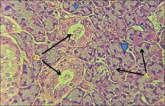
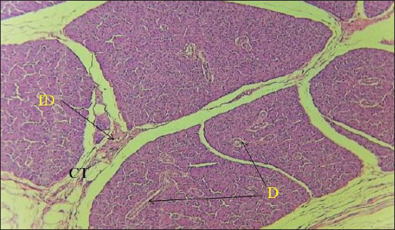
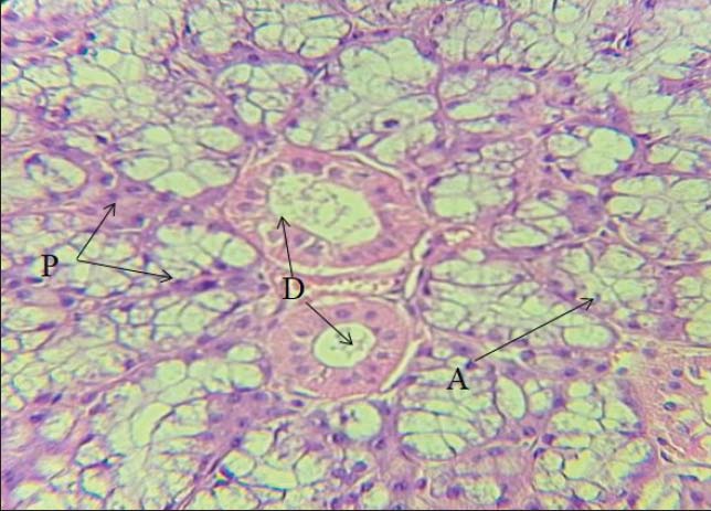
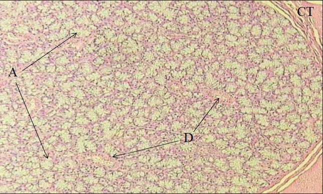
ABSTRACTBackground: Methotrexate (MTX), a potent chemotherapeutic agent, is known for its efficacy in the treatment of various malignancies. However, its use is often accompanied by adverse effects on non-target tissues, including the salivary glands. Aim: This study aimed to identify the histological events and effects of MTX on the salivary glands of the experimental animals (rats). Methods: This study used albino rats (Rattus norvegicus). The patients were divided into two groups: a control group that received normal saline and a treatment group that received a therapeutic dose of MTX. The animals were euthanized after the specified treatment duration, and their submandibular glands were harvested for histological examination. Tikrit University, College of Veterinary Medicine, from October 2023 to December 2023. Results: Histological analysis revealed significant alterations in the submandibular gland tissue of the MTX-treated animals compared with the control group. These histological alterations underscore the potential cytotoxic effects of MTX on the salivary glands, which may contribute to the development of xerostomia (dry mouth) and other oral complications in patients undergoing MTX therapy. Conclusion: MTX administration in albino rats induces histological changes in the submandibular salivary glands. Therefore, further studies using special dyes are recommended to detect more specific changes, as well as the use of some medicinal herbs to reduce the harmful effects of the drug. Keywords: Albino rat, Histology, Histopathological changes, Methotrexate, Submandibular glands. IntroductionAt the start of the 20th century, surgery was the sole treatment available to patients with solid tumors, which led to a high death rate. Over the past 40 years, chemotherapy has steadily increased the survival rates of individuals with tumors. Methotrexate (MTX), a cytotoxic chemotherapy medication, is used to treat some autoimmune disorders and malignant tumors. MTX exerts cytotoxic effects in cancer cells (Fathi et al., 203). Regretfully, it not only targets malignant cells but also destroys healthy tissues, particularly those with a high rate of proliferation, such as the hematopoietic cells of the bone marrow and the actively proliferating cells of the intestinal mucosa. Mucositis is one of the most common toxic adverse effects of chemotherapy. It is also one of the most common adverse reactions of MTX. In addition to the above, the drug affects taste, swallowing, and speech. Furthermore, oral mucositis causes discomfort in the pharynx, oral tissue inflammation, tooth decay, and tooth loss. The infection may spread to the bloodstream and cause sepsis. All of these conditions delay recovery from illness by affecting the nutrition and psychological state of patients during and after treatment (Pickett and Terezhalmy, 2009). Due to its multiple components, saliva, which is secreted by the acinar cells of the salivary glands and controlled by their duct cells, plays a pivotal role in maintaining the health and integrity of oral tissues; it also has numerous immunological properties against both microbes and infections (Al-Moula et al., 2020). The main salivary glands release 90% of saliva daily. When it malfunctions, salivary flow and composition are altered, leading to xerostomia (dry mouth), which is clinically diagnosed as oral mucositis (Taha et al., 2024; Abd et al., 2025). Chemotherapy causes this dysfunction, which heightens the secondary mucosal toxic effects of the drug (Singh et al., 2018). Chemotherapy medications have many side effects; thus, avoiding their toxicity is important. Systemic oxidative stress is one of the primary mechanisms of MTX-induced toxicity. It significantly reduces Glutathione levels, affecting the antioxidant defense system. Similarly, MTX decreases nucleic acid metabolism (El Emam et al., 2019; Abd, 2024). MTX has been a widely used chemotherapeutic medication for many years. It is used to treat autoimmune illnesses, including psoriasis, lichen planus, and rheumatoid arthritis, as well as a variety of malignancies, such as acute leukemia and osteosarcoma. The primary mechanism of action of MTX is to inhibit DNA synthesis, thereby reducing the growth of these aberrant cells (Fathi et al., 2023). Oral mucositis is one of the most common side effects of chemotherapy. Chemotherapy causes direct damage or atrophy of mucosal epithelial cells, which may manifest as widespread or localized ulcers. MTX has a cytotoxic effect on the epithelial cell layer, leading to DNA damage in the basal progenitor cells. This results in cell death and malfunction (Al-Refai et al., 2014). Materials and MethodsThe study included 10 adult male rats (Rattus norvegicus), each weighing about 160–180 g. To exclude the influence of sex differences, only male rats were selected. The experiment was conducted from October 2023 to December 2023 at the Animal House Facility of the College of Veterinary Medicine at Tikrit University. The animals were housed in individual cages in healthy, controlled conditions and provided with food and water according to international standards. The animals were housed in well-ventilated plastic cages with stainless steel surfaces and wood shaving bedding. The temperature was regulated at 25℃ ± 2℃. Laboratory conditions allowed an acclimatization period of 28 days before the animals were used in the experiments. Design of experimentThe experimental animals were divided into two groups: Group I: The control group (G1). Five animals received normal saline and a balanced diet for 28 days. Group II: MTX-treated group (G2). Five animals were injected intraperitoneally weekly as a single dose of MTX (20 mg/kg body weight) for 28 days. After the treatment period, the patients were transported to the veterinary anatomy laboratory at Tikrit University. All animals were exposed to chloroform on the day of general anesthesia. The midventral incision was extended from the inguinal region to the mandibular symphysis. For preservation, the submandibular salivary gland was fixed in 10% neutral buffered formalin. The stained slides were examined using an ALTAY light microscope at the Department of Anatomy, Faculty of Veterinary Medicine, Tikrit University. Ethical approvalThis study was prepared based on the ethical instructions of the College of Veterinary Medicine, Tikrit University (Approval No. Tu. 95). ResultsControl group: The results of the current study showed the normal condition of the tissues of the submandibular salivary gland for both acini regions (Figs. 1 and 2) and the serous acini region (Figs. 3 and 4), where the normal condition of the interlobular ducts and the intralobular ducts was observed, as was the normal condition of the secretory acini.
Fig. 1. (G1), control group, shows the normal state of the serous region of the submandibular gland (G1). Intralobular (D) duct, acini (A), pyramidal cell (H &E. ×60).
Fig. 2. (G1), control group, shows the normal state of the serous region of the submandibular gland, interlobular connective tissue (CT), interlobular duct (ID), and intralobular duct (D) (H &E. ×20).
Fig. 3. (G1), control group, shows the normal state of the submandibular gland mucus region. Intralobular duct (D), acini (A), and pericytes (P) (H &East ×60).
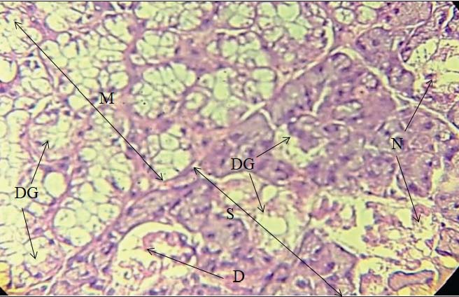
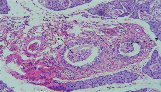
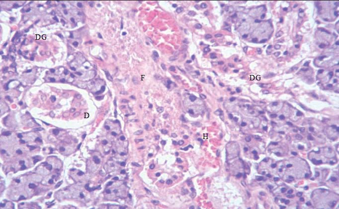
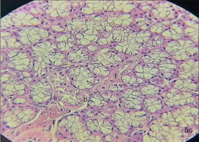
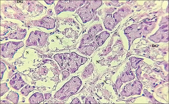
Fig. 4. (G1), control group, shows the normal state of the submandibular gland mucus region. Intralobular duct (D), interlobular connective tissue (CT), and acini (A) (H &East ×20). Treated groups: While these results showed the occurrence of several changes and the appearance of lesions at the histological level in the entire tissues of the submandibular salivary glands in animals treated with MTX, the drug's intensity appears to affect the serous area more than the mucous area. The mucous area was characterized by necrosis and degeneration of the salivary ducts. The interlobular and intralobular ducts are shown in Figure 5. Inflammatory cells and blood clots infiltrated the capillaries (Fig. 6). Hemorrhage was also noted in the gland tissue between the serous acini, with desquamation of the epithelial lining of the intralobular ducts, and saliva clotting in some others. Fibrosis appeared between the acini and around the intralobular ducts, and its expansion within the interlobular connective tissue (Figs. 7–10).
Fig. 5. (G2) shows the effect of the drug severity on the serous region more than the mucus region and the mucus region (M). Serous region (S). Desquamated intercalated duct (D), necrotic acini (N), and degeneration (DG) (H &East ×60). Fig. 6. (G2), treated group shows the presence and infiltration of inflammatory cells (IF), blood clot (B), degenerating duct (D), and general degeneration (DG) (H &E. ×60).
Fig. 7. (G2), treated group, shows a serous region and the presence of interlobular fibrosis (F), inflammatory cells (IF), hemorrhage (H), and thickened saliva (C). (H &East ×20).
Fig. 8. (G2), treated group, shows a serous region and the presence of intralobular fibrosis (F), desquamated epithelial cells (D), hemorrhage (H), and degeneration (DG) (H &E. ×60).
Fig. 9. (G2) shows the mucus region and the presence of intralobular fibrosis (F), desquamated epithelial cells (D), and degeneration (DG) in the treated group (H &E. ×40).
Fig. 10. (G2), treated group, shows the serous region and the presence of necrotic (N), and degenerated ducts (DG) (H &East ×60). DiscussionThe salivary glands have been used in research to understand fundamental pharmacological issues and the close relationship between oral health and the health of the whole body, thanks to saliva produced by the salivary glands (Howard et al., 2016). Dosing with the chemotherapy drug MTX leads to a systematic loss of salivary gland tissue function, with a marked decrease in saliva production (Bancroft et al., 2012). The submandibular gland is considered the 2nd-largest salivary gland in the body; therefore, it was chosen. This gland produces more than 40% of the body’s saliva. Mucosal inflammation, extensive dental caries, pharyngitis, and swallowing, speaking, and tasting difficulties are manifestations of salivary insufficiency that may affect patients undergoing treatment for malignant tumors or radiation therapy (Ayoub et al., 2006). The action mechanism of MTX, a folate antagonist, is to lower the action of a number of enzymes depending on folate, inhibit DNA synthesis, and promote cell division and renewal. Patients with cancer benefit from MTX as a chemotherapy drug given to different age groups. Simultaneously, the main reason for its toxicity is the increase in systemic oxidative stress in the body (Mohan and Huneke, 2019). Current study results indicate: MTX can damage the submandibular gland tissue in experimental animals by halting and/or inhibiting protein synthesis as a result of folate cofactor depletion, which may cause the formation of cytosolic particles, a marker of apoptotic reactions throughout the time of MTX treatment, and the damage to the submandibular gland may result from oxidative stress caused by free radicals (Fadare and O, 2003). The onset of apoptosis or oncotic cell death is always thought to be followed by necrosis, with the term “oncosis” being the opposite of cell death and bulge. Previous research explains why localized alterations, such as oncosis, were detected in acinar cells of the Submandibular gland in this study (Cizkova et al., 2021). In addition, a hemorrhage between the glandular tissue cells was observed, with obvious degradation, diapedesis of inflammatory cells, and bloody congestion in the acinar vessels. These results have been confirmed. They are explained by methotrexa-induced oxidative stresste. It has been proven that the metabolism of drugs within the cell cavity depletes antioxidants, whose action depends on glutathione, and generates oxygen-free radicals (Mahmoud and Mahmoud, 2017). Lipid peroxidation and subsequent lysis of organelles and plasma membranes result from this imbalance between oxidants and antioxidants.tS The goal of these inflammatory responses may be to increase blood flow to the damaged areas of the body (Mahmoud and Mahmoud, 2017). Significant increases in collagen fibers in the ileum, liver, and kidney after MTX exposure (Doostan et al., 2019). This was supported by our results, which showed glandular degeneration, a prominent increase in connective tissue between acini and around ducts, and a reduction in parenchymal structures. ConclusionIn conclusion, the administration of MTX in albino rats induces histological changes in the salivary glands, characterized by parenchymal degeneration, stromal fibrosis, and vascular alterations. AcknowledgmentsWe are thankful for the help and support provided by the Veterinary Medicine College/University of Tikrit in conducting this research work. Conflict of interestThe authors have no conflicts of interest to declare. FundingThis study received no grant; it was supported by self-funding. Authors' contributionsAssi. Prof. Dr. Mahmood Nawfal Mustafa and Assi Prof. Dr. Bader Khatlan Hameed were the corresponding author for this article and was responsible for animal care, dissecting laboratory animals, and obtaining tissue samples. Assi. Prof. Dr. Shaymaa Abdalqader Mahdy was responsible for study observations and research management. Article writing was done equally by the authors. Data availabilityAll data are provided in the manuscript. ReferencesAbd, A.A. 2024. Impact of sulpiride and PMSG hormone treatment on estrus and number of resulted offsprings in female rats. Assiut. Vet. Med. J. 70(183), 1–6. Abd, A.A., Al-Juhaishi, O.A. and Jumma, Q.S. 2025. Effects of sulpiride on the reproductive system of male rats after puberty. World Vet. J. 15(1), 42–48. Al-Moula, A.D., Ahmed, M.H. and Ahmed, M.H. 2020. Histological study on the effect of methotrexate on the oral tissues of adult male rabbits. Al-Rafidain Dent. J. 13(3), 432–441. Al-Refai, A.S., Khaleel, A.K. and Ali, S. 2014. The effect of green tea extract on submandibular salivary gland of methotrexate treated albino rats: immunohistochemical study. J. Cytol. Histol. 5(2), 1. Ayoub, R.S., Hadi, A.D. and Al-Shandok, T.A. 2006. Effect of natural apple cider vinegar on morphometric changes of salivary glands in hydroxyurea treated mice. J. Edu. Sci. 18(3), 62–77. Bancroft, J.D., Kim, S.S. and Christopher, L. 2012. Bancroft's theory and practice of histological techniques, Cizkova, K., Foltynkova, T., Gachechiladze, M. and Tauber, Z. 2021. Comparative analysis of immunohistochemical staining intensity determined by light microscopy, ImageJ and QuPath in placental Hofbauer cells. Acta Histoch. Cytoch. 54(1), 21–29. Doostan, F., Mesgari Abbasi, M., Khordadmehr, M., Fallah, F. and Behrouzy, A. 2019. Effects of pomegranate seed and peel methanolic extracts on methotrexate-induced hepatotoxicity in rats. Pharm. Sci. 25(2), 111–117. El Emam, H., Abd El Salam, N. and Ghazy, S. 2019. Impact of bone marrow stem cells application on the methotrexate induced submandibular salivary gland degenerative changes in albino rats (histological and immuno-histochemical study). Egypt. Den. J. 65(2), 1751–1762. Fadare, O. 2003. Digital histology: an interactive CD atlas with review text. Yale. J. Biol. Med. 76(4-6), 194–195. Fathi, R., Abd Allah, D. and Helmy, I. 2023. Effect of green tea and black seed on methotrexate induced cytotoxicity of the oral mucosa, tongue and the submandibular salivary gland of albino rats. Egypt. Dent. J. 69(1), 413–426. Howard, S.C., Mccormick, J., Pui, C.H., Buddington, R.K. and Harvey, R.D. 2016. Preventing and managing toxicities of high-dose methotrexate. Oncologist 21(12), 1471–1482. Mahmoud, E. and Mahmoud, M. 2017. Effect of pomegranate peel extract on submandibular salivary glands of streptozotocin-induced diabetes in rats: histological, immunohistochemical and ultrastructural study. J. Advan. Biol. Biotechnol. 13(1), 1–15. Mohan, S. and Huneke, R. 2019. The role of IACUCs in responsible animal research. ILAR J. 60(1), 43–49. Pickett, F.A. and Terezhalmy, G.T. 2009. Basic principles of pharmacology with dental hygiene applications. Philadelphia, PA: Wolters Kluwer Health/Lippincott Williams & Wilkins. Singh, K., Bhori, M., Kasu, Y.A., Bhat, G. and Marar, T. 2018. Antioxidants as precision weapons in war against cancer chemotherapy induced toxicity–exploring the armoury of obscurity. Saudi. Pharm. J. 26(2), 177–190. Taha , R.M., Abdel-Latif, G.A. and Said, R.H. 2024. The prospective effect of green tea versus pomegranate peels extracts on submandibular salivary glands of albino rats after methotrexate administration (histological and immunohistochemical study). Int. J. Den. 3290187, 1–12. | ||
| How to Cite this Article |
| Pubmed Style Hameed BK, Mustafa MN, Mahdy SA. Histological effect of methotrexate on submandibular salivary glands in albino rats (Rattus norvegicus). Open Vet. J.. 2026; 16(5): 3052-3059. doi:10.5455/OVJ.2026.v16.i5.47 Web Style Hameed BK, Mustafa MN, Mahdy SA. Histological effect of methotrexate on submandibular salivary glands in albino rats (Rattus norvegicus). https://www.openveterinaryjournal.com/?mno=291203 [Access: June 26, 2026]. doi:10.5455/OVJ.2026.v16.i5.47 AMA (American Medical Association) Style Hameed BK, Mustafa MN, Mahdy SA. Histological effect of methotrexate on submandibular salivary glands in albino rats (Rattus norvegicus). Open Vet. J.. 2026; 16(5): 3052-3059. doi:10.5455/OVJ.2026.v16.i5.47 Vancouver/ICMJE Style Hameed BK, Mustafa MN, Mahdy SA. Histological effect of methotrexate on submandibular salivary glands in albino rats (Rattus norvegicus). Open Vet. J.. (2026), [cited June 26, 2026]; 16(5): 3052-3059. doi:10.5455/OVJ.2026.v16.i5.47 Harvard Style Hameed, B. K., Mustafa, . M. N. & Mahdy, . S. A. (2026) Histological effect of methotrexate on submandibular salivary glands in albino rats (Rattus norvegicus). Open Vet. J., 16 (5), 3052-3059. doi:10.5455/OVJ.2026.v16.i5.47 Turabian Style Hameed, Bader Khatlan, Mahmood Nawfal Mustafa, and Shaymaa Abdalkader Mahdy. 2026. Histological effect of methotrexate on submandibular salivary glands in albino rats (Rattus norvegicus). Open Veterinary Journal, 16 (5), 3052-3059. doi:10.5455/OVJ.2026.v16.i5.47 Chicago Style Hameed, Bader Khatlan, Mahmood Nawfal Mustafa, and Shaymaa Abdalkader Mahdy. "Histological effect of methotrexate on submandibular salivary glands in albino rats (Rattus norvegicus)." Open Veterinary Journal 16 (2026), 3052-3059. doi:10.5455/OVJ.2026.v16.i5.47 MLA (The Modern Language Association) Style Hameed, Bader Khatlan, Mahmood Nawfal Mustafa, and Shaymaa Abdalkader Mahdy. "Histological effect of methotrexate on submandibular salivary glands in albino rats (Rattus norvegicus)." Open Veterinary Journal 16.5 (2026), 3052-3059. Print. doi:10.5455/OVJ.2026.v16.i5.47 APA (American Psychological Association) Style Hameed, B. K., Mustafa, . M. N. & Mahdy, . S. A. (2026) Histological effect of methotrexate on submandibular salivary glands in albino rats (Rattus norvegicus). Open Veterinary Journal, 16 (5), 3052-3059. doi:10.5455/OVJ.2026.v16.i5.47 |


