





| Research Article | ||
Open Vet. J.. 2026; 16(5): 2685-2695 Open Veterinary Journal, (2026), Vol. 16(5): 2685-2695 Research Article Magnesium oxide nanoparticles: A novel shield against CCl₄-induced hepatic damage via caspase-3 and TNF-α in ratsHarseen M. Rahim1*, Nadia A. Salih1 and Bruska Azhdar21Department of Basic Sciences, College of Veterinary Medicine and Surgery, University of Sulaimani, Sulaymaniyah, Iraq 2Department of Physics, Nanotechnology Research Laboratory, College of Science, University of Sulaimani, Sulaymaniyah, Iraq *Corresponding Author: Harseen M. Rahim. Department of Basic Sciences, College of Veterinary Medicine and Surgery, University of Sulaimani, Sulaymaniyah, Iraq. Email: harseen.rahim [at] univsul.edu.iq Submitted: 22/03/2026 Revised: 19/04/2026 Accepted: 23/04/2026 Published: 31/05/2026 © 2025 Open Veterinary Journal
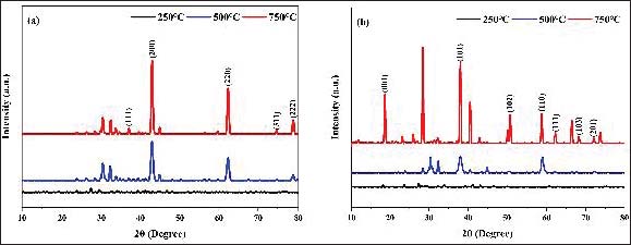
AbstractBackground: Carbon tetrachloride (CCl₄) is a widely used hepatotoxin that induces liver injury through apoptosis and inflammation. Magnesium oxide (MgO) nanoparticles have shown potential hepatoprotective effects, which may depend on synthesis conditions such as pH. Aim: This study aimed to investigate the hepatoprotective effects of biosynthesized MgO nanoparticles from Ornithogalum cuspidatum against CCl₄-induced liver injury in rats and compare the efficacy of nanoparticles synthesized at pH 5 and pH 9. Methods: MgO nanoparticles were biosynthesized at pH 5 and pH 9 and characterized for crystalline structure (X-ray diffraction), elemental composition (energy-dispersive spectroscopy), and thermal stability (thermogravimetry). Male Wistar rats were divided into six groups: control negative, control positive (CCl₄), MgO pH 5, MgO pH 5 + CCl₄, MgO pH 9, and MgO pH 9 + CCl₄. Liver sections were immunohistochemically evaluated for caspase-3 and Tumor necrosis factor—alpha (TNF-α) expression using immunohistochemistry. Staining was assessed semiquantitatively based on intensity and percentage of positive cells, and a final immunoreactivity score was calculated as Intensity × % positive cells. Results: Characterization confirmed the successful synthesis and structural features of MgO nanoparticles. CCl₄ exposure increased caspase-3 and TNF-α immunoexpression, with higher scores observed in the CCl₄ group (caspase-3=12; TNF-α=10) compared with the control group (score=0 for both markers). MgO nanoparticles reduced caspase-3 and TNF-α expression in a pH-dependent manner. The pH 5 formulation showed moderate reduction (caspase-3=6; TNF-α=6), whereas the pH 9 formulation showed greater reduction (caspase-3=4; TNF-α=2), indicating a stronger protective trend. Conclusion: MgO nanoparticles mitigate apoptosis and inflammation in CCl₄-induced liver injury, with higher efficacy observed for nanoparticles synthesized at pH 9, highlighting the importance of synthesis conditions for optimizing hepatoprotective activity. Keywords: Carbon tetrachloride, Caspase-3, Immunohistochemistry, Magnesium oxide nanoparticles, TNF-α. IntroductionNanoparticles are materials with unique properties, such as high surface area, tunable size, and enhanced bioavailability, which make them highly useful in medicine (Wang et al., 2020; Altammar, 2023; Abbasi et al., 2023). Recently, they have been applied in drug delivery (Altammar, 2023), antimicrobial therapy, cancer treatment, and organ protection (Mitchell et al., 2021) because of their ability to modulate cellular processes and reduce side effects (Karnwal et al., 2024). Metal oxide nanoparticles, including magnesium oxide (MgO), have antioxidant, anti-inflammatory, and cytoprotective effects (Sirelkhatim et al., 2020), highlighting their potential as therapeutic agents (Shahid et al., 2022). MgO nanoparticles were synthesized using Ornithogalum caspidatum extract at pH 5 and pH 9, as previous studies have demonstrated that synthesis pH significantly influences nanoparticle structure and functional properties (Yin et al., 2024), and plant-mediated methods ensure environmentally friendly production with confirmed structural characteristics by X-ray diffraction (XRD), energy-dispersive spectroscopy (EDS), and thermogravimetry (TGA) (Proniewicz et al., 2024). MgO nanoparticles (NPs) produced from O. caspidatum at pH 5 and pH 9 represent an emerging field of study in biomedical applications due to their remarkable therapeutic properties (Heydary et al., 2015; Yin et al., 2024). They also demonstrate considerable antibacterial activity, which results from their ability to generate reactive oxygen species (ROS) (Kermanizadeh et al., 2012; Rotti et al., 2023). Recent investigations have highlighted the wound healing capabilities of MgO NPs and their neuroprotective potential by protecting dopaminergic neurons from 1-methyl-4-phenylpyridinium toxicity (Senthil Kumaran et al., 2015; Adhikari et al., 2016). Additionally, these nanoparticles have proven effective against the toxicity of amino-polycyclic hydrocarbons and picloram (Yasmin Nisa et al., 2023). The biosynthesized MgO nanoparticles were characterized to confirm their properties. XRD was used to determine the crystalline structure (Al-Gaashani et al., 2020), while EDS was used to verify the elemental composition. Thermal stability was assessed by thermogravimetric analysis (TGA) (Altammar, 2023). These analyses confirmed the quality of the nanoparticles and supported their potential hepatoprotective activity (Proniewicz et al., 2024). However, despite the recognition of the protective effects of MgO NPs, research regarding their efficacy against Carbon tetrachloride (CCl4)-induced hepatic injury is lacking, revealing a significant literature gap (Ranjbar et al., 2018; Yue et al., 2018). Therefore, this study aims to address the research gap regarding the hepatoprotective potential of MgO nanoparticles synthesized under varying pH conditions. Materials and MethodsBiosynthesis of MgO-NPs using O. caspidatumPreparation of O. caspidatum leaf extractOrnithogalum cuspidatum was collected from the Piramagrwn mountain in Iraq. The plant was washed with distilled water, dried at room temperature for 3 days, and ground using an electric grinder. The extract was prepared at a 1:20 (w/v) ratio by mixing 50 g of powdered leaves with 1,000 ml of deionized water. The mixture was ultrasonicated for 15 minutes at a frequency of 60 kHz and 400 W and then cooled to room temperature. Filter paper (11 µm, Thomas Baker, India) was used to obtain a clear extract and centrifuged at 10,000 rpm for 10 minutes (Ozturk et al., 2022). Plant-mediated synthesis of MgO-NPsMgO nanoparticles were biosynthesized by mixing 2 volumes of O. cuspidatum extract with 1 volume of magnesium (Mg) nitrate hexahydrate solution under continuous stirring at 50°C for 1 hour. Ammonia solution (1M) was added dropwise to adjust the pH using a pH meter, and the mixture was allowed to condense before oven-drying overnight. The powder was divided into 3 batches, and annealed at 250°C, 500°C, and 750°C, and ultrasonicated in absolute ethanol for 10 minutes. The final pure powder was dried overnight. Characterization of the MgO nanoparticlesXRD analysisThe crystalline structure of the synthesized MgO nanoparticles was determined using XRD analysis. The powdered samples obtained at pH 5 and pH 9 were analyzed using an X-ray diffractometer equipped with Cu Kα radiation (λ=1.5406 Å). Data were recorded over a 2θ range of 10°–80° at a scanning rate of approximately 0.02°/s. The diffraction peaks were compared with standard reference patterns from the Joint Committee on Powder Diffraction Standards database to confirm the crystalline phase of MgO. The Debye–Scherrer equation was used to estimate the average crystallite size of the nanoparticles. Energy-dispersive X-ray spectroscopyThe elemental composition of the synthesized MgO nanoparticles was examined using energy dispersive X-ray spectroscopy (EDS) coupled with a scanning electron microscope. Small quantities of the powdered samples prepared at pH 5 and pH 9 were mounted onto conductive carbon tape and coated with a thin layer of gold, if necessary, to improve the conductivity. The EDS spectra were recorded under the appropriate accelerating voltage conditions. The obtained spectra were analyzed to determine the presence and relative abundance of Mg and oxygen (O) elements, confirming the formation and purity of MgO nanoparticles. Thermogravimetric analysisThe thermal stability and weight loss behavior of the synthesized MgO nanoparticles were investigated using TGA. Approximately 5–10 mg of each dried sample (pH 5 and pH 9) was placed in an alumina crucible and heated from room temperature to approximately 800°C at a heating rate of 10°C/min under a nitrogen atmosphere. The changes in weight during heating were recorded as a function of temperature. The obtained thermograms were used to evaluate the moisture loss, decomposition of organic residues from plant extracts, and thermal stability of the MgO nanoparticles. Animal preparation and groupingThe animals were divided into 6 groups of 10 rats each. Group 1 (Control): received a standard diet only. Group 2 (CCl₄): received CCl₄ alone. Group 3 (MgO NPs pH 5): received MgO NPs at pH 5 only. Group 4 (MgO NPs pH 9): received MgO NPs at pH 9 only. Group 5 (CCl₄ + MgO NPs pH 5): received CCl₄ along with MgO NPs at pH 5. Group 6 (CCl₄ + MgO NPs, pH 9): received CCl₄ along with MgO NPs at pH 9. MgO NPs were orally administered at 75 mg/kg/day. The MgO NP doses were selected based on previous in vivo studies showing biological activity in rats without acute toxicity. Lower doses (~75 mg/kg) have demonstrated protective effects, allowing the evaluation of dose-dependent impacts on liver function and histology (Mazaheri et al., 2019; Majeed et al., 2023; Shafiq et al., 2024). Then CCl4 was injected weekly at 0.5 ml/kg. The rats were anesthetized by intramuscular injection of ketamine (75 mg/kg body weight) and xylazine (2 mg/kg body weight) (Oh and Narver, 2024) on day 22. The abdominal cavity was carefully opened, and liver tissue was collected for analysis. Immunohistochemistry (Caspase -3+ TNF α)Immunohistochemical analysis was performed according to Elhabashy et al. (2022). Liver tissue sections were subjected to antigen retrieval using citrate buffer (10 mM, pH 6.0) at 95°C–100°C for 20 minutes in a microwave oven, followed by blocking of endogenous peroxidase activity for 15 minutes. Nonspecific binding was blocked using 5% bovine serum albumin and 0.1% Triton X-100 in Phosphate-buffered saline (PBS). Sections were then incubated overnight at 4°C with primary antibodies against Tumor necrosis factor—alpha (TNF-α) (1:600, DAKO, Denmark) and Caspase-3 (1:400, DAKO, Denmark). Sections were incubated with biotinylated secondary antibody for 30 minutes, followed by streptavidin-horseradish peroxidase for 60 minutes after washing in PBS. Immunoreactivity was visualized using diaminobenzidine, and sections were counterstained with hematoxylin. Immunohistochemical evaluation was performed using AHSQ image analysis software. Caspase-3 and TNF-α expression were assessed semiquantitatively based on staining intensity and percentage of positive cells. Staining intensity was graded as 0=none, 1=weak, 2=moderate, and 3=strong. The percentage of positive cells was scored as 0=0%–5%, 1=6%–20%, 2=21%–40%, 3=41%–65%, and 4=>65%. A final immunoreactivity score was calculated by multiplying the intensity by the percentage score (intensity × % positive cells) (García Iglesias et al., 2020). This scoring system was applied to both Caspase-3 and TNF-α immunostaining. Only these two markers were quantitatively analyzed in this study. Statistical analysisGraphical presentation and data plotting were performed using the Origin software (OriginLab Corporation, USA). Inferential statistical analyses (e.g., ANOVA or post hoc tests) were not performed in this study. Data were analyzed using descriptive statistics. The immunohistochemical results were expressed as semiquantitative scores based on staining intensity and percentage of positive cells (Intensity × percentage score). The results were presented descriptively as mean-based scores and observed staining patterns. Ethical approvalAuthorization for the research was obtained from the local ethical council for animal experiments at the College of Veterinary Medicine, University of Sulaimani (permission VMUS.EC). Doc 14,2025 in May 2025 in Sulaymaniyah, Iraq. ResultsCharacterization of MgO NPStructural analysis (XRD)The XRD patterns of MgO NPs synthesized at pH 5 and pH 9 showed characteristic diffraction peaks corresponding to the cubic periclase structure. Major peaks were observed at approximately 2θ ≈ 36.9°, 42.9°, 62.3°, 74.7°, and 78.6°, corresponding to the (111), (200), (220), (311), and (222) planes, confirming high crystallinity and phase purity, respectively. The NPs synthesized at pH 9 exhibited sharper and more intense diffraction peaks than those synthesized at pH 5 (Fig. 1), indicating improved crystallinity and larger crystallite size. The average crystallite size of the pH 9 sample was slightly higher than that of the pH 5 sample.
Fig. 1. XRD pattern of MgO NPs at different pH values and annealing temperatures: (a) pH 5 and (b) pH 9. The crystallite size (D) was calculated using the Scherrer equation:
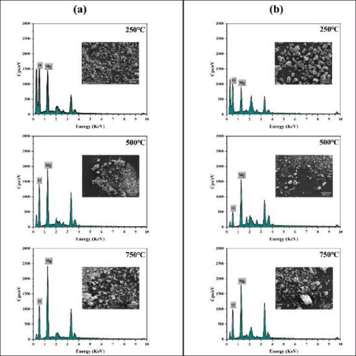
Elemental analysisEDS analysis confirms that MgO nanoparticles are composed of Mg and O (Fig. 2). The pH level during synthesis affects the surface chemistry but not the overall elemental composition. Higher synthesis temperatures (250°C, 500°C, and 750°C) enhance crystallinity and phase purity, resulting in sharp, impurity-free EDS peaks for Mg and O. Organic impurities may be green introduced by green synthesis methods, but none were detected in this study, indicating the high elemental purity of the MgO nanoparticles. This consistency holds across different pH levels during synthesis, indicating that pH does not significantly change the elemental composition, although it affects surface chemistry. The MgO nanoparticles synthesized at higher temperatures show well-defined, sharp EDS peaks for Mg and O, indicating high crystallinity and phase purity. The absence of additional peaks suggests minimal impurities, favoring the formation of MgO nanoparticles that are purer and more crystalline.
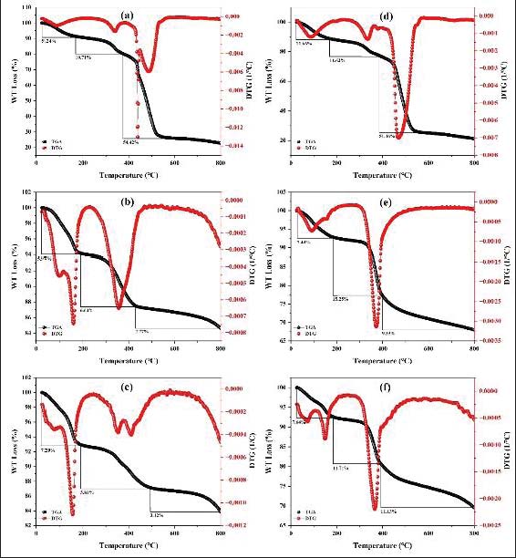
Fig. 2. EDS analysis of MgO nanoparticles at different pH value and annealing temperatures: (a) pH 5 and (b) pH 9. Thermogravimetric analysisThe TGA spectra of MgO nanoparticles synthesized under different conditions provided valuable insights into their thermal stability, composition, and presence of organic or inorganic impurities. We can monitor the weight loss of the nanoparticles as they are heated by studying the TGA spectrum, revealing how they decompose and at what temperatures these changes occur. The pH during the synthesis process can significantly influence the amount of organic matter and water content in MgO nanoparticles. This effect is reflected in the observed weight loss during TGA. For instance, at higher pH levels, there tends to be more organic content on the NPs, leading to greater weight loss during heating. Conversely, at lower pH levels, the surface of the NPs has fewer hydroxyl groups and organic compounds, resulting in weight loss primarily due to the evaporation of moisture and the loss of any adsorbed water. At higher pH levels, specifically pH 9, more hydroxyl groups and organic compounds are present on the surface of the MgO nanoparticles (Fig. 3). This increased surface content leads to higher weight loss because, in addition to moisture evaporation, the decomposition of these organic compounds and hydroxyl groups occurs. The higher weight loss observed at pH 9 indicates that the nanoparticles have more surface-bound species that decompose upon heating, reflecting their increased organic content.
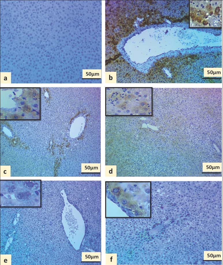
Fig. 3. TGA analysis of MgO nanoparticles at various annealing temperatures (a-c PH=5℃, 250°C, 500°C, and 750°C) and (d-f pH=9,250°C, 500°C, and 750°C). Immunohistochemical assessment of MgO nanoparticle-induced liver tissueCaspase-3 immunoexpressionCaspase-3 immunoexpression varied among the experimental groups (Fig. 4). The negative control group exhibited no detectable staining (score=0), indicating normal hepatic architecture without apoptotic activity. In contrast, the CCl₄-treated group showed strong diffuse expression of caspase-3 (score=12), reflecting marked hepatocyte apoptosis.
Fig. 4. Immunohistochemical sections of rat liver showing immunoexpression of caspase-3 in different experimental groups: a. Negative control: no immunostaining (score=0). b. CCl₄ group-diffuse strong staining (score=12). c. MgO pH 5 group: -moderate focal staining (score=6). d. CCl₄ + MgO pH 5 group: - moderate diffuse staining (score=8). e. MgO pH 9 group: - weak focal staining (score=2). f. CCl₄ + MgO (pH 9) group: -focal weak staining (score=4). MgO nanoparticles administered alone reduced caspase-3 expression compared with the CCl₄ group. The pH 5 group showed moderate expression (score=6) and the pH 9 group showed weak focal expression (score=2), indicating low basal apoptotic activity. In CCl₄-treated animals, coadministration of MgO nanoparticles reduced caspase-3 expression compared with CCl₄ alone. The CCl₄ + MgO pH 5 group showed moderate diffuse staining (score=8), while the CCl₄ + MgO pH 9 group showed lower focal staining (score=4), suggesting a potential protective effect, with a stronger reduction observed at pH 9. TNF-α immunoexpressionTNF-α expression also varied among the experimental groups, as illustrated in Figure 5. The negative control group showed no immunoreactivity (score=0), whereas the CCl₄ group exhibited strong diffuse staining (score=10), indicating a marked inflammatory response.
Fig. 5. Immunohistochemical sections of rat liver showing TNF-α immunoexpression in different experimental groups: a. Negative control group: - no immunostaining (score=0). b. CCl₄ group-diffuse strong staining (score=10). c. MgO pH 5 group: - moderate focal staining (score=6). d CCl₄ + MgO pH 5 group: - moderate diffuse staining (score=8). e MgO pH 9 group: - no detectable staining (score=0). f. CCl₄ + MgO (pH 9) group: - weak focal staining (score=2). MgO nanoparticles alone induced low to moderate TNF-α expression depending on the pH conditions. The pH 5 group showed moderate staining (score=6), whereas the pH 9 group showed no detectable expression (score=0). In CCl₄-treated groups, MgO co-treatment reduced TNF-α expression compared with CCl₄ alone. The CCl₄ + MgO pH 5 group showed moderate staining (score=8), whereas the CCl₄ + MgO pH 9 group showed weak focal expression (score=2), suggesting a more pronounced anti-inflammatory effect at pH 9. DiscussionHepatotoxic injury is commonly associated with oxidative stress-mediated damage, leading to lipid peroxidation, mitochondrial dysfunction, hepatocyte necrosis, and inflammatory activation. These pathological processes are closely linked with the activation of apoptotic and inflammatory markers, including caspase-3 and TNF-α, which are widely recognized indicators of liver injury progression (Wang et al., 2020; Flessa et al., 2022; Cohen et al., 2023). Exposure to CCl₄ in experimental models is known to induce severe hepatic injury through free radical generation, resulting in increased expression of caspase-3 and TNF-α. In the present study, elevated immunoexpression of these markers reflected activation of apoptosis and inflammatory responses in liver tissue, consistent with previous reports describing oxidative stress-mediated hepatotoxicity (Elkhamesy et al., 2022; Abdelnaser et al., 2025). MgO NPs demonstrated a protective trend against hepatic injury, as evidenced by reduced caspase-3 and TNF-α immunoreactivity in treated groups. This suggests that MgO nanoparticles may attenuate hepatocyte apoptosis and inflammatory responses associated with toxic injury (Dong et al., 2016). The variation observed between pH 5 and pH 9 formulations indicates that the synthesis conditions may influence the biological activity and therapeutic potential of MgO nanoparticles (Mansha et al., 2023; Altammar, 2023; Shafiq et al., 2024). Caspase-3 plays a central role as an executioner of apoptosis, while TNF-α is a key proinflammatory cytokine involved in hepatic inflammation and tissue injury. The reduction of both markers following MgO treatment suggests a potential hepatoprotective effect through the modulation of apoptotic and inflammatory pathways (Mitchell et al., 2021; Shi et al., 2024; Algefare et al., 2024). Nanoparticles have gained significant attention in biomedical applications due to their ability to modulate oxidative stress, inflammation, and cellular signalling pathways (Kashef and Ibrahim, 2023). Metal oxide nanoparticles, including MgO, have been reported to exhibit cytoprotective effects in various experimental models; however, their role in chemically induced liver injury remains limited, highlighting the importance of the present findings (Sirelkhatim et al., 2020; El-Sherbiny et al., 2021). TNF-α is rapidly induced during hepatic injury and contributes to the progression of inflammation and tissue damage. Previous studies have demonstrated sustained TNF-α expression following toxic liver exposure, although its relationship with apoptosis may vary depending on injury severity and timing (Farid et al., 2023; Zhang et al., 2020). Although the present findings suggest a protective trend of MgO nanoparticles against hepatic injury, the study is limited to semiquantitative immunohistochemical analysis in male Wistar rats. Future studies should include biochemical assays, molecular investigations, both sexes, and long-term toxicity evaluation to further clarify the hepatoprotective mechanisms and safety profile of MgO nanoparticles. ConclusionBiosynthesized MgO nanoparticles from O. cuspidatum exhibit significant hepatoprotective effects against CCl₄-induced liver injury in rats. MgO nanoparticle treatment reduced apoptosis and inflammation, as reflected by decreased caspase-3 and TNF-α expression. Nanoparticles synthesized at pH 9 provided greater protection than those synthesized at pH 5, highlighting the critical influence of synthesis conditions on therapeutic efficacy. These results demonstrate that optimizing nanoparticle properties can enhance hepatoprotective activity, suggesting the potential of MgO nanoparticles as a novel intervention for liver injury. Further studies are warranted to elucidate the molecular mechanisms underlying their protective effects and to explore their clinical applications. AcknowledgmentSpecial thanks to the Nanotechnology Research Laboratory, Department of Physics, and the Research Center, College of Veterinary Medicine and Surgery, University of Sulaimani/Iraq, for their laboratory support. Conflict of interestThe authors confirm that they have no competing financial interests or personal relationships that could have influenced the work described in this paper. This research did not receive funding from any public, commercial, or nonprofit organization. FundingThis work is personally funded. Authors' contributionsConceptualization, B.A.; Methodology, H.R. and B.A.; Validation, H.R. and B.A.; Formal Analysis, H.R.; Investigation, H.R. and B.A.; Data Curating, H.R. and B.A.; Writing—Original Draft Preparation, H.R.; Writing—Review & Editing, B.A. and N.S.; Visualization, H.R. and B.A.; Supervision, N.S. and B.A.; Project Administration, N.S. and B.A. Data availabilityThe data used to support the findings of this study are included within the manuscript. ReferencesAbbasi, R., Shineh, G., Mobasheri, A. and Avci, P. 2023. Structural parameters of nanoparticles affecting their toxicity. Nanomaterials 13(5), 1234; doi:10.3390/nano13051234 Abdelnaser, M., Attya, M.E., El-Rehany, M.A. and Fathy, M. 2025. Mitigation of sepsis-induced liver injury by clemastine via modulating GSDMD/NLRP-3/Caspase-1/NF-κB signalling pathways. Eur. J. Med. Res. 30(1), 715; doi:10.1186/s40001-025-02982-w Adhikari, A., Polley, N., Darbar, S., Bagchi, D. and Pal, S.K. 2016. Citrate functionalized Mn3O4 in nanotherapy of hepatic fibrosis by oral administration. Future Sci. Open Access 2(4), 146; doi:10.4155/fsoa-2016-0030 Al-Gaashani, R., Radiman, S., Tabet, N. and Daud, A.R. 2020. X-ray diffraction analysis of ZnO nanoparticles. Ceram. Int. 46(2), 2866–2871; doi:10.1016/j.ceramint.2019.09.190 Algefare, A.I., Alfwuaires, M., Famurewa, A.C., Elsawy, H. and Sedky, A. 2024. Geraniol prevents CCl4-induced hepatotoxicity via suppression of hepatic oxidative stress, pro-inflammation and apoptosis in rats. Toxicol. Rep. 12, 128–134; doi:10.1016/j.toxrep.2024.01.016 Altammar, K.A. 2023. A review on nanoparticles: characteristics, synthesis, applications, and challenges. Front. Microbiol. 14, 1155622; doi:10.3389/fmicb.2023.1155622 Cohen, S.M., Bevan, C., Gollapudi, B. and Klaunig, J.E. 2023. Evaluation of the carcinogenicity of carbon tetrachloride. J. Toxicol. Environ. Health B Crit. Rev. 26(6), 342–370; doi:10.1080/10937404.2023.2246623 Dong , Y., Liu, Y., Kou, X., Jing, Y., Sun, K., Sheng, D., Yu, G., Yu, D., Zhao, Q., Zhao, X., Li, R., Wu, M. and Wei, L. 2016. The protective or damaging effect of tumor necrosis factor α in acute liver injury is concentration dependent. Cell Biosci. 6, 8; doi:10.1186/s13578-016-0077-9 Elhabashy, M., Kashef, S., AbuoHashish, N., Mahmoud, H. and Fathi, F. 2022. Evaluating the prophylactic anti-inflammatory and antioxidant effects of galantamine hydrobromide through α-7 nicotinic receptor on lithium carbonate-induced thyroid dysfunction in rats. J. Adv. Med. Med. Res. 34(21), 406–417; doi:10.9734/jammr/2022/v34i2131574 Elkhamesy, A., Refaat, M., Gouida, M.S.O., Alrdahe, S.S. and Youssef, M.M. 2022. Diminished CCl4-induced hepatocellular carcinoma, oxidative stress and apoptosis by co-administration of curcumin or selenium in mice. J. Food. Biochem. 46(4), e13845; doi:10.1111/jfbc.13845 El-Sherbiny, M., Eldosoky, M., El-Shafey, M. and Othman, E. 2021. Protective effects of natural antioxidants against CCl₄-induced liver injury via modulation of inflammatory and apoptotic markers. J. Biochem. Mol. Toxicol. 35(6), e22768; doi:10.1002/jbt.22768 Farid, A., Michael, V. and Safwat, G. 2023. Melatonin-loaded poly (lactic-co-glycolic acid) nanoparticles reduce inflammation, inhibit apoptosis and protect rat liver from the hazardous effects of CCl4. Sci. Rep. 13, 17432; doi:10.1038/s41598-023-44466-4 Flessa, C.M., Nasiri-Ansari, N., Kyrou, I., Chatzigeorgiou, A., Tsatsanis, C., Stamatakis, K., Tsilimigras, D.I. and Iliopoulos, D. 2022. Genetic and diet-induced animal models for non-alcoholic fatty liver disease research. Int. J. Mol. Sci. 23(24), 15791; doi:10.3390/ijms232415791 García Iglesias, M.J., Cuevas Higuera, J.L., Bastida Sáenz, A., Polledo, L., González Fernández, J. and Pérez Martínez, C. 2020. Immunohistochemical detection and scoring of protein expression in canine hemangiomas and hemangiosarcomas. BMC Vet. Res. 16, 123; doi:10.1186/s12917-020-02338-3 Heydary, V., Navaei-Nigjeh, M., Rahimifard, M., Farhadi, N., Hosseini, S., Jafari, H., Mahmoudi, M. and Rahimi, H.R. 2015. Biochemical and molecular evidences on the protection by magnesium oxide nanoparticles of chlorpyrifos-induced apoptosis in human lymphocytes. Drug Chem. Toxicol. 38(4), 473–480; doi:10.3109/01480545.2014.928092 Karnwal, A., Sharma, V., Kumar, G., Jassim, A.Y., Dohroo, A. and Sivanesan, I. 2024. Transforming medicine with nanobiotechnology: nanocarriers and their biomedical applications. Pharmaceutics 16(9), 1114; doi:10.3390/pharmaceutics16091114 Kashef, S. and Ibrahim, M. 2023. Role of Ashwagandha extract in ameliorating the histological and immunohistochemical changes in parotid gland induced by hypothyroidism in adult male rat. Egypt. J. Histol. 46(2), 506–517. Kermanizadeh, A., Gaiser, B.K., Hutchison, G.R. and Stone, V. 2012. An in vitro liver model: assessing oxidative stress and genotoxicity following exposure of hepatocytes to a panel of engineered nanomaterials. Part. Fibre Toxicol. 9, 28. Majeed, S.I., Mohammed, A. and Mohammad, N. 2023. Bioaccumulation and evaluation of MgO nanoparticle toxicity (62.5–500 mg/kg) with hepatic oxidative stress and histopathology in male rats. Kurdistan J. Appl. Res. 8(1), 1–15; doi:10.24017/Science.2023.1.1 Mansha, M.T., Maqbool, A., Nadir, N., Ahmed, S., Khan, R. and Ali, F. 2023. Functional assessment of MgO nanoparticle supplementation in an acute liver injury rat model. Haya. Saudi. J. Life. Sci. 8(11), 233–241. Mazaheri, N., Naghsh, N., Karimi, A. and Salavati, H. 2019. In vitro toxicity of magnesium oxide nanoparticles in Wistar rats (62.5–500 µg·mL⁻¹ i.p.), affecting liver histology and biochemistry. Iran. J. Biotechnol. 17(1), 1543; doi:10.21859/ijb.1543 Mitchell, M.J., Billingsley, M.M., Haley, R.M., Wechsler, M.E., Peppas, N.A. and Langer, R. 2021. Engineering precision nanoparticles for drug delivery. Nature Rev. Drug Discovery 20(2), 101–124; doi:10.1038/s41573-020-0090-8 Oh, S.S. and Narver, H.L. 2024. Mouse and rat anesthesia and analgesia. Curr. Protoc. 4(2), 995; doi:10.1002/cpz1.995 Ozturk, D., Ozguven, A., Yonten, V. and Ertas, M. 2022. Green synthesis, characterization and antimicrobial activity of silver nanoparticles using Ornithogalum narbonense L. Inorg. Nano-Met. Chem. 52(3), 329–341; doi:10.1080/24701556.2022.2031985 Proniewicz, E., Vijayan, A.M., Surma, O., Szkudlarek, A. and Molenda, M. 2024. Plant-assisted green synthesis of MgO nanoparticles as a sustainable material for bone regeneration: spectroscopic properties. Int. J. Mol. Sci. 25(14), 7801; doi:10.3390/ijms25147801 Ranjbar, A., Ghasemi, H., Abedian, A. and Kheiripour, N. 2018. Cerium oxide nanoparticle modulates hepatic damage, inflammatory and oxidative stress biomarkers in a dose-dependent manner: an in vitro study of rat liver. J. Trace. Elem. Med. Biol. 49, 273–280; doi:10.1016/j.jtemb.2018.02.005 Rotti, R.B., Sunitha, D.V., Manjunath, R., Roy, A., Borehalli Mayegowda, S., Gnanaprakash, A.P., Alghamdi, S., Almehmadi, M., Abdulaziz, O., Aljuaid, A., Alsaiari, A.A., Ashgar, S.S., Babalghith, A.O., El-Lateef, A.E.A. and Khidir, E.B. 2023. Green synthesis of MgO nanoparticles and its antibacterial properties. Front. Chem. 11, 1143614; doi:10.3389/fchem.2023.1143614 Senthil Kumaran, R., Choi, Y.K., Singh, V. and Song, H.J. 2015. In vitro cytotoxic evaluation of MgO nanoparticles and their effect on the expression of ROS genes. J. Nanomater. 2015, 535406; doi:10.1155/2015/535406 Shafiq, A.A., Luaib, N.M., Mahdi, R.H. and Alsaimary, I.E. 2024. Oral toxicity of magnesium oxide nanoparticles (MgO NPs) on liver in male rats. Iraqi. J. Sci. 65, 6323–6335; doi:10.24996/ijs.2024.65.11.11 Shahid, S., Ejaz, A., Javed, M., Mansoor, S., Iqbal, S., Elkaeed, E.B., Alzhrani, R.M., Alsaab, H.O., Awwad, N.S., Ibrahium, H.A., Fatima, U., Zaman, S. and Nazim Sarwar, M. 2022. The anti-inflammatory and free radical scavenging activities of bio-inspired nano-magnesium oxide. Front. Mater. 9, 875163; doi:10.3389/fmats.2022.875163 Shi, C., Li, Y., You, Z., Tian, Y., Zhu, X., Xu, H., Yang, M., Zhang, Y., Dong, R., Quan, H., Shang, Y. and Li, X. 2024. Mangiferin ameliorates CCl4-triggered acute liver injury by inhibiting inflammatory response and oxidative stress involving the Nrf2-ARE pathway. J. Inflamm. Res. 17, 7081–7097; doi:10.2147/JIR.S378725 Sirelkhatim, A., Mahmud, S., Seeni, A., Kaus, N.H.M., Ann, L.C., Bakhori, S.K.M., Hasan, H. and Mohamad, D. 2020. Review on zinc oxide nanoparticles: antibacterial activity and toxicity mechanism. Nanomicro. Lett. 12(11), 11; doi:10.1007/s40820-019-0345-8 Wang, Y., Pi, C., Feng, X., Hou, Y., Zhao, L. and Wei, Y. 2020. The influence of nanoparticle properties on oral bioavailability of drugs. Int. J. Nanomed. 15, 6295–6310; doi:10.2147/IJN.S248580 Yasmin Nisa, F., Atiar Rahman, M., Saha, S., Karim, R., Chowdhury, S. and Ahmed, T. 2023. Unraveling Tamarindus indica pulp-derived green magnesium oxide nanoparticles for cardioprotective potential against doxorubicin-induced cardiomyopathy: a comprehensive biochemical and gene expression study. Biomed. Pharmacother. 164, 114995; doi:10.1016/j.biopha.2023.114995 Yin, Z., Li, S., Li, X., Shi, W., Wang, A., Yu, C., Shi, X., Carroll, D.L. and Ma, C. 2024. Effect of pH on the microstructure and antibacterial properties of MgO nanoparticles by microwave-assisted solution combustion. J. Alloys. Compd. 945, 171123; doi:10.1016/j.jallcom.2023.171123 Yue, Z., Zhang, X., Yu, Q., Li, Y., Liu, H. and Wang, S. 2018. Cytochrome P450-dependent reactive oxygen species production contributes to Mn3O4 nanoparticle-caused liver injury. Chem. Res. Toxicol. 31(12), 1283–1292; doi:10.1021/acs.chemrestox.8b00279 Zhang, C.Y., Yuan, W.G., He, P., Lei, J.H. and Wang, C.X. 2020. Liver fibrosis and hepatic stellate cells: etiology, pathological hallmarks and therapeutic targets. World J. Gastroenterol. 26(10), 105–119. | ||


| How to Cite this Article |
| Pubmed Style Rahim HM, Salih NA, Azhdar B. Magnesium oxide nanoparticles: A novel shield against CCl₄- induced hepatic damage via caspase-3 and TNF-α in rats. Open Vet. J.. 2026; 16(5): 2685-2695. doi:10.5455/OVJ.2026.v16.i5.11 Web Style Rahim HM, Salih NA, Azhdar B. Magnesium oxide nanoparticles: A novel shield against CCl₄- induced hepatic damage via caspase-3 and TNF-α in rats. https://www.openveterinaryjournal.com/?mno=314767 [Access: June 26, 2026]. doi:10.5455/OVJ.2026.v16.i5.11 AMA (American Medical Association) Style Rahim HM, Salih NA, Azhdar B. Magnesium oxide nanoparticles: A novel shield against CCl₄- induced hepatic damage via caspase-3 and TNF-α in rats. Open Vet. J.. 2026; 16(5): 2685-2695. doi:10.5455/OVJ.2026.v16.i5.11 Vancouver/ICMJE Style Rahim HM, Salih NA, Azhdar B. Magnesium oxide nanoparticles: A novel shield against CCl₄- induced hepatic damage via caspase-3 and TNF-α in rats. Open Vet. J.. (2026), [cited June 26, 2026]; 16(5): 2685-2695. doi:10.5455/OVJ.2026.v16.i5.11 Harvard Style Rahim, H. M., Salih, . N. A. & Azhdar, . B. (2026) Magnesium oxide nanoparticles: A novel shield against CCl₄- induced hepatic damage via caspase-3 and TNF-α in rats. Open Vet. J., 16 (5), 2685-2695. doi:10.5455/OVJ.2026.v16.i5.11 Turabian Style Rahim, Harseen M., Nadia A. Salih, and Bruska Azhdar. 2026. Magnesium oxide nanoparticles: A novel shield against CCl₄- induced hepatic damage via caspase-3 and TNF-α in rats. Open Veterinary Journal, 16 (5), 2685-2695. doi:10.5455/OVJ.2026.v16.i5.11 Chicago Style Rahim, Harseen M., Nadia A. Salih, and Bruska Azhdar. "Magnesium oxide nanoparticles: A novel shield against CCl₄- induced hepatic damage via caspase-3 and TNF-α in rats." Open Veterinary Journal 16 (2026), 2685-2695. doi:10.5455/OVJ.2026.v16.i5.11 MLA (The Modern Language Association) Style Rahim, Harseen M., Nadia A. Salih, and Bruska Azhdar. "Magnesium oxide nanoparticles: A novel shield against CCl₄- induced hepatic damage via caspase-3 and TNF-α in rats." Open Veterinary Journal 16.5 (2026), 2685-2695. Print. doi:10.5455/OVJ.2026.v16.i5.11 APA (American Psychological Association) Style Rahim, H. M., Salih, . N. A. & Azhdar, . B. (2026) Magnesium oxide nanoparticles: A novel shield against CCl₄- induced hepatic damage via caspase-3 and TNF-α in rats. Open Veterinary Journal, 16 (5), 2685-2695. doi:10.5455/OVJ.2026.v16.i5.11 |


