





| Research Article | ||
Open Vet. J.. 2026; 16(6): 3717-3730 Open Veterinary Journal, (2026), Vol. 16(6): 3717-3730 Research Article An enhanced deep learning approach for the automated identification of lumpy skin disease in cattleIthaya Rani Panneer Selvam1*, Saziya Tabbassum1, Abdul Jainul Fathima2, Selvi Shanmuga Pandian3, Gul Shira Banu4, Shalini Divya Prasanna Anbalagan5 and Hari Prasath Thinakaran61Department of CSE, Koneru Lakshmaiah Education Foundation, Vaddeswaram, India 2Department of AI & DS, Francis Xavier Engineering College, Tirunelveli, India 3Department of CSE, Sethu Institute of Technology, Madurai, India 4Department of CSE, PMC TECH, Hosur, India 5Division of Digital Sciences, Karunya Institute of Technology and Sciences, Coimbatore, India 6Department of EEE, Kamaraj College of Engineering and Technology, Virudhunagar, India *Corresponding Author: Ithaya Rani Panneer Selvam. Department of CSE, Koneru Lakshmaiah Education Foundation, Vaddeswaram, India. Email: muhilrani [at] gmail.com Submitted: 23/02/2026 Revised: 05/05/2026 Accepted: 16/05/2026 Published: 12/06/2026 © 2025 Open Veterinary Journal
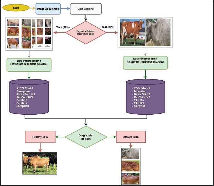
AbstractBackground: The rapid spread of infectious cattle diseases, particularly lumpy skin disease (LSD), has created serious challenges for animal health and livestock-based economies. Conventional diagnostic methods mainly depend on clinical examination, laboratory analysis, and expert interpretation, which are often time-consuming, expensive, and prone to subjective variations. Aim: This study aims to develop an automated and accurate framework for early identification of LSD in cattle by combining histogram-based image enhancement techniques with deep learning models. Methods: Initially, cattle skin images were pre-processed using Contrast-Limited Adaptive Histogram Equalization (CLAHE) to improve image contrast and enhance lesion visibility under uneven illumination conditions. The enhanced images were then classified using multiple deep learning architectures, including Convolutional Neural Networks, DenseNet121, ResNet50V2, InceptionV3, VGG16, VGG19, and Xception. The proposed framework was evaluated on three datasets: Mendeley, Kaggle, and an Indian veterinary research dataset. Results: The proposed method achieved high classification accuracies of 96.5%, 96%, and 95% on the Mendeley, Kaggle, and Indian Journal of Research datasets, respectively. Comparative analysis demonstrated that the CLAHE-integrated deep learning models outperformed several existing approaches in identifying infected and healthy cattle images. Conclusion: The integration of CLAHE with deep learning models provides a reliable, scalable, and cost-effective solution for automated detection of LSD in cattle. The proposed framework can support veterinarians and farmers in early disease diagnosis, improving livestock health management and clinical decision-making. Keywords: Convolutional neural network, DenseNet121, ResNet50V2, InceptionV3, VGG16. IntroductionCattle production plays a vital role in food systems and rural economies worldwide, contributing significantly to nutrition, employment, and income generation. Lumpy skin disease (LSD) is a highly contagious viral disease that adversely affects livestock productivity through reduced milk yield, skin damage, and trade restrictions, often delaying timely intervention and facilitating further spread. This highlights the need for reliable, affordable, and automated identification methods. Conventional diagnostic practices rely on visual examination, laboratory testing, and expert veterinary assessment, which are time-consuming, costly, and require specialized infrastructure, making them less feasible in remote or underdeveloped regions. Delays in diagnosis may lead to uncontrolled infection and increased livestock mortality. With the rapid advancement of artificial intelligence, deep learning—particularly Convolutional Neural Networks (CNNs)—has become a dominant approach for image-based cattle disease identification. However, the effectiveness of different CNN architectures varies depending on the visual characteristics of LSD, which typically presents as irregular lesions with diverse sizes, textures, and nonuniform color distributions. These characteristics require models capable of capturing both fine-grained texture details and multiscale spatial patterns. Despite promising results, existing studies reveal inconsistencies in model selection, feature extraction strategies, and real-world applicability, indicating the need for comprehensive and hybrid frameworks. A substantial body of research has focused on transfer learning using pretrained CNN architectures. Abdullah et al. (2025) evaluated models such as DenseNet121, InceptionV3, MobileNetV2, and Xception for LSD identification and reported the superior performance of DenseNet121 owing to its efficient feature reuse mechanism. Dense architectures, such as DenseNet121, are particularly suitable for LSD detection because their dense connectivity preserves low-level texture features while enabling deeper semantic understanding, which is essential for identifying subtle lesion patterns. While deep architectures improve classification accuracy, their high computational complexity limits their deployment in resource-constrained environments. This highlights a key trade-off between accuracy and efficiency that remains unresolved in the current literature. Shakeel et al. (2024) employed data augmentation techniques to improve model robustness, enhancing performance under varying illumination and background conditions. Although effective, such approaches depend on artificially generated variations and may not fully represent real-world complexities. Similarly, Saha (2024) and Senthilkumar et al. (2024) emphasized the importance of lightweight architectures, such as MobileNetV2, which offer reduced computational cost and suitability for real-time applications. However, Saha (2024) reported that the effectiveness of lightweight architectures is context-dependent, as these models prioritize computational efficiency over deep feature representation. Although they perform well in real-time scenarios, they may not be optimal for LSD detection, where lesions exhibit complex textures, irregular shapes, and multiscale variations. In contrast, Abdullah et al. (2025) demonstrated that deeper architectures, such as DenseNet121, achieved higher accuracy by effectively capturing these fine-grained and multiscale lesion characteristics. Therefore, while lightweight models are advantageous for efficiency, deeper architectures are more suitable when classification accuracy is the primary objective. Hybrid feature extraction approaches have been explored to enhance deep learning (DL) models. In the case of LSD detection, Reza et al. (2026) produced an image classification system based on a CNN system that utilized cattle mouth and hoof images. Their findings demonstrated that DL models could be used effectively in discriminating between infected and healthy cows in the absence of manual feature extraction. Olorunshola et al. (2025) improved the LSD classification by adding CNN features to Haralick texture descriptors, and the results were better than those of CNN-only models. Khandait et al. (2025) demonstrated that a combination of multiple pretrained CNN models enhances classification performance across diverse datasets. Similarly, Goddeti and Narayana (2024) integrated CNN feature extraction with Extreme Learning Machines, achieving faster training and high diagnostic accuracy. Although these methods improve reliability, their increased architectural complexity can hinder scalability and real-time deployment. Recent research has explored the use of DL models for LSD identification, highlighting the effectiveness of various CNN architectures. Saqib et al. (2024) demonstrated that MobileNetV2 provides a good balance between accuracy and computational efficiency, making it suitable for practical applications. Sentoso et al. (2025) reported that VGG16 performs well on moderately sized datasets, indicating that dataset characteristics influence model performance. Architectures such as Inception and Xception are particularly effective because of their ability to capture multiscale lesion patterns. However, these studies do not establish a clear consensus on the optimal model architecture. More advanced approaches, such as ensemble CNN models and hybrid frameworks such as YOLO-EfficientNet, have achieved high accuracy but often focus on specific architectures without providing a unified evaluation across multiple models. Despite these advancements, several research gaps remain. The integration of histogram-based enhancement techniques with deep CNN models is limited; there is a lack of systematic comparison of multiple architectures within a consistent framework, and there is insufficient attention to balancing accuracy with computational efficiency for practical use. To address these limitations, we propose a histogram-based preprocessing approach integrated with multiple deep CNN architectures. This framework enhances feature representation, particularly for texture-rich and multiscale lesion patterns, and enables a comprehensive comparison of model performance, ultimately improving LSD identification accuracy. The main contributions of this work were centered on the development of an automated framework for LSD by integrating Contrast-Limited Adaptive Histogram Equalization (CLAHE)-based preprocessing with DL models. To ensure fair evaluation, a systematic comparison of multiple architectures, including CNN, DenseNet121, ResNet50V2, InceptionV3, VGG16, VGG19, and Xception, was conducted under a unified pipeline. This study demonstrated that CLAHE significantly enhanced lesion visibility and consistently improved classification accuracy compared with other preprocessing techniques (Gaussian filtering and Z-score normalization) and raw images, highlighting the importance of preprocessing–model interaction. The proposed approach achieved high accuracy across three independent datasets, confirming its robustness and generalizability. Additionally, the findings revealed that a baseline CNN outperformed more complex models when combined with CLAHE, offering a cost-effective and practical solution for real-world livestock health monitoring. Materials and MethodsFigure 1 illustrates the proposed enhanced deep learning-based image classification system, which transforms raw cattle images into meaningful class labels. The workflow begins with image acquisition, followed by image resizing to a uniform dimension and preprocessing using CLAHE to enhance contrast and highlight disease-affected regions. During this stage, data augmentation techniques are also applied to increase dataset diversity and improve model generalization. The enhanced images are then provided as input to DL models, enabling stable training and improved classification performance.
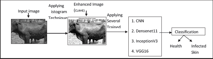
Fig. 1. Proposed LSD identification framework using histogram-enhanced deep learning. Several pre-trained neural networks, including CNN, DenseNet121, Inception V3, VGG16, VGG19, Xception, and ResNet50V2, are employed to independently process the input images, each contributing distinct strengths. CNNs identify hierarchical patterns in images for classification tasks through their core components, namely convolutional, pooling, flattening, and fully connected layers, which work together to extract features from low- to high-level representations, reduce computational complexity, and perform final classification. DenseNet121 improves information flow through dense connections, and Inception V3 captures multi-scale features using parallel filters. VGG16 and VGG19 extract deep hierarchical features using simple and uniform kernels, Xception utilizes efficient depthwise separable convolutions, and ResNet50V2 mitigates vanishing gradients in deep networks using residual connections. The final output categorizes the cattle images into two classes: healthy and LSD-infected. Pre-processing using histogram techniquesPre-processing is essential for improving image quality and optimizing model performance. Raw images may contain noise, low contrast, and uneven illumination, which can reduce the efficiency of feature extraction. To address these issues, adaptive histogram techniques, such as CLAHE, are applied, which redistribute the intensity values of pixels locally to enhance contrast across different regions of the image. Unlike standard global histogram equalization, CLAHE operates on small image tiles, ensuring that regions with varying illumination are enhanced without over-amplifying noise, thereby improving the skin lesions visibility. The transformation can be expressed as follows:
where L is the number of gray levels, nj is the number of pixels at intensity j, and N is the total number of pixels. This enhancement improves the visibility of features and supports better model accuracy, as shown in Figure 2.
Fig. 2. System diagram of the proposed enhancement technique. Multimodal architectureConvolutional neural networkA CNN is a deep learning model used for image analysis, as proposed by Dip Kumar Saha (2024). It automatically learns the spatial hierarchies of features from the input through convolution layers, which use filters to extract patterns such as edges, textures, and shapes. These features are down-sampled using pooling layers to reduce computational complexity and prevent overfitting. For the final classification, the features that are extracted are mapped for flattening and passing it to fully connected layers. CNNs use activation functions, such as ReLU, to introduce nonlinearity and softmax for binary-class prediction. CNNs efficiently manage large image data due to weight sharing and local connectivity and are widely applied in medical imaging, object detection, and disease diagnosis. Hence, CNN is used for cattle disease identification, with improved input quality, training on diverse data, and evaluation on a separate test set to ensure generalization and prevent overfitting, as defined in the corresponding equation [2]. For input layer:
Convolutional layer 1: H1=Relu (W1X + b1)(2) where W1 and b1 represent weights and biases for the first convolutional layer, which represents the convolution operation. H1 represents the output feature map after applying a 32 filter of size (3 × 3). It consisted of three consecutive convolution networks with the number of filters slowly varying (32, 64, 128) and (2 × 2) max-pooling operations to extract discriminative features and reduce spatial dimensions. The resulting feature maps were flattened and passed to a fully connected layer with ReLU activation of 128 neurons. To reduce overfitting, a dropout rate was applied before the output layer. The softmax activation function was used in the last layer to produce class probability distributions among the two categories, namely, healthy and LSD. Max Pooling Layer 1:
In this case, P1 reduces the spatial resolution of H1 by 2. The network was optimized using the Adam algorithm, where the loss function was categorical cross-entropy. The training was conducted over 20 epochs, and the classification accuracy was monitored during that period to evaluate convergence behaviour and possible overfitting. Convolutional layer 2:
DenseNet121DenseNet121 is a deep convolutional architecture proposed by Huang et al. (2017) which contains 121 layers organized through densely connected blocks. In this structure, each layer obtains inputs from all previous layers within the same block, enabling continuous feature reuse and improved information flow. Compared to a conventional CNN model, this connectivity pattern strengthens gradient propagation, mitigates vanishing gradient issues, and reduces redundant parameters. The conventional convolution and pooling operations are performed using a network comprising of number of dense blocks, followed by a final classification stage. Owing to its compact design and strong feature extraction capability, DenseNet121 is extensively used in image analysis and medical diagnostic tasks. The input of the model is a batch of images in the form of tensors (B, H, W), where B is the size of the batch, H and W are the image height and width, respectively. The DenseNet121 backbone can be stated as follows:
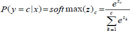
In the expression, X0, X1, ....Xn−1 denote the feature maps produced by all preceding layers, and the channel-wise concatenation of these feature maps, rather than addition, is represented by the bracket notation [.]. The function Fl (.) denotes a composite transformation comprising batch normalization, ReLU activation, and convolution. This formulation highlights the dense connectivity pattern, where each layer receives inputs from all previous layers, promoting feature reuse and improved gradient flow. These metrics reveal the model’s strengths and limitations. Global average pooling (GAP) For each channel c,
Vector Form
where H and W represent the spatial dimensions of the last feature map, and c is the channel index. ResNet50V2ResNet50V2 is a 50-layer residual network introduced by Goceri (2019). Identity shortcut connections bypass intermediate layers, enabling improved gradient flow and preventing degradation in deep networks. The V2 variant adopts a pre-activation structure in which batch normalization and activation precede convolution, thereby enhancing training stability. ResNet50V2 is widely used in image classification and medical imaging tasks because of its strong feature extraction and efficient optimization. Feature extraction with ResNet50V2 (pre-activation)
where x is the input feature map from the previous layer, BN is the batch normalization, and σ is the ReLU activation non-linearity. W is the activation preceding the convolution filter. ResNet50V2 was chosen because its deep residual design uses skip connections to allow gradients to propagate more effectively through many layers, avoiding vanishing gradient issues common in deep CNNs. Transfer learning was applied by starting the network with ImageNettrained weights, allowing it to reuse general visual features. After freezing the base convolutional layers, a new classification head with GAP, a dense ReLU layer, dropout (0.5), and a final softmax output for two classes was included. Global average poolingGAP is applied after the convolution base of the ResNet50V2 to reduce the spatial dimensions of feature maps while preserving important information. Unlike fully connected layers, GAP significantly reduces the number of parameters and helps prevent overfitting
where H and W stand for the height and width of the feature map, respectively. Fki, j is the activation at position (i, j) in the Kth feature map. Fully connected layers: The first dense layer is given by:
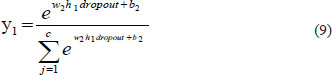
Dropout Layer (Regularization): h1dropout=Dropout(0.5) The output layer (Softmax activation) is given by the following equation:
Here, C is the number of classes (two in our case: healthy and infected skin), and Y1 represents the predicted probability for class j. InceptionV3InceptionV3 was proposed by Rony et Alzubi (2024). The model uses inception modules that perform parallel convolutions with different filter sizes to efficiently extract multi-scale features. It also incorporates factorized convolutions, batch normalization, and auxiliary classifiers to reduce the computational cost and enhance the convergence of training. This preparation helps the model learn distinct features and reduces overfitting for more accurate classification, owing to its optimized structure and strong feature learning capability. 1. Standard Convolution
where M is the number of input channels, Wk,m(u, v) represents the convolution kernel, and bk is the bias term. 2. Factorized Convolution (InceptionV3) (a) 5×5 Factorization(11)
where M is the input channel and N is output channel. (b) Asymmetric factorization
1×1 Convolution (Dimensionality Reduction)
Used for reducing feature depth before expensive convolutions. Softmax classification
where zc is the logit (raw output) for class c, C represents the total number of classes, and zk represents the logit for class k. The model was trained for 20 epochs with a batch size of 32 using the Adam optimizer and categorical cross-entropy loss, while accuracy was tracked as the evaluation metric. The validation data helped monitor generalization and detect overfitting during training. The trained model was saved for later use on new images. VGG16VGGNet, introduced by Simonyan and Zisserman (2015)is a deep CNN known for its uniform architecture. It stacks small (3 × 3) convolution layers with periodic max-pooling are stacked, followed by fully connected layers for classification. The straightforward design enables effective hierarchical feature extraction, making Visual Geometry Group widely used in image analysis tasks. Hence, the VGG16-based classifier is used to sort cattle images into healthy and LSD. 1. Convolution Operation (3×3 Kernel)
where Xm represents the input feature map, Wk,m denotes (3 × 3) convolution kernel, bk represents bias, M represents number of input channels, and Yk denotes output feature map. 2. ReLU Activation
3. Max Pooling (2×2)
4. Softmax classifier
where C states the number of classes, and zc are logit for class c. VGG19The VGG19 is a deep CNN with 19 weight layers, developed by Simonyan and Zisserman (2015). It is built using repeated (3 × 3) convolutional layers with ReLU activation, followed by periodic max-pooling for spatial reduction and fully connected layers for classification. The VGG19 model employs the same fundamental mathematical operations as VGG16, including convolution, activation, pooling, and softmax classification. Compared with VGG16, VGG19 has greater depth, enabling the learning of more complex and hierarchical feature representations; however, it also increases the number of parameters, resulting in higher computational cost and a greater risk of overfitting, particularly when applied to limited datasets such as LSD images. XceptionChollet (2017) proposed an Xception, a deep convolutional architecture that relies entirely on depth-wise separable convolutions. This approach divides standard convolution into channel-wise spatial filtering followed by point-wise convolution for feature integration, which reduces computational overhead while maintaining strong representational power. The network also incorporates residual connections to enhance the gradient flow and training stability. Xception is extensively applied in image classification tasks, including disease detection in cattle. ReLU activation:Xception is chosen because it uses separable depth-wise convolutions to break standard convolutions into lightweight spatial and pointwise steps, reducing computation while still capturing detailed features for image classification. Transfer learning with ImageNetpretrained weights enables the reuse of learned visual features for cattle disease detection. On top of the frozen base, custom layers—GAP 2D, the ReLU-activated dense layer with dropout, and a softmax output—produce probabilities for the two classes: healthy and lumpy skin. 1. Standard Convolution
2. Depth-wise separable convolution (Xception) (a) Depth-wise convolution
(b) Point-wise Convolution (1×1)
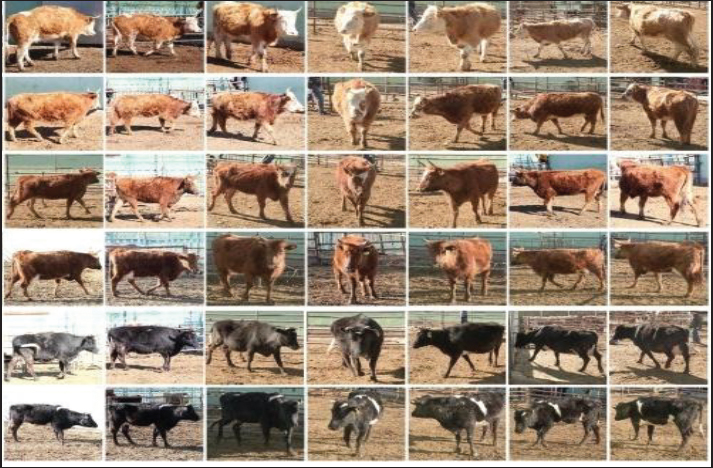
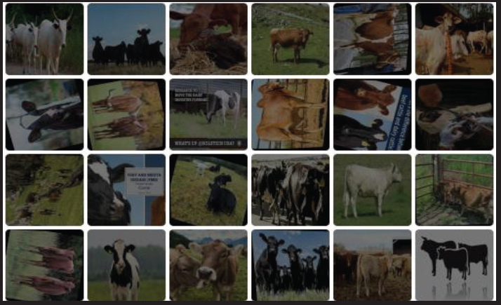
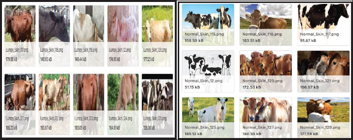
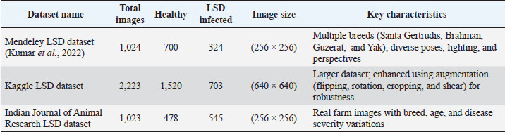
where X states the input feature map, K represents depth-wise kernel, M states number of input channels, and Y is the output feature map. Model.evaluate ( ) is used to measure the performance of the trained network on previously unseen test samples by calculating the loss value and classification accuracy, thereby assessing its generalization capability. Randomly selected test images are visualized with their predicted categories and associated probability scores for further validation, which assists in spotting incorrect predictions. The saved model can also be loaded again to perform inference on new input images, enabling its effective application in cattle disease diagnosis. ResultsThis section provides an overview to introduce the findings and discussion of the proposed scheme. This study employed three publicly available datasets for experimental evaluation: the Mendeley LSD image dataset proposed by Kumar et al. (2022) the Kaggle LSD Images dataset, and the Indian Journal of Animal Research LSD dataset (collected from publicly available veterinary sources and prior published studies). These datasets are used to evaluate the proposed framework’s performance. Figures 3–5 show sample images from the three datasets. Their detailed descriptions are presented in Table 1. For computational efficiency, the image resolutions in all datasets were uniformly resized to (256 × 256) pixels. The study was conducted on a personal computer equipped with an Intel Core i5 processor running at 3.20 GHz with 8 GB of RAM. All experiments were performed using MATLAB R2013a.
Fig. 3. Sample images from the Mendeley LSD dataset.
Fig. 4. Sample images from the Kaggle LSD dataset.
Fig. 5. Sample images from the LSD dataset of the Indian Journal of Animal Research. Table 1. Overview of LSD image datasets and characteristics.
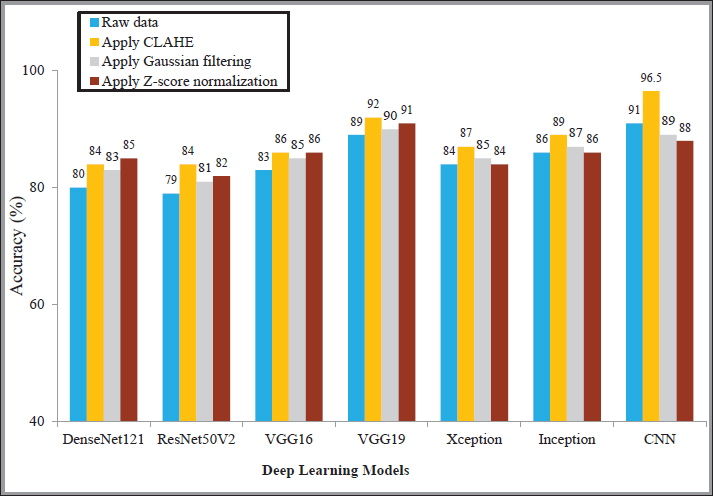
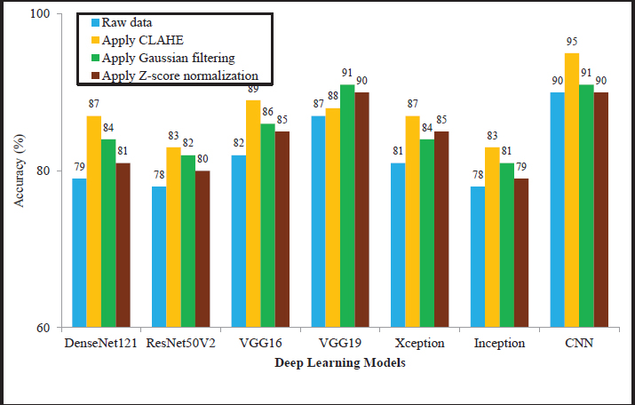
The models were trained using the Adam optimizer with a learning rate of 0.001 and a batch size of 32. Training was conducted for 50–100 epochs, with early stopping applied based on validation loss (patience=10) using an 80:20 training–testing split. Overfitting was controlled using dropout and data augmentation techniques, including rotation, flipping, and scaling. As shown in Table 1, the class imbalance was addressed by applying data augmentation to the minority (infected) class, along with class weighting during training to balance the loss contribution. The use of these three datasets—Mendeley, Kaggle, and the LSD dataset of the Indian Journal of Animal Research—is significant for several reasons. They provide diversity in data sources, which helps reduce dataset bias and improves the model’s reliability. Performance analysis of preprocessing methods for LSD identificationIn this experiment, raw cattle images were initially enhanced using the histogram-based CLAHE technique, along with Gaussian filtering and Z-score normalization to improve image quality and feature representation. Then, the processed images were supplied as input to several pre-trained architectures. The models were independently evaluated on the Mendeley, Kaggle, and Indian Journal of Animal Research datasets, with the results presented in Figures 6–8, respectively, for the classification of healthy and infected skin. The findings indicate that CLAHE consistently improved classification accuracy across all models compared with other pre-processing methods, whereas models trained on raw images showed lower performance. Among the architectures, the CNN achieved the highest accuracies of 96.5%, 96%, and 95% when combined with CLAHE in three datasets. Overall, the results demonstrate that histogram-based enhancement significantly improves model performance for cattle image classification across different architectures.
Fig. 6. Comparison of the accuracy of deep learning models with various preprocessing techniques for LSD identification on the Mendeley dataset.
Fig. 7. Comparison of the accuracy of deep learning models with various preprocessing techniques for LSD identification on the Kaggle dataset.
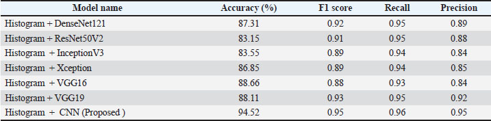
Fig. 8. Comparison of the accuracy of deep learning models with various preprocessing techniques for LSD identification on the Indian Journal of Animal Research dataset. Performance evaluation of several models on LSD in the Indian Journal of Animal ResearchThe Indian Journal of Animal Research LSD dataset includes 1,023 images (478 healthy and 545 infected), split into an 80:20 ratio for training and testing. The performance evaluation of the models showed varied results, as shown in Table 2. Among them, the proposed Histogram + CNN model achieves the best performance with an accuracy of 94.52%, and a high F1-score (0.95), recall (0.96), and precision (0.95). Other models, such as DenseNet121, ResNet50V2, InceptionV3, Xception, VGG16, and VGG19, produce comparatively lower accuracies, generally ranging between 83.15% and 88.66%, while maintaining F1-scores between 0.88 and 0.93. Overall, the findings confirm that integrating histogram-based enhancement with deep CNN architectures improves cattle disease detection performance, making the histogram technique the most effective pre-processing strategy in this study. Table 2. Performance of the proposed method in the LSD dataset of the Indian Journal of Animal Research.
Table 2 summarizes the classification results for this dataset. Based on these results, Table 3 presents the corresponding confusion matrix. Of the 545 infected images, 523 were correctly identified (true positives), whereas 22 were misclassified as healthy (false negatives). Similarly, among the 478 healthy images, 450 were correctly classified (true negatives), and 28 were incorrectly predicted as infected (false positives). These results yield an overall accuracy of 0.945, precision of 0.95, recall of 0.96, and F1-score of 0.95, demonstrating the strong performance of the proposed method despite the slight class imbalance in the dataset. Table 3. Confusion matrix for LSD detection on the LSD dataset of the Indian Journal of Animal Research Dataset.
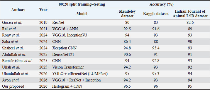
This study focuses on balancing precision and recall, as both are essential for effective LSD detection. High recall ensures that most infected cattle are correctly identified, thereby minimizing false negatives. High precision reduces false positives and avoids unnecessary interventions. The experimental results show that the proposed models achieve a strong balance between these metrics, with consistently high F1-scores (approximately 0.95–0.96). Although minor variations exist across architectures, some models favor recall slightly over precision, indicating a focus on disease detection. Overall, the framework effectively balances these metrics, making it suitable for practical applications requiring both early detection and reduced false alarms. The model evaluation was conducted using an 80:20 training–testing split, and the following statistical measures were included: ✓ Cohen’s kappa coefficient was computed to assess the agreement between the predicted and actual classifications beyond chance. The obtained value of κ=0.90 indicates an almost perfect agreement. ✓ A 95% confidence interval for the kappa statistic was calculated as (0.87–0.93), demonstrating the robustness and consistency of the model’s performance. ✓ McNemar’s test was performed to evaluate the statistical significance of misclassification differences. The results indicated no significant discrepancy (p > 0.05), confirming that the classification was balanced. ✓ A paired t-test was conducted using multiple experimental runs under the same 80:20 data split to compare the proposed model with baseline approaches, confirming statistically significant improvement (p < 0.05). ✓ Additionally, one-way ANOVA analysis was performed to compare multiple models, and the results showed statistically significant differences among them (p < 0.05). Performance comparison of state-of-the-art models for LSD identificationThe performance comparison of LSD detection methods using an 80:20 training–testing split demonstrates a clear improvement in model effectiveness, as shown in Table 4. Earlier research by Goceri (2019) based on a ResNet architecture, reported accuracy values of about 80%, 83%, and 82.6%, respectively. Subsequent work by Rai et al. (2021) using a hybrid VGG16 + ANN model significantly improved results, achieving 92.5%, 91.6%, and 89% accuracy. Further advancements were observed in later studies, where Rony et al. (2024) attained approximately 94%, 93%, and 93% using VGG16 and InceptionV3, while Saha (2024)reported accuracy levels between 86.4%, 88% and 90% with a CNN-based approach. Similarly, Shakeel et al. (2024) using Xception CNN and Abdullah et al. (2025) with DenseNet121 maintained consistent performance in the range of 90%–94% accuracy. More advanced techniques, including the Vision Transformer proposed by Ullah (2025) and the YOLO + EfficientNet (LUMPNet) model by Ubaidullah et al. (2026)achieved accuracy levels up to 95.3%, while ensemble-based methods by Ayon et al. (2026) reported around 94% accuracy. In contrast, the proposed approach integrating Histogram features and CNN achieves superior results, reaching an accuracy of 0.965 on the Mendeley dataset, 0.96 on the Kaggle dataset, and 0.95 on the Indian Journal of Animal Research dataset, indicating its improved capability for accurate LSD classification. Table 4. Comparison of state-of-the-art models for LSD identification with proposed methodology.
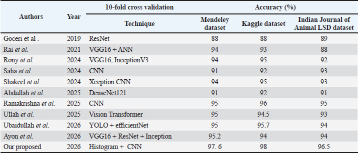
Moreover, a 10-fold cross-validation strategy was adopted to ensure the robust evaluation of the proposed model. Table 5 summarizes the existing methods for LSD identification, with most approaches achieving accuracies between 88% and 95% across the datasets. In comparison, the proposed Histogram + CNN model achieved higher accuracies of 97.6%, 98%, and 96.5% on the Mendeley, Kaggle, and Indian Research LSD datasets, respectively, demonstrating improved performance over existing methods. Table 5. Comparison of the existing LSD detection approaches with the proposed methodology.
DiscussionDiscussion of the experimental results indicates that the proposed CLAHE-based deep learning framework provides improved performance for automated detection of LSD in cattle images. The relatively lower performance of DenseNet121, ResNet50V2, InceptionV3, VGG16, VGG19, and Xception is mainly due to their mismatch with the dataset properties and their architectural characteristics. Very deep models, such as DenseNet121 and ResNet50V2, tend to overfit when trained on relatively limited or less diverse datasets, thereby reducing the generalization performance. VGG16 and VGG19, which are parameter-heavy and lack modern optimization strategies, are less effective in capturing fine-grained and texture-specific lesion features. InceptionV3 and Xception rely on complex multiscale feature extraction, which may not effectively emphasize subtle lesion boundaries in the images. In contrast, the proposed approach integrates histogram-based preprocessing, which enhances contrast and highlights lesion regions that are discriminative. This preprocessing step improves feature separability, allowing the CNN model to learn more relevant patterns and achieve superior classification performance compared to the aforementioned architectures. ConclusionThe increasing incidence of cattle diseases, such as LSD, highlights the urgent need for advanced and reliable detection techniques. Traditional methods, including manual examination and laboratory analysis, are time-consuming, costly, and susceptible to human error. In this study, a robust framework combining histogram-based preprocessing with deep learning models is proposed to address these challenges. The use of CLAHE effectively enhances image contrast and highlights skin lesions, enabling improved feature representation. These enhanced images are then processed by various deep learning architectures, including CNN, DenseNet121, ResNet50V2, InceptionV3, VGG16, VGG19, and Xception, to accurately classify the cattle as healthy or infected. The framework was evaluated on the LSD datasets of Mendeley, Kaggle, and the Indian Journal of Animal Research, where it consistently outperformed conventional deep learning approaches. The results demonstrate that the integration of image enhancement techniques with deep learning significantly improves identification accuracy. Among the models, CNN-based approaches combined with CLAHE showed reliable and consistent performance across all datasets. However, certain limitations exist, including dataset constraints, image quality, and background variations, and the absence of ensemble or hybrid feature extraction methods, which may affect generalizability. Future work will focus on incorporating real-time field datasets, standardizing image acquisition to ensure consistent capture conditions (lighting, angle, distance, and image quality), and exploring advanced modeling techniques to further enhance detection performance. AcknowledgmentsThe authors thank all the veterinarians, farm owners, and technical staff who assisted in data collection and provided valuable insights. We also acknowledge the support of the Koneru Lakshmaiah Education Foundation for providing resources for this research. Conflict of interestThe authors declare that there is no conflict of interest. FundingThis research received no specific grant. Authors' contributionsAll authors participated in data analysis, manuscript drafting, and revisions. Consent to assume responsibility for all aspects of this study. Data availabilityAll data were provided in the manuscript. ReferencesAbdullah, W., Tanwar, S. and Abouhawwash, M. 2025. Deep learning based detection of lumpy skin disease in livestock using CNNs. Sustain. Mach. Intell. J. 11(1), 1–10. Abdullah, W. 2025. Transfer learning with DenseNet121 for automated livestock disease classification. IEEE. Access. 13, t1–t12. Alzubi, A.A. 2024. Lumpy skin disease detection in cattle by a robust approach using advanced convolutional neural networks. Indian J. Anim. Res. 58(12), 2146–2153. Ayon, N.B., Rahman, M.S. and Islam, M.R. 2026. Ensemble deep learning approach for the automated detection of lumpy skin disease and foot-and-mouth disease in cattle. Artif. Intell. Agric. 12, 2345–2353. Chollet, F.2017. Xception: deep learning with depthwise separable convolutions. IEEE Conf. Comp. Vision and Pattern Recognition. 1251–1258. Goddeti, M. and Narayana, V.A. 2024. Utilizing extreme learning machine for the diagnosis of lumpy skin disease in cattle. Inter. J. Eng. Trends Technol. 72(9), 67–77. Goceri, E. 2019. Deep learning based classification of skin lesions using ResNet architectures. Biomed. Signal. Process. Control. 51(1), 1–8. Huang, G. and Liu, Z. 2017. Densely connected convolutional networks. IEEE Conf. Comp. Vision and Pattern Recog. 4700–4708. Khandait, P.D., Lanjewar, K.K., Patle, S. and Meshram, R. 2025. Deep learning-based cattle disease detection: a CNN approach for Identifying lumpy skin disease and foot-and-mouth disease. Int. J. Innov. Res. Technol. (IJIRT). 11(10), 11. Kumar, S. and Shastri, S. 2022. Lumpy skin images dataset. Mendeley Data. Available via https://data.mendeley.com/datasets/w36hpf86j2/1 Olorunshola, E. and Lakan, N. 2025. Hybrid convolutional neural network-haralick framework for foot-and-mouth disease classification in cattle. Asian J. Res. Comp. Sci. 18(6), 390–402. Rai Latha, S. and Sravanthi, S. 2021. Hybrid VGG16 and ANN-based classification of animal diseases. Procedia Comput. Sci. 192, 2950–2959. Ramakrishna, P. 2025. Convolutional neural network-based classification of cattle diseases. Inter.J. Advan. Comp. Sci. and Appl.16 (2), 345–354. Reza, M.R., Sahriani, P. and Mukjizat, S. 2026. Clinical image based classification of foot and mouth disease in cattle using convolutional neural networks. JurnalSistem. Dan TeknologiInformasi. Indonesia (JUSTINDO). 6(1), 22–31. Rony, M.A.T. 2024. Comparative analysis of vgg16 and inception v3 for livestock disease detection. Multimed. Tools Appl. 83(4), 10141–10163. Saha, D.K. 2024. Extensive investigation of convolutional neural network architectures for the diagnosis of lumpy skin disease in dairy cattle. Halcyon 10(4), e27893. Saqib, S., Khan, M., Sharif, A. and Rehman, A. 2024. Lightweight CNN based detection of lumpy skin disease in cattle images. Comput. Biol. Med. 168, 107692. Senthilkumar, C., Sindhu, C., Vadivu, G. and Neethirajan, S. 2024. Early detection of lumpy skin disease in cattle using deep learning—a comparative analysis of pretrained models. Vet. Sci. 11(10), 510. Sentoso, F., Ardiansyah, R. and Wangsa, S. 2025. Performance evaluation of VGG and ResNet architectures for cattle disease image classification. Sistemasi. J. Syst. Inf. 14(1), 89–98. Shakeel, M., Tauheed, Z., Javaid, N. and Aslam, T. 2024. A deep learning tool for early detection and control of lumpy skin disease using convolutional neural networks. J. Comput. Biomed. Informat. 7(2), 547. Shakeel, M.Z., Tauheed, N., Javaid, M.T. and Aslam, T. 2024. A deep learning tool for early detection and control of lumpy skin disease using convolutional neural networks. J. Comp. Biomedical Informatics, 5(1). 1–14. Simonyan, K., Zisserman, A. 2015. Very deep convolutional networks for large-scale image recognition. arXiv preprint arXiv:1409.1556. Ubaidullah, A., Hussain, H. and Jafri, M. 2026. LUMPNet: a hybrid YOLO–EfficientNet framework for lumpy skin disease detection. Comput. Biol. Med. 180, 107694. Ullah, A. 2025. Vision transformer-based automated classification of cattle skin diseases. Expert Syst. Appl. 2025, 235. | ||











 Computational Cost can be given as follows:












| How to Cite this Article |
| Pubmed Style Selvam IRP, Tabbassum S, Fathima AJ, Pandian SS, Banu GS, Anbalagan SDP, Thinakaran HP. An enhanced deep learning approach for the automated identification of lumpy skin disease in cattle. Open Vet. J.. 2026; 16(6): 3717-3730. doi:10.5455/OVJ.2026.v16.i6.44 Web Style Selvam IRP, Tabbassum S, Fathima AJ, Pandian SS, Banu GS, Anbalagan SDP, Thinakaran HP. An enhanced deep learning approach for the automated identification of lumpy skin disease in cattle. https://www.openveterinaryjournal.com/?mno=311467 [Access: June 26, 2026]. doi:10.5455/OVJ.2026.v16.i6.44 AMA (American Medical Association) Style Selvam IRP, Tabbassum S, Fathima AJ, Pandian SS, Banu GS, Anbalagan SDP, Thinakaran HP. An enhanced deep learning approach for the automated identification of lumpy skin disease in cattle. Open Vet. J.. 2026; 16(6): 3717-3730. doi:10.5455/OVJ.2026.v16.i6.44 Vancouver/ICMJE Style Selvam IRP, Tabbassum S, Fathima AJ, Pandian SS, Banu GS, Anbalagan SDP, Thinakaran HP. An enhanced deep learning approach for the automated identification of lumpy skin disease in cattle. Open Vet. J.. (2026), [cited June 26, 2026]; 16(6): 3717-3730. doi:10.5455/OVJ.2026.v16.i6.44 Harvard Style Selvam, I. R. P., Tabbassum, . S., Fathima, . A. J., Pandian, . S. S., Banu, . G. S., Anbalagan, . S. D. P. & Thinakaran, . H. P. (2026) An enhanced deep learning approach for the automated identification of lumpy skin disease in cattle. Open Vet. J., 16 (6), 3717-3730. doi:10.5455/OVJ.2026.v16.i6.44 Turabian Style Selvam, Ithaya Rani Panneer, Saziya Tabbassum, Abdul Jainul Fathima, Selvi Shanmuga Pandian, Gul Shira Banu, Shalini Divya Prasanna Anbalagan, and Hari Prasath Thinakaran. 2026. An enhanced deep learning approach for the automated identification of lumpy skin disease in cattle. Open Veterinary Journal, 16 (6), 3717-3730. doi:10.5455/OVJ.2026.v16.i6.44 Chicago Style Selvam, Ithaya Rani Panneer, Saziya Tabbassum, Abdul Jainul Fathima, Selvi Shanmuga Pandian, Gul Shira Banu, Shalini Divya Prasanna Anbalagan, and Hari Prasath Thinakaran. "An enhanced deep learning approach for the automated identification of lumpy skin disease in cattle." Open Veterinary Journal 16 (2026), 3717-3730. doi:10.5455/OVJ.2026.v16.i6.44 MLA (The Modern Language Association) Style Selvam, Ithaya Rani Panneer, Saziya Tabbassum, Abdul Jainul Fathima, Selvi Shanmuga Pandian, Gul Shira Banu, Shalini Divya Prasanna Anbalagan, and Hari Prasath Thinakaran. "An enhanced deep learning approach for the automated identification of lumpy skin disease in cattle." Open Veterinary Journal 16.6 (2026), 3717-3730. Print. doi:10.5455/OVJ.2026.v16.i6.44 APA (American Psychological Association) Style Selvam, I. R. P., Tabbassum, . S., Fathima, . A. J., Pandian, . S. S., Banu, . G. S., Anbalagan, . S. D. P. & Thinakaran, . H. P. (2026) An enhanced deep learning approach for the automated identification of lumpy skin disease in cattle. Open Veterinary Journal, 16 (6), 3717-3730. doi:10.5455/OVJ.2026.v16.i6.44 |


