





| Research Article | ||
Open Vet. J.. 2026; 16(6): 3631-3639 Open Veterinary Journal, (2026), Vol. 16(6): 3631-3639 Research Article Proliferative effects of glial cell-derived neurotrophic factor on putative spermatogonial stem cells derived from Zebu crossbred (Bos indicus X Bos taurus) malesSunil Kumar1*, Arumugam Kumaresan1, Kaustubh K. Saraf1, Subhash Gahalot1, Utkarsh Kumar Tripathi1 and Tushar Kumar Mohanty21Department of Animal Husbandry and Dairying, Haryana; Animal Reproduction, ICAR-National Research Centre on Pig, Rani; Theriogenology Laboratory, Southern Regional Station, ICAR- National Dairy Research Institute, Bengaluru; Faculty of Veterinary and Animal Sciences, Mirzapur Campus, BHU 2Pr. Scientist (Retd.), ABRC, ICAR- National Dairy Research Institute, Karnal, India *Corresponding Author: Sunil Kumar. Department of Animal Husbandry and Dairying, Haryana; Animal Reproduction, ICAR-National Research Centre on Pig, Rani; Theriogenology Laboratory, Southern Regional Station, ICAR- National Dairy Research Institute, Bengaluru; Faculty of Veterinary and Animal Sciences, Mirzapur Campus, BHU. Email: skicar2200 [at] gmail.com Submitted: 17/02/2026 Revised: 14/04/2026 Accepted: 21/04/2026 Published: 09/06/2026 © 2025 Open Veterinary Journal
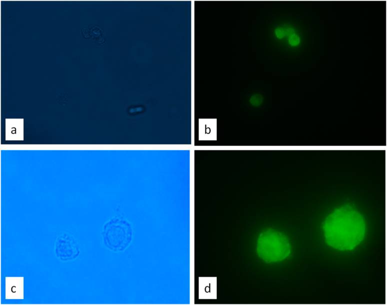
AbstractBackground: Reports on the effects of glial cell-derived neurotrophic factor (GDNF) on spermatogonial stem cells (SSCs) of Zebu or crossbred bovine are lacking. Proliferative effects were reported in ovine and caprine, whereas non-proliferative effects were only observed in Bos taurus. Aim: Understanding the roles of GDNF in maintaining germ cell proliferation may be crucial for reducing the high rate of reproductive failure in crossbred (Bos indicus X Bos taurus) bovine. Methods: In the present study, the in vitro culture effects of GDNF on SSCs of crossbred bovine were assessed. In the treatment groups, SSCs were cultured in a medium supplemented with two different doses of GDNF (T1-10 ng/ml, T2-40 ng/ml) and co-cultured with the Sertoli cell layer (T3). In the control group, SSCs were grown without GDNF or without co-culture (C). The proliferative effects in terms of the number of cells and colonies were determined on days 4, 7, 10, and 13 of in vitro culture. Results: There was a significant (p < 0.05) increase in the number of cells in the T2and T3 groups compared with the control group on days 4, 7, 10, and 13 of culture. There was no significant (p < 0.05) difference observed in the number of colonies between the control and GDNF (T1 and T2)-supplemented groups. However, the number of colonies was significantly (p < 0.05) higher in the T3 group than in the T1, T2, and control groups on days 10 and 13. Supplementation of GDNF (T2) and co-culture of SSCs with Sertoli cells significantly increased the surface area of SSCs colony during the entire period of in vitro culture compared with the control and T1 groups. However, on day 7, a significant difference (p < 0.05) in the surface area of the colonies was observed between the T2 and T3groups. Conclusion: The supplementation of GDNF at 40 ng/ml and co-culture of SSCs with Sertoli cells enhanced both the multiplication and growth of SSCs during in vitro culture, indicating their possible role in SSC proliferation and self-renewal in crossbred bovine. Keywords: Bovine, GDNF, Sertoli cells, Spermatogonial stem cells. IntroductionThe aim of spermatogonial stem cell (SSC) transfer technology is to transplant cultured SSCs to yield functional gametes with altered genotypes. However, a suitable stem cell culture system for the effective growth and multiplication of SSCs is a major limitation. Several growth factors have been studied to improve the yield of SSCs during in vitro culture (Oatley et al., 2009; Lee et al., 2013). Indeed, there are inconsistencies in the reports describing the effects of growth factors on the propagation of SSCs from domestic animals (Aponte et al., 2006; Aponte et al., 2008; Kuijk et al., 2009; Zhu et al., 2012; Kadam et al., 2013; Zheng et al., 2013). Glial cell line-derived neurotrophic factor (GDNF) plays a critical role in bovine fertility by regulating the self-renewal, proliferation, and maintenance of SSCs in the testes (Meng et al., 2000; Hofmann et al., 2005; Parekh et al., 2019). GDNF may be a key molecular factor in the regulation of spermatogenesis in the context of crossbreeding between Bos taurus (European) and Bos indicus (Zebu), and its signaling pathways are linked to the sub-fertility often observed in crossbred bulls. Crossbred bulls often exhibit high rates of sub-fertility, characterized by low sperm concentration and poor motility. Studies have suggested that dysregulation of signaling pathways contributes to this sub-fertility (Elango et al., 2020; Kumaresan et al., 2021), where one of the factors may be the involvement of GDNF-related pathways in maintaining proper SSC proliferation in crossbred bulls. The effects of GDNF on B. taurus SSCs have been reported to enhance self-renewal and increase the survival rates of SSCs rather than inducing proliferation (Aponte et al., 2006). GDNF has been reported to work along with other factors for the in vitro proliferation of SSCs in ewes (Binsila et al., 2020) and goats (Sharma et al., 2020). Hence, it may be confirmed from previous studies that GDNF is responsible for self-renewal, survival, and colony formation in B. taurus. However, no such studies have been reported to date with respect to proliferation and colony formation in B. indicus and crossbred (Bos indicus X Bos taurus). Furthermore, GDNF dose-dependently controlled the self-renewal and differentiation of mouse SSCs in vivo (Sariola and Immonen, 2008). For the culture of SSCs, a system must be devised that not only supports their survival but also supports their self-renewing proliferation. Co-culture of SSCs with Sertoli cells (as an endogenous source of GDNF) mimics the dynamics of SSCs and Sertoli cells in the testis. Assessment of the effects of different doses of GDNF during in vitro culture of SSCs could provide a means for expanding stem cell lines and investigating the biology of SSCs related to fertility or sub-fertility in crossbred Zebu cattle. However, the usefulness of GDNF in the proliferation of crossbred bovine SSCs is not well understood. Therefore, this study aimed to investigate the dose-dependent proliferative effects of GDNF supplementation on putative SSC culture in crossbred bovine SSCs. Materials and MethodsLocation of the study siteThis study was conducted at the Theriogenology Laboratory, ICAR-National Dairy Research Institute, Karnal, India. Experimental groupsIn the present study, the effects of GDNF supplementation on the number of SSCs and colony characteristics (number and surface area of the colonies) during in vitro culture in triplicate were assessed. In the treatment groups, SSCs were cultured in a medium supplemented with two different doses of GDNF (T1-10 ng/ml, T2-40 ng/ml) and co-cultured with the Sertoli cell layer (T3). In the control group, SSCs were grown without GDNF or without co-culture (C). Castration of the experimental malesTestes of crossbred bovine (Holstein Friesian X Tharparkar) males (n–7; 2–4 months age) were obtained from castration. Castration was performed using xylazine hydrochloride (0.1 mg/kg b.wt.; Xylazin, Indian Immunologicals, Hyderabad, India) and 2% lignocaine (Cadila Healthcare Ltd., Ahmedabad, India) as per the standard veterinary procedures. An incision was made at the level of the spermatic cord to expose the testis, and the spermatic cord was tightly ligated using catgut. After ligation, the intact testis was removed and placed in individual sterile containers containing normal saline with penicillin. The cells were processed in the laboratory for stem cell isolation. Due postoperative care was given to the animals after castration as per the standard veterinary practices. Isolation of spermatogonial stem cellsSpermatogonial stem cells were isolated from the testes as per the standardized procedures described by Izadyar et al. (2002, 2011) and Tripathi et al. (2014) with minor modifications. Briefly, after washing with Dulbecco’s phosphate-buffered saline (DPBS) and Dulbecco’s minimum essential medium (DMEM), the seminiferous epithelial cells were dispersed and suspended in DMEM containing 1 mg/ml collagenase (Sigma Aldrich,USA), 1 mg/ml hyaluronidase type II (Sigma Aldrich, USA), 5 μg/ml DNase (Sigma Aldrich, USA), and 1 mg/ml trypsin (Sigma Aldrich, USA) in a shaker incubator at 37°C for 45 minutes. The dispersed tissue was centrifuged at 1,000 rpm for 2 minutes, and the collected tissue pellet was washed once with DMEM. For the secondenzymatic digestion, the dispersed tissue was suspended in DMEM containing collagenase, hyaluronidase type II, and DNase ina shaker incubator at 37°C for 30 minutes. After this, 10% fetal bovine serum (Hyclone, Canada) was immediately added. The supernatant was filtered through a 60 μm (Merck Millipore, India) and then a 41 μm (Merck Millipore, India) nylon mesh filter. The filtered cell suspension was then transferred to a 1.5 ml tube (Nunc, Denmark) and centrifuged at 10,000 rpm for 1 minute. The pellet cell mass was collected and dissolved in 0.5 ml DMEM for further fractionation of cells using the Percoll discontinuous density gradient method. The Percoll density gradient was carried out according to the procedure given in previous reports (Kala et al., 2012). An iso-osmotic Percoll suspension containing 82.2% Percoll (Sigma Aldrich, USA) in DMEM (without additives) with 0.6% BSA and 45 μg/ml DNase was prepared. A discontinuous density gradient was prepared by diluting the iso-osmotic Percoll suspension with DMEM, 0.7% bovine serum albumin (Sigma Aldrich, USA), and 50 μg/ml DNase. The gradients were created by layering 1 ml of 65%, 60%, 50%, 45%, 40%, and 20% Percoll into a 15-ml centrifuge tube. The cell suspension was layered on top of the gradient in 500-μl MDEM, 0.7 % BSA, and 50 μg/ml DNase. This gradient was for 30 minutes at 18°C. Cells found in the interface between the different densities (45%–60%) suspensions were collected. Differential platingThe isolated SSCs were cultured in vitro in the presence or absence of GDNF (Sigma Aldrich, USA) to assess the effect of GDNF on SSC culture. The SSC culture groups were as follows: (1) control group, in which SSCs were grown without the exogenous addition of GDNF; (2) SSCs were cultured with exogenous GDNF supplementation (two subgroups: 10 and 40 ng/ml). GDNF was added on the day of seeding and on the day of media change during culture. (3) SSCs were co-cultured with Sertoli cells layer as an endogenous GDNF source. GDNF was added on the day of seeding, 4th, 7th and 10th day post seeding. The SSC-enriched cells obtained after Percoll density gradient centrifugation were subjected to differential plating by Kala et al. (2012) with minor modifications. Briefly, 15 × 106 cells per dish (3 ml of suspension per dish) were seeded onto DSA-lectin-coated dishes, prepared by coating 35 mm Petri dishes with lectin from Datura stramonium agglutinin (5 μg/ml in DPBS), DPBS washed, and blocked by adding 0.6% BSA for 1 hour at 37°C in a humidified atmosphere of 5% CO2 in air. Before the addition of isolated cells, phosphate buffered saline was replaced with DMEM + 10% fetal bovine serum. After overnight incubation, the non adhering cells were seeded into the culture groups described above at a concentration of 1 × 10⁶ cells/ml in each culture group. From differential plating, a Sertoli cell feeder monolayer was prepared and made mitotically inactive by mitomycin-C treatment, 7 days prior to seeding of enriched spermatogonial stem cells in respective culture groups. Cultures were carried out at 38.2°C in 5% CO2 in air, and the media was changed twice weekly. The number of cells and colony characteristics (surface area and number of colonies) were estimated on days 4, 7, 10, and 13 after culture seeding. Cell viability was determined using the Trypan blue exclusion test. Immunocytochemistry of the isolated cellsAfter isolation, the cells were subjected to immunocytochemistry using the Anti-UCHL1 Antibody (Sigma Aldrich, USA) marker. Isolated cells were washed in DPBS and then incubated for 1 hour after the addition of diluted (1:300) primary (Anti-UCHL1) antibody. The cells were then washed five times with DPBS, after which a secondary antibody was added in the dark, kept in a humidified box for 4 hours, and mounted by overlaying DABCO (1,4-diazabicyclo [2.2.2] octane; Sigma Aldrich, USA). The cells were observed for fluorescence (Fig. 1) under a green filter and a bright field microscope (SMZ 100/SMZ 800, Nikon, Tokyo, Japan).
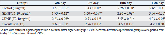
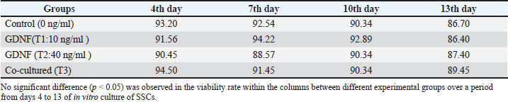
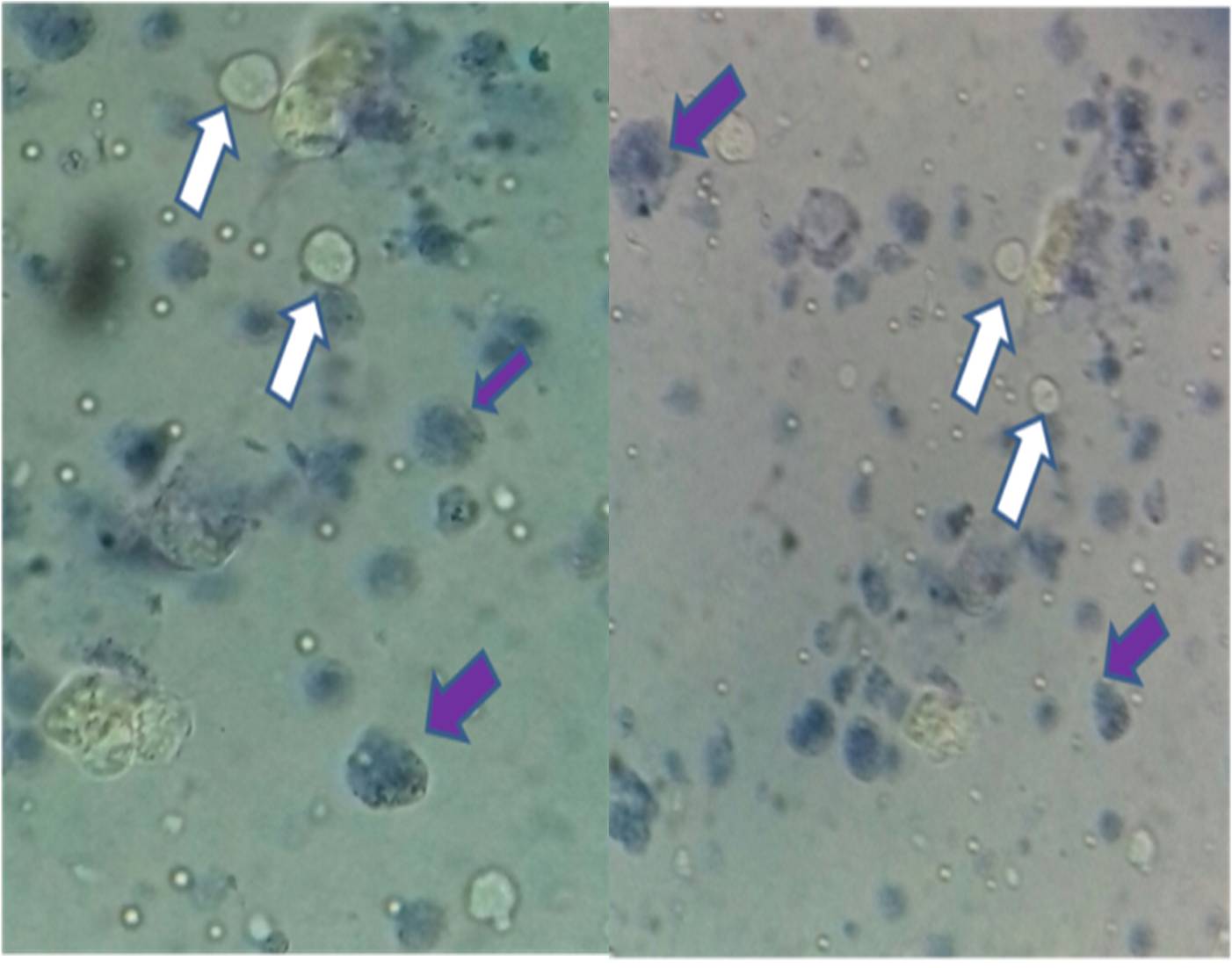
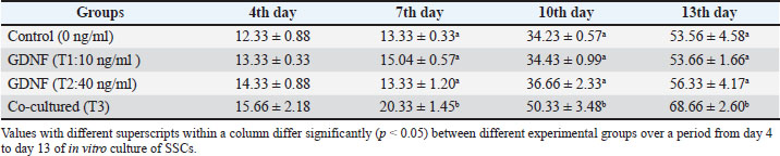
Fig. 1. Representative image of immunocytochemistry of isolated SSCs; a) SSCs under phase contrast at 400 X; b) SSCs under fluorescence at 400 X; c: SSCs under phase contrast at 1000 X; d: SSCs under fluorescence at 400 X. Statistical analysisAll statistical analyses were performed using SPSS 22 (IBM, USA). The values were expressed as Mean ± S.E. The number of SSCs and surface area of colonies under culture conditions (GDNF and Sertoli cell co-culture) and at different days of in vitro culture were repeated independently and analyzed statistically using repeated measure analysis of variance (ANOVA) design. Post ANOVA, Tukey’spost hoc tests were conducted to identify exactly the group means difference. Power analysis and assumption checks were performed using open-access statistical tools (clincalc.com). Differences were considered significant when p < 0.05. Ethical approvalAll experimental protocols and procedures were approved by the Institute Animal Ethics Committee (IAEC No. 84/16). ResultsIsolated putative spermatogonial stem cells were cultured to estimate the proliferative effects of GDNF supplementation in crossbred bovine males. Effect of GDNF on SSC number and viabilityTable 1 shows the number of SSCs during different days of culture in GDNF-supplemented (endogenous and exogenous) and control groups. On all the days of assessment, a significantly (p < 0.05) higher number of SSCs was observed in both the GDNF (40 ng/ml)-supplemented and SSC-Sertoli cell co-culture groups compared with the other two groups. There was no significant difference in the SSC count between the control and GDNF (10 ng/ml)-supplemented groups on any day of assessment. On any day of assessment, the proportion of viable cells (Fig. 2) ranged between 86.4% and 94.50% (Table 2). Although there was no significant difference in cell viability between the different groups on different days of culture, the proportion of viable cells decreased on day 13 of culture. Table 1. Spermatogonial stem cell numbers (X 106/ml) (mean ± SE) during in vitro culture with or without GDNF.
Table 2. Spermatogonial stem cell viability rate (%) during in vitro culture with or without GDNF.
Fig. 2. Representative images of SSCs isolated after enzymatic digestion for trypan blue exclusion. The white arrow indicates live cells, and the blue arrow indicates dead cells. Effect of GDNF on the number of SSC coloniesOn day 4 of culture, no significant difference was observed in the number of SSC colonies among the groups. However, on days 7, 10, and 13 of culture, the SSC-Sertoli cell co-cultured group had significantly (p < 0.05) higher number of colonies than the other three groups (Table 3). No significant differences were observed in the number of SSC colonies among the control, GDNF 10 ng/ml, and GDNF 40 ng/ml groups on any given day of culture. Table 3. Spermatogonial stem cell colonies (mean ± SE) during in vitro culture with or without GDNF.
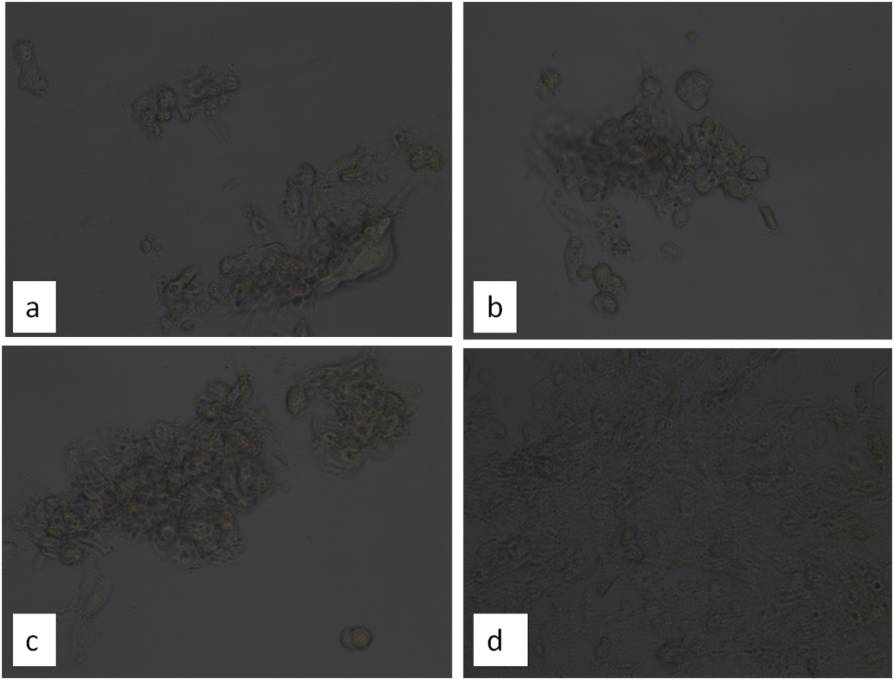
Distinct or particularly defined morphology of the colonies was observed from day 4 of culture. On day 7, colonies appeared as mixed aggregates of loosely connected cells in the GDNF-supplemented and co-cultured groups. On day 7, the colonies appeared as small low-density cell masses in the control group (Fig. 3a), whereas in GDNF-treated cultures, they appeared more compact and flattened (Fig. 3b and c). In SSC-Sertoli cell co-culture group, the colonies were small low-density cell masses that were not firmly attached to the Sertoli cell layer (Fig. 3d). These colonies continued to grow and condense until day 13 of culture.
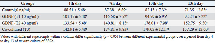
Fig. 3. Representative images of SSCs cultured colonies in different groups. a) Control where no GDNF was added; b) Colonies in the 10 ng/ml GDNF supplemented group; c) Colonies in 40 ng/ml GDNF supplemented group; d) Colonies in the co-cultured group. Effect of GDNF on the surface area of the SSC coloniesOn day 4 of culture, the mean surface area of SSC colonies was similar between the control and GDNF 10 ng/ml-supplemented groups (Table 4). Both the GDNF 40 ng/ml supplemented group and the SSC-Sertoli cell co-cultured group had colonies with significantly (p < 0.05) greater surface area than the other 2 groups. Similar results were also observed on day 10 of culture. On day 7 of culture, there were significant differences in the mean surface area of SSC colonies among the four groups with the highest surface area of SSC colonies in the SSC-Sertoli cell co-cultured group. On day 13, similar findings were observed; however, no significant difference was observed between the GDNF 40 ng/mL supplemented group and the SSC-Sertoli cell co-cultured group. Table 4. Surface area of spermatogonial stem cell colonies (mm2; mean ± SE) during in vitro culture with or without GDNF.
DiscussionRecently, India has registered the B. indicus X B. taurus crossbreed as a high-yielding synthetic cattle breed, namely Karan Fries. However, crossbred bulls often display lower fertility, higher rates of subfertility, and higher semen rejection rates than purebred B. taurus bulls. Crossbreds frequently suffer from poor spermiograms, often requiring strict screening as over 50% of crossbreds may fail to meet freezing standards (Elango et al., 2020).The differences in testicular composition, sperm phenotypic characteristics, molecular composition, environmental influence, and other details between purebred and crossbred males have been described (Kumaresan et al., 2021). These differences may be responsible for the differential fertility or sub-fertility between Bos indicus and B. taurus or their crossbreeds. Similarly, the biology of SSCs and the underlined proliferative effects of GDNF may be the differential features in B. indicus. In B. taurus, GDNF promotes self-renewal but not SSC proliferation (Aponte et al., 2006). Self-renewal maintains the stem cell pool by producing at least one daughter cell that remains undifferentiated. Proliferation is the general increase in cell number through division, which may result in either self-renewal or differentiation of progenitors. Self-renewal ensures lifelong fertility by preventing the depletion of SSCs, while proliferation provides cells for spermatogenesis. Hence, this difference needs to be explored further for a better understanding of SSC transplant technology for fertility improvement in crossbred bulls. A suitable stem cell culture system for the effective growth and multiplication of SSCs has been sought for the effective utilization of SSC transplant technology in ovine, caprine, and murine. Recently, in mouse models, it has been shown that GDNF dose-dependently controls the self-renewal and proliferation of SSCs in vivo. However, the proliferative role of GDNF during in vitro culture of SSCs of B. indicus and its crosses is not well understood or reported. Here, we report the proliferative effect of exogenous (T1 and T2) or endogenous (T3) GDNF on the growth and multiplication of SSCs during in vitro culture. In the present study, a significant (p < 0.05) increase in the number of SSCs was observed in the GDNF (T2: 40 ng/ml) and SSC-Sertoli cell co-cultured group (T3) compared with the control group on different days of culture. However, there was no significant difference in the number of cells between the control and GDNF (T1: 10 ng/ml) groups,indicating a dose-dependenteffect of GDNF on SSC proliferation. The current studyindicated that GDNF influenced the SSC growth rate in a dose-dependent manner, inducing a higher cell proliferation rate at higher concentrations. There was no significant (p < 0.05) difference was observed in the number of colonies in the control and exogenous GDNF (10 and 40 ng/ml)-supplemented groups. However, a significant difference (p < 0.05) was observed between the control and co-cultured SSC-Sertoli cells on days7, 10, and 13 of culture. After day 4, spermatogonial colonies increased in size in the GDNF-supplemented and SSC-Sertoli cell co-culturedgroups, but the number of colonies did not differ significantly between the control and GDNF-supplemented groups. These results indicated that endogenous GDNF from co-cultured Sertoli cells significantly increased the number of colonies compared with exogenous GDNF supplementation. In the GDNF-supplemented group, the daughter cells of SSCs undergoing self-renewing division apparently did not migrate away from each other to form separate colonies but remained inside the colonies to increase the surface area of the colonies, thus increasing the total number of cells while maintaining the number of colonies without any significant increase. This is further supported by our findings on the colonies’ surface area. The surface area of the SSC colonies increased with the dose of GDNF. On days 7 and 13 of culture, a significant difference was observed between the control and GDNF (10 ng/ml)-supplemented groups, but the difference on days 4 and 10 was not significant. GDNF at a 10 ng/ml concentration had an inconsistent effect on the surface area of colonies compared to the control group. The GDNF (40 ng/ml) group showed a significant difference in the surface area of SSC colonies compared with the control groupon all days of culture. The interpretation of these observations may be correlated with the colony number on the respective days. Therefore, it may be inferred that GDNF at a concentration of 40 ng/ml increased the surface area of the colonies by increasing the self-renewal of cells inside the colony. The mean surface area of the colonies observed in this study was less than that observed by Aponte et al. (2006). This may be due to the supplementation of a lesser concentration of GDNF in this study as compared to earlier studies. In every culture, some cells (scattered and within the colonies) showed morphological signs of apoptosis as evidenced by shrinkage between days 4 and 13. On day 13 of culture, cells in the colonies appeared to be smaller, scattered, and less interconnected in all groups. However, it was observed that the number of scattered individual cells was lower than that observed by Aponte et al. (2006). This indicated that the differentiation of stem cells was more effectively controlled in the present study, providing evidence of the more proliferative effects of GDNF. In earlier studies, GDNF was evaluated for SSC renewal in vivo (Meng et al., 2000; Yomogida et al., 2003) and in vitro (Nagano et al., 2003; Kubota et al., 2004). Normally, Sertoli cells secrete GDNF in the seminiferous epithelium, whereas the receptors for this growth factor, GFR-alfa 1 and c-ret, are present on SSCs (Meng et al., 2000; Tadokoro et al., 2002; Oatley et al., 2004). Furthermore, follicle stimulating hormone was found to stimulate GDNF production by Sertoli cells (Tadokoro et al., 2002). The current findings are in agreement with those reported earlier, as exogenous GDNF supplementation as well as SSCs-Sertoli cell co-culture increased the number of SSC in vitro by stimulating their proliferation. GDNF acts as a self-renewal factor for SSCs, promotes cell division (proliferation), and prevents differentiation or apoptosis. Daughter cells tended to stay attached to the parent colony, and limited migration or dispersal was observed. Colony initiation often requires physical separation + niche support, not just proliferation. Sertoli cells provide a functional niche, not just growth signals. They contribute to adhesion molecules (help cells anchor and reorganize), extracellular matrix components, additional paracrine factors beyond GDNF, and signals that may promote cell detachment, migration, or reaggregation. Colony formation depends on cell motility, the microenvironment (niche signals), and cell–cell and cell–matrix interactions, whereas the total number of SSCs depends mainly on the proliferation rate. Hence, GDNF boosts the proliferation of Sertoli cells, enabling colony initiation and structuring. In summary, GDNF increases SSC proliferation, but new colony formation requires niche-driven spatial reorganization, which was provided by Sertoli cells not GDNF alone. However, transplant assay, migration, or clonal tracking analysis, along with a broader dose–response curve, will strengthen the claim of future studies. Hence, supplementation of GDNF at a concentration of 40 ng/ml or co-culture of SSCs with Sertoli cells enhanced the growth and multiplication of SSCs during in vitro culture due to its involvement in the proliferation of crossbred bovine SSCs. AcknowledgmentsThe authors would like to thank the Director, ICAR-NDRI, Karnal, Labmates (Dr Sreela L, Dr Shivani Chillar, and Smt. Samiksha Nayak), Staff of Animal Health Complex and Livestock Research Station, ICAR-NDRI, Karnal for providing necessary facilities and support in the conducted work. Conflict of interestThe authors have no conflicts of interest to declare. FundingThe authors are grateful to the Indian Council of Agricultural Research, Department of Agricultural Research and Education, Govt. of India, New Delhi, for providing the necessary facilities at ICAR-National Dairy Research Institute, Karnal. Authors 'contributionsSK and AK conceived the idea. KKS, SG, and UT helped SK and AK in the experimental study. TK supervised the study. Data availabilityAll data were provided in the manuscript. ReferencesAponte, P.M., Soda, T., Teerds, K.J., Mizrak, S.C., Van De Kant, H.J.G. and De Rooij, D.G. 2008. Propagation of bovine spermatogonial stem cells in vitro. Reproduction 136, 543–557. Aponte, P.M., Soda, T., Van De Kant, H.J. and De Rooij, D.G. 2006. Basic features of bovine permatogonial culture and effects of glial cell line-derived neurotrophic factor. Theriogenology 65, 1828–1847. Binsila, B.K., Selvaraju, S., Ghosh, S.K., Ramya, L., Arangasamy, A., Ranjithkumaran, R. and Bhatta, R. 2020. EGF, GDNF and IGF-1 influence the proliferation and stemness of ovine spermatogonial stem cells in vitro. J. Assist. Reprod. Genet. 37, 2615–2630. Elango, K., Kumaresan, A., Sharma, A., Nag, P., Prakash, M.A., Sinha, M.K., Manimaran, A., Peter, E.S.K.J., Jeyakumar, S., Selvaraju, S., Ramesha, K.P. and Datta, T.K. 2020. Sub-fertility in crossbred bulls: deciphering testicular level transcriptomic alterations between zebu (Bos indicus) and crossbred (Bos taurus × Bos indicus) bulls. BMC. Genomics 21, 502. Hofmann, M.C., Braydich-Stolle, L. and Dym, M. 2005. Isolation of male germ-line stem cells; influence of GDNF. Dev. Biol. 279, 114–124. Izadyar, F., Spierenberg, G.T., Creemers, L.B., Den Ouden, K. and De Rooij, D.G. 2002. Isolation and purification of type A spermatogonia from the bovine testis. Reproduction 124, 85–94. Izadyar, F., Wong, J., Maki, C., Pacchiarotti, J., Ramos, T., Howerton, K., Yuen, C., Greilach, S., Zhao, H.H., Chow, M., Chow, Y.C., Rao, J., Barritt, J., Bar-Chama, N. and Copperman, A. 2011. Identification and characterization of repopulating spermatogonial stem cells from the adult human testis. Hum. Reprod. 26, 1296–1306. Kadam, P.H., Kala, S., Agrawal, H., Singh, K.P., Singh, M.K., Chauhan, M.S., Palta, P., Singla, S.K. and Manik, R.S. 2013. Effects of glial cell line-derived neurotrophic factor, fibroblast growth factor 2 and epidermal growth factor on proliferation and gene expression in buffalo (Bubalus bubalis) spermatogonial cells. Reprod. Fertil. Dev. 25, 1149–1157. Kala, S., Kaushik, R., Singh, K.P., Kadam, P.H., Singh, M.K., Manik, R.S., Singla, S.K., Palta, P. and Chauhan, M.S. 2012. In vitro culture and morphological characterization of prepubertal buffalo (Bubalus bubalis) putative spermatogonial stem cells. J. Assist. Reprod. Genet. 29, 1335–1342. Kubota, H., Avarbock, M.R. and Brinster, R.L. 2004. Culture conditions and single growth factors affect fate determination of mouse spermatogonial stem cells. Biol. Reprod. 71, 722–731. Kuijk, E.W., Colenbrander, B. and Roelen, B.A.J. 2009. The effects of growth factors on in vitro-cultured porcine testicular cells. Reproduction 138, 721–731. Kumaresan, A., Elango, K., Datta, T.K. and Morrell, J.M. 2021. Cellular and molecular insights into the etiology of subfertility/infertility in crossbred bulls (Bos taurus × Bos indicus): a review. Front. Cell Dev. Biol. 9, 696637. Lee, K., Lee, W., Kim, J., Yoon, M., Kim, N., Kim, J., Uhm, S., Kim, D., Chung, H. and Song, H. 2013. Characterization of GFRα-1-positive and GFRα-1-negative spermatogonia in neonatal pig testis. Reprod. Domest. Anim. 48, 954–960. Luo, J., Megee, S., Rathi, R. and Dobrinski, I. 2006. Protein gene product 9.5 is a spermatogonia-specific marker in the pig testis. Mol. Reprod. Dev. 73, 1531–1540. Meng, X., Lindahl, M., Hyvönen, M.E., Parvinen, M., De Rooij, D.G., Hess, M.W., Raatikainen-Ahokas, A., Sainio, K., Rauvala, H., Lakso, M., Pichel, J.G., Westphal, H., Saarma, M. and Sariola, H. 2000. Regulation of cell fate decision of undifferentiated spermatogonia by GDNF. Science 287, 1489–1493. Nagano, M., Ryu, B.Y., Brinster, C.J., Avarbock, M.R. and Brinster, R.L. 2003. Maintenance of mouse male germ line stem cells in vitro. Biol. Reprod. 68, 2207–2214. Oatley, J.M., De Avila, D.M., Reeves, J.J. and McLean, D.J. 2004. Testis tissue explant culture supports survival and proliferation of bovine spermatogonial stem cells. Biol. Reprod. 70, 625–631. Oatley, J.M., Oatley, M.J., Avarbock, M.R., Tobias, J.W. and Brinster, R.L. 2009. Colony Stimulating Factor 1 Is An Extrinsic Stimulator Of Mouse Spermatogonial Stem Cell Self-Renewal. Development 136, 1191–1199. Parekh, P.A., Garcia, T.X. and Hofmann, M.C. 2019. Regulation of GDNF expression in Sertoli cells. Reproduction 157, R95–R107. Sariola, H. and Immonen, T. 2008. GDNF maintains mouse spermatogonial stem cells in vivo and in vitro. Methods Mol. Biol. 450, 127–135. Sharma, A., Shah, S.M., Tiwari, M., Roshan, M., Singh, M.K., Singla, S.K., Palta, P., Manik, R.S. and Chauhan, M.S. 2020. Propagation of goat putative spermatogonial stem cells under growth factor defined serum-free culture conditions. Cytotechnology 72, 489–497. Tadokoro, Y., Yomogida, K., Ohta, H., Tohda, A. and Nishimune, Y. 2002. Homeostatic regulation of germinal stem cell proliferation by the GDNF/FSH pathway. Mech. Dev. 113:29–39. Tripathi, U.K., Aslam, M.K., Pandey, S., Nayak, S., Chhillar, S., Srinivasan, A., Mohanty, T.K., Kadam, P.H., Chauhan, M.S., Yadav, S. and Kumaresan, A. 2014. Differential proteomic profile of spermatogenic and Sertoli cells from peripubertal testes of three different bovine breeds. Front. Cell. Dev. Biol. 2, 24. Yomogida, K., Yagura, Y., Tadokoro, Y. and Nishimune, Y. 2003. Dramatic expansion of germinal stem cells by ectopically expressed human glial cell line-derived neurotrophic factor in mouse Sertoli cells. Biol. Reprod. 69, 1303–1307. Zhu, H., Liu, C., Li, M., Sun, J., Song, W. and Hua, J. 2012. Optimization of the conditions of isolation and culture of dairy goat male germ line stem cells (mGSC). Anim.Reprod. Sci. 137, 45–52. | ||
| How to Cite this Article |
| Pubmed Style Kumar S, Kumaresan A, Saraf KK, Gahalot S, Tripathi UK, Mohanty TK. Proliferative effects of glial cell-derived neurotrophic factor on putative spermatogonial stem cells derived from Zebu crossbred (Bos indicus X Bos taurus) males. Open Vet. J.. 2026; 16(6): 3631-3639. doi:10.5455/OVJ.2026.v16.i6.35 Web Style Kumar S, Kumaresan A, Saraf KK, Gahalot S, Tripathi UK, Mohanty TK. Proliferative effects of glial cell-derived neurotrophic factor on putative spermatogonial stem cells derived from Zebu crossbred (Bos indicus X Bos taurus) males. https://www.openveterinaryjournal.com/?mno=310852 [Access: June 26, 2026]. doi:10.5455/OVJ.2026.v16.i6.35 AMA (American Medical Association) Style Kumar S, Kumaresan A, Saraf KK, Gahalot S, Tripathi UK, Mohanty TK. Proliferative effects of glial cell-derived neurotrophic factor on putative spermatogonial stem cells derived from Zebu crossbred (Bos indicus X Bos taurus) males. Open Vet. J.. 2026; 16(6): 3631-3639. doi:10.5455/OVJ.2026.v16.i6.35 Vancouver/ICMJE Style Kumar S, Kumaresan A, Saraf KK, Gahalot S, Tripathi UK, Mohanty TK. Proliferative effects of glial cell-derived neurotrophic factor on putative spermatogonial stem cells derived from Zebu crossbred (Bos indicus X Bos taurus) males. Open Vet. J.. (2026), [cited June 26, 2026]; 16(6): 3631-3639. doi:10.5455/OVJ.2026.v16.i6.35 Harvard Style Kumar, S., Kumaresan, . A., Saraf, . K. K., Gahalot, . S., Tripathi, . U. K. & Mohanty, . T. K. (2026) Proliferative effects of glial cell-derived neurotrophic factor on putative spermatogonial stem cells derived from Zebu crossbred (Bos indicus X Bos taurus) males. Open Vet. J., 16 (6), 3631-3639. doi:10.5455/OVJ.2026.v16.i6.35 Turabian Style Kumar, Sunil, Arumugam Kumaresan, Kaustubh K. Saraf, Subhash Gahalot, Utkarsh Kumar Tripathi, and Tushar Kumar Mohanty. 2026. Proliferative effects of glial cell-derived neurotrophic factor on putative spermatogonial stem cells derived from Zebu crossbred (Bos indicus X Bos taurus) males. Open Veterinary Journal, 16 (6), 3631-3639. doi:10.5455/OVJ.2026.v16.i6.35 Chicago Style Kumar, Sunil, Arumugam Kumaresan, Kaustubh K. Saraf, Subhash Gahalot, Utkarsh Kumar Tripathi, and Tushar Kumar Mohanty. "Proliferative effects of glial cell-derived neurotrophic factor on putative spermatogonial stem cells derived from Zebu crossbred (Bos indicus X Bos taurus) males." Open Veterinary Journal 16 (2026), 3631-3639. doi:10.5455/OVJ.2026.v16.i6.35 MLA (The Modern Language Association) Style Kumar, Sunil, Arumugam Kumaresan, Kaustubh K. Saraf, Subhash Gahalot, Utkarsh Kumar Tripathi, and Tushar Kumar Mohanty. "Proliferative effects of glial cell-derived neurotrophic factor on putative spermatogonial stem cells derived from Zebu crossbred (Bos indicus X Bos taurus) males." Open Veterinary Journal 16.6 (2026), 3631-3639. Print. doi:10.5455/OVJ.2026.v16.i6.35 APA (American Psychological Association) Style Kumar, S., Kumaresan, . A., Saraf, . K. K., Gahalot, . S., Tripathi, . U. K. & Mohanty, . T. K. (2026) Proliferative effects of glial cell-derived neurotrophic factor on putative spermatogonial stem cells derived from Zebu crossbred (Bos indicus X Bos taurus) males. Open Veterinary Journal, 16 (6), 3631-3639. doi:10.5455/OVJ.2026.v16.i6.35 |


