





| Research Article | ||
Open Vet. J.. 2026; 16(6): 3518-3527 Open Veterinary Journal, (2026), Vol. 16(6): 3518-3527 Research Article Molecular characterization, expression profiling, and phylogenetic analysis of key adhesion-associated genes in Mycoplasma gallisepticum isolated from poultrySaeed Mohammed Hasan1* and Alaa Abdulaziz Abed21Department of Veterinary Microbiology, College of Veterinary Medicine, University of Al-Qadisiyah, Al Diwaniyah, Iraq 2Department of Pathology and Poultry Diseases, College of Veterinary Medicine, University of Al-Qadisiyah, Al Diwaniyah, Iraq *Corresponding Author: Saeed Mohammed Hasan. Department of Veterinary Microbiology, College of Veterinary Medicine, University of Al-Qadisiyah, Al Diwaniyah, Iraq. Email: vet.post23.36 [at] qu.edu.iq Submitted: 04/02/2026 Revised: 05/05/2026 Accepted: 18/05/2026 Published: 05/06/2026 © 2025 Open Veterinary Journal
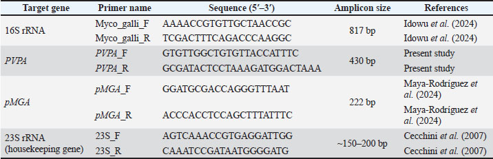
AbstractBackground: Considerable economic and productivity losses result from the impact of Mycoplasma gallisepticum on the poultry industry, causing increased mortality and expenses from control practitioner implementations. Phenomena associated with the cytoadherence and adaptive variation of the Putative Variable Protein A (PVPA) and Mycoplasma gallisepticum hemagglutinin protein (pMGA) multigene families are notable; however, there is limited information on the expression, molecular characteristics, and genetic variation of local poultry isolates. Aim: The aim of the study was to examine and analyze the genomic organization, molecular characteristics, expression patterns, and phylogenetic relationships for PVPA and pMGA from M. gallisepticum isolates from poultry to pathogenicity and their genetic variation. Methods: Clinical poultry specimens (tracheal swabs) were incubated with pleuropneumonia-like organism broth and agar media with selective supplements for Mycoplasma. Isolates were diagnosed using standard cultural characteristics, Diene's staining, and molecular methods. polymerase chain reaction and reverse transcription polymerase chain reaction were applied to extracted genomic Deoxyribonucleic acid (DNA) (16S rRNA, PVPA, and pMGA genes) and total RNA (PVPA and pMGA genes). Agarose gel products were subjected to sequencing analysis of the 16S rRNA gene. Their sequence was analyzed and compared to international databases. Local and global 16S rRNA sequences were used to perform a phylogenetic analysis. Results: All isolates showed similar growth characteristics of M. gallisepticum, including the fried egg colony morphology (as observed in Diene's staining). Analysis of total cultured genomic DNA verified the 16S rRNA gene, as well as the presence of the specified amplified fragments of the PVPA (430 bp) and pMGA (222 bp) genes. Analysis of the transcriptome showed that the PVPA and pMGA genes were actively expressed during the infection. Analysis of the local isolates revealed strong sequence homogeneity, though the local isolates showed unique, distinct clustering from neighboring countries in the phylogenetic trees. A few local sequences were deposited in the National Center for Biotechnology Information database, which serves the data as the first record of local, regional genomic information. Conclusion: This study provides molecular characterization of M. gallisepticum isolates at genomic, transcriptional, and phylogenetic levels, focusing on the PVPA and pMGA adhesion-associated genes. The results demonstrate the genetic relationships among local poultry isolates and confirm the transcriptional presence of these genes. These findings contribute to a better understanding of the molecular epidemiology of M. gallisepticum and may support improved monitoring and control strategies in poultry production. Keywords: Antigenic variation, Avian respiratory disease, Cytoadherence, Phylogenetics, Surface proteins. IntroductionMycoplasma gallisepticum has gained recognition as one of the most significant economically damaging pathogens threatening the poultry industry around the globe. It is mainly linked to the chronic respiratory disease complex, which is associated with increased mortality, decreased feed efficiency, reduced egg production, and exacerbation of other infections (superinfection). The organism's persistence in commercial flocks is aided by a combination of factors, including a minimal genome, lack of a cell wall, and a significant dependence on host cell environments. These features make the organism difficult to diagnose and cumbersome to control. Outbreaks in both backyard and commercial poultry operations remain a documented phenomenon, and gaps in the understanding of the organism's molecular pathogenesis and adaptive mechanisms exemplified by the available vaccines and antimicrobials (Ferguson-Noel et al., 2025; Chen et al., 2026). A hallmark of M. gallisepticum pathogenicity is its ability to tightly bind to the respiratory epithelium and escape the host’s immune system through antigenic variation. This is primarily accomplished by surface-exposed proteins, which are a product of the M. gallisepticum gene complex and are responsible for cyto-adherence and hemagglutination. For instance, the Mycoplasma gallisepticum hemagglutinin protein (pMGA) multigene family, also called vlhA, has been documented to have numerous occurrences of phase and size variation. Thus, a given microorganism can escape immune detection by altering its antigenic expression. Recent molecular research has shown that field strains of M. gallisepticum exhibit substantial sequence variation of pMGA genes, which is supportive of their role in immune evasion and the organism’s ability to persist in each flock for a prolonged period of time (Idowu et al., 2024; Maya-Rodríguez et al., 2024). Additionally, there has been renewed interest in other adhesion-associated genes which, in combination with or in a modulating manner to pMGA proteins, may serve pMGA proteins. Cyto-adhesins are thought to help in establishing stable attachments to host epithelial cells, thereby aiding in colonization (Idowu et al., 2024). While there are some genomic and experimental studies indicating that the Putative Variable Protein A (PVPA) gene plays some biologically significant roles in the course of infection, studies describing the gene's distribution, expression patterns, and sequence variation in the geographically distinct populations of the poultry are almost non-existent. The most recent molecular detection tools, namely polymerase chain reaction (PCR) and Clustered regularly interspaced short palindromic repeats-based assays that are highly sensitive, have been used to study field isolates of M. gallisepticum and some of the associated virulence genes (Hu et al., 2026). Genomic and phylogenetic analyses have provided further evidence that M. gallisepticum populations are structured by geography and host management, with proof of cross-border strain circulation due to the global poultry trade. Analyses of comparative sequences in conserved targets such as the 16S rRNA gene and virulence-associated loci have been useful in mapping evolutionary pathways and local lineages (Miller et al., 2024). Recent studies conducted on multiple continents employing whole genome and targeted sequencing have highlighted the need for molecular surveillance and the choice of vaccines and control (Forero-Marin et al., 2025). Mycoplasma gallisepticum control using vaccines is a dynamic process. Live, recombinant, and subunit vaccines have had varying success in the control of clinical disease and bacterial load. Strain antigenic variation, particularly the difference in the surface protein expression of vaccine strains and the field isolate, is a reason for the inadequate control. The need for local strain information to support vaccine construct design is paramount (Miller et al., 2024; Kamathewatta et al., 2025; Sabir et al., 2026). The findings indicate that M. gallisepticum pathogenesis cannot be understood without knowledge of the adhesion-associated genes, their expression, and their interrelatedness. Hence, the current study focused on the molecular attributes, expression patterns, and phylogenetic analysis of the PVPA gene and the pMGA multigene family in M. gallisepticum isolates from poultry, and on the comparison of local strains to globally documented strains to provide better coverage of the strains' epidemiology and pathogenicity. Materials and MethodsStudy designThe research involves a description of basic molecular research procedures in a laboratory setting. A range of clinical specimens was collected from M. gallisepticum isolates from poultry with clinical respiratory disease. Birds were non-vaccinated; they displayed varying degrees of respiratory disease with clinical signs of nasal discharge, cough, and tracheal rales. Aseptic sampling was done during the period of October–November 2024 in poultry farms of Al-Diwaniyah and Al-Najaf governorates, Iraq. A total of 22 tracheal swab samples from different age groups were collected with sterile cotton swabs from the trachea and upper respiratory tract. These samples were subsequently processed in sterile transport tubes. Isolation and cultivation of Mycoplasma gallisepticumInitial isolation was carried out using pleuropneumonia-like organism (PPLO) agar and PPLO broth media (Difco™, USA) and PPLO broths supplemented with 10% (v/v) horse serum, yeast extract, and selective antibiotics to inhibit the growth of bacteria and fungi. Following inoculation, broths were incubated at 37°C for 3–5 days in microaerophilic conditions. Cultures with a positive result and mild turbidity or color change after the 3–5-day incubation were considered positive, and only turbidity or color changes occurring in the first 24 hours were excluded as positive results as a precaution to contamination concerns. Positive broth cultures were then subcultured onto PPLO agar plates and incubated for 5–15 days until the colonies became visible. Selected for further confirmation are typical colonies of M. gallisepticum that display a fried-egg morphology with a central dense zone. Microscopic confirmationUsing Diene’s staining technique, presumptive Mycoplasma colonies were confirmed. Briefly, colonies are contained in agar blocks, which are then stained and examined under a light microscope, where the blue-stained colonies in the Mycoplasma spp. characteristically with darker central regions are indicative of Mycoplasma (Miller et al., 2024). Sufficient biomass for molecular analysis is obtained by transferring confirmed colonies to fresh PPLO broth and incubating for an additional 3–5 days. Molecular techniquesExtraction of Deoxyribonucleic acid (DNA)Using a DNA extraction kit from Geneaid Biotech Ltd. in Taiwan, genomic DNA was extracted from cultures of M. gallisepticum, which was previously identified. The manufacturer’s protocol was followed. A NanoDrop spectrophotometer (Thermo Fisher Scientific, USA) was used to determine the quantity and quality of the extracted DNA. DNA samples were kept for further analysis at −20°C. Extraction of genomic DNA and total Ribonucleic acid (RNA) and complementary DNA (cDNA) synthesisGenomic DNA and total RNA were extracted from M. gallisepticum cultured in the logarithmic phase. Isolation of genomic DNA was done using a DNA extraction kit (Geneaid Biotech Ltd., Taiwan) as guided by the manufacturer. Total RNA was extracted using an RNA extraction kit (Geneaid Biotech Ltd., Taiwan) as the manufacturer instructed. The RNA extracted was treated to eliminate any contamination by genomic DNA. Thereafter, from the purified RNA, cDNA was synthesized using an RNA-to-cDNA conversion kit (Geneaid Biotech Ltd., Taiwan) as instructed by the manufacturer. The synthesized cDNA was kept at 20°C for use in the subsequent gene expression studies. Polymerase chain reaction assays Detection of Mycoplasma gallisepticum (16S rRNA Gene)Mycoplasma gallisepticum species confirmation was carried out by PCR of the 16S rRNA gene with the primer sequences in Table 1. For PCR reactions, a 2 × PCR Master Mix (abmGood®, Applied Biological Materials Inc., Richmond, BC, Canada) that had Taq polymerase, reaction buffer, and 1.5 mM MgCl₂, and 0.2 mM of each of the deoxynucleoside triphosphates (dNTPs). Each reaction was 25 µl in volume, and comprised 12.5 µl of 2 × PCR Master Mix, 1.0 µl of forward and 1.0 µl of reverse primer (10 pmol/µl each), supplemented with 2.0 µl of template DNA (50–100 ng) and with nuclease-free water to the final volume. Each PCR had a negative control reaction that had only nuclease-free water to check for the presence of contamination. Table 1. Primers used for the target genes.
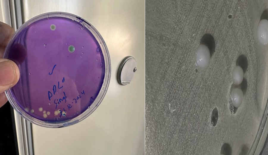
The PCR reactions were run on a programmable thermal cycler (Applied Biosystems™, USA) under these conditions, Initial denaturation at 95°C for 5 minutes, and 35 cycles of the following, 95°C for 30 seconds (denaturation), 58°C for 30 seconds (annealing), and 72°C for 60 seconds (extension), a final extension of 72°C for 7 minutes, and then held at 4°C. Detection of the pvpA GeneThe study used the primer sequences in Table 1 to amplify the pvpA gene. The preparation of the PCR reaction mixture was identical to the preparation described for the 16S rRNA assay. The thermal cycling conditions were the same as for 16S rRNA amplification, except the annealing temperature was 56°C and the extension time was reduced to 45 seconds to account for the smaller expected amplicon size. Detection of the pMGA/vlhA GeneThe primers listed in Table 1 were used to perform PCR detection of the gene pMGA/vlhA. The reaction mixture and the setup of the PCR were the same as for the previous assays. The thermal cycling conditions were the same as for the previous assays except for the extension time, which was 30 seconds, and the annealing temperature, which was 57°C. These conditions reliably generated amplification products of the anticipated fragment size. Gene expressionmRNA levels were analyzed for gene expression, and reverse transcription polymerase chain reaction (RT-PCR) was utilized to do so. To extract total RNA, an RNA extraction kit (Geneaid Biotech Ltd., Taiwan) was used, and the manufacturer's claims were followed to extract total RNA from log phase M. gallisepticum cultures. For measurement of concentration and purity of total RNA, a NanoDrop™ (Thermo Fisher Scientific, USA) was used for spectrophotometric measurement, and an A260/A280 ratio ranging from 1.8 to 2.1 was deemed appropriate for use in further procedures. Using a commercially available reverse transcription kit (Geneaid Biotech Ltd., Taiwan), cDNA was synthesized from 1 µg of total RNA. Reverse transcription was performed in a total volume of 20 µl containing buffer specific to reverse transcription, enzyme specific to reverse transcriptase, random primers (hexamers), a mixture of dNTP, and water that has been treated to remove enzymes (nuclease-free). The reaction was done for 10 minutes at 25°C in order to allow the primers to anneal, and for 60 minutes at 42°C to allow for the synthesis of cDNA. To inactivate the enzyme, the reaction was done at 70°C for 10 minutes. The next step was the use of the synthesized cDNA as a template for RT-PCR amplification of the PVPA and pMGA genes. The same primer and the same reaction that were used for the genomic PCR assays were used here to prepare the amplification reactions. The primer sequences for amplification of the target genes and the internal reference gene are shown in Table 1. To determine the transcription levels, the 23S rRNA gene was chosen to be the internal housekeeping reference gene, as it has been shown to have stable expression in M. gallisepticum (Cecchini et al., 2007). The amplification of the 23S rRNA gene was done in parallel to the target genes under the same experimental conditions. The relative expression of the PVPA and pMGA genes was determined relative to the expression of the 23S rRNA reference gene, measured under the identical reaction conditions. The pMGA amplification primers focus on a specific conserved area within the pMGA (vlhA) multigene family locus, producing a partial fragment of about 222 bp, which is typically utilized for the identification of the constituents of this gene family. To confirm reproducibility of the gene expression results, all RT-PCR reactions were done in triplicate. DNA sequencing and constructing phylogenetic treesThe PCR products were purified, and then, using the designed primers, the products were sequenced in both the forward and reverse directions. The sequence data were aligned and edited using the BioEdit software, version 7.0.5. The multiple sequence alignment was done using the ClustalW algorithm in the MEGA software, version 7.0. Using the reference strains retrieved from the National Center for Biotechnology Information (NCBI) database, the partial 16S rRNA gene sequence-based phylogenetic trees were constructed. The generated sequences were assigned accession numbers and deposited in the GenBank of NCBI. Statistical analysisStatistical calculations were made using GraphPad Prism software, version 9.0 (GraphPad Software Inc., San Diego, CA, USA). Data from the PCR detection assays were analyzed descriptively and stated as frequencies and proportions. For gene expression analysis, the RT-PCR data relating to the PVPA and pMGA genes were determined and compared to the reference gene, and the new values were processed in the software and analyzed. The presence of the pMGA and PVPA genes was evaluated using RT-PCR. Target genes were amplified using the same conditions applied for genomic PCR on the synthesized cDNA. For the purpose of transcriptional detection normalization, the 23S rRNA gene was selected as the internal housekeeping reference gene, given its stable expression in M. gallisepticum. The genes of interest and the reference gene were amplified in parallel. As for the expression, detection of the PCR products at the presumed molecular weights for the genes in reference to the gene of the housekeeper was established. All reactions were done in triplicate to ensure that the results are reproducible. All assays were executed in triplicate, and results were described as average values with variability where appropriate. A p value of less than 0.05 was interpreted to be statistically significant. Ethical approvalThe Committee for Ethical Scientific Research, College of Veterinary Medicine, University of Al-Qadisiyah, Iraq, reviewed and approved the study protocol (Approval No. 4914, dated 14 November 2024). All the sampling procedures were done in compliance with the institutional guidelines and the national regulations for the ethical treatment of animals in scientific research. The study was conducted with biosafety and biosecurity measures to minimize the risks. ResultsIsolation and phenotypic identification of Mycoplasma gallisepticumThe current study isolated Mycoplasma from the clinical samples collected from the poultry, demonstrating respiratory signs that were grown on the PPLO broth and agar media. Some broth turbid cultures were seen after 3–5 days, which could suggest Mycoplasma growth. Meanwhile, cultures that were exhibiting rapid turbidity within 24 hours were excluded as probable contaminants. Subculturing on the PPLO agar demonstrated characteristic colony appearance after incubation for 5–15 days. The colonies exhibited the most common bacterial colony, pale-yellow egg empyema colonies with a dense central zone and translucent surrounding zone. Other colonies were noted to have center zones that were reduced and/or difficult to see. Microscopic confirmation utilizing Diene’s stain demonstrated colonies with central areas that stained intensely blue, confirming the M. gallisepticum. These positive microscopically stained colonies are shown in Figure 1.
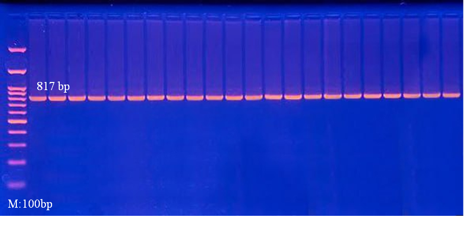
Fig. 1. Mycoplasma gallisepticum phenotypical identification. Both images are PPLO agar plates M. gallisepticum with colonies characteristic of fried-egg colonies after 5–15 days incubation at 37°C. Central zones with darker margins show the Gram-positive Mycoplasma colonies. PCR16S rRNA genePhenotypically confirmed isolates of genomic DNA were subjected to PCR. Amplification was completed for the 16S rRNA gene of M. gallisepticum. Every isolate that was tested showed amplification products of the M. gallisepticum gene, with an expected product size of 817 base pairs. Negative control reactions showed no amplification to confirm that no contamination occurred. The consistent amplification of the 16S rRNA gene for each isolate gave positive molecular confirmation of the species and success of the isolation. The recovered isolates were confirmed to be M. gallisepticum and were thus qualified for subsequent gene-specific studies (Fig. 2).
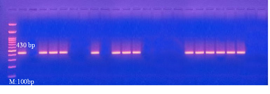
Fig. 2. PCR Amplification of Mycoplasma gallisepticum 16S rRNA Gene. PCR product and agarose (1.5%) M. gallisepticum band 16S rRNA primer amplifications. M: 100 bp DNA ladder. 1–n: all positive samples and 817 bp. Negative control shows no amplification. Assertions show assay specificity. PVPA geneSuccessful PCR amplification of the PVPA gene was evident in most of the positive M. gallisepticum isolates. Using electrophoresis, bands of the expected size (430 bp) were observed, clear and specific. Most of the isolates had similar band intensity, suggesting the presence of the PVPA gene in most of the isolates (Fig. 3).
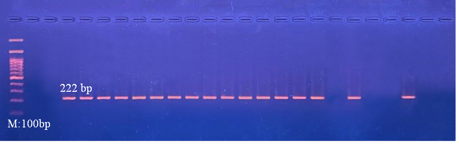
Fig. 3. Agarose 1.5% PCR PVPA Mycoplasma gallisepticum gene isolates. Electrophoresis M: 100 bp DNA ladder. Clear bands: positive isolates show specific bands (430 bp). No bands: negative no amplification. pMGAThe majority of the isolated analyzed showed correct expected size-specific PCR products (222 bp) after amplification of the pMGA multigene family. Therefore, finding pMGA-specific bands demonstrates the presence of this multigene family within the local population of the M. gallisepticum strains. Some isolates showed varying band intensity, which may indicate differing amplification efficiency or gene copy number (Fig. 4).
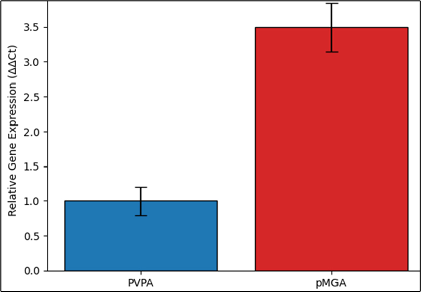
Fig. 4. PCR amplification of the 5.1. The 1.5% agarose gel shows the PCR products of the pMGA multigene family and the various isolates. M is the 100bp DNA (M). Clear bands: positive results with the 222 bp bands. No bands: negative no amplification. Gene expression of PVPA and pMGAThe pMGA multigene family exhibited significantly higher relative expression levels compared with the PVPA gene across most isolates (Fig. 5)
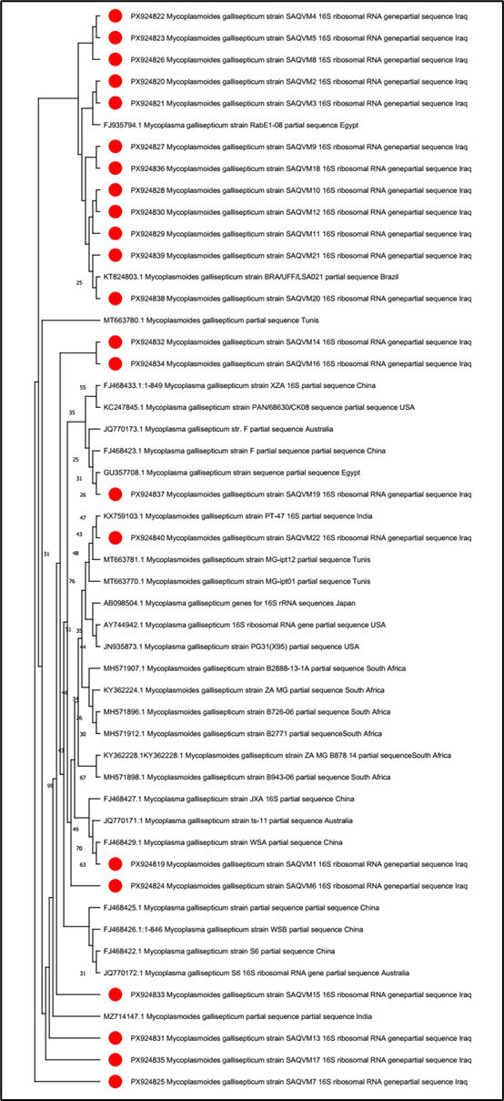
Fig. 5. Relative gene expression levels of PVPA and pMGA genes in Mycoplasma gallisepticum isolates. Phylogenetic analysis of Mycoplasma gallisepticum based on the 16S rRNA geneMost local isolates are genetically close to previously reported M. gallisepticum strains from neighboring Middle Eastern countries, especially Egypt and Tunisia (Accession No. in Fig. 6), implying possible regional spread or similar evolutionary origins. Several isolates clustered with reference strains from Asia, Europe, Africa, and Australia, demonstrating the worldwide distribution and evolutionary conservation of M. gallisepticum 16S rRNA gene. Despite this overall conservation, minor sequence variations were enough to separate local isolates into different branches, indicating genetic divergence within the isolates (Fig. 6).
Fig. 6. Phylogenetic relationships of Mycoplasma gallisepticum isolates based on partial 16S rRNA gene sequences. The tree was constructed using the Neighbor-Joining method implemented in MEGA software version 11.0 with 1,000 bootstrap replicates to assess the reliability of the tree topology. DiscussionThe current analysis sustains the predominant impact of surface-associated genes in the pathogenicity and adaptability of M. gallisepticum. Local isolates with confirmed presence and active transcription of the PVPA gene, as well as the pMGA multigene family, strengthen the theory of the close association between adhesion and antigenic variation in M. gallisepticum infection. Prior research has noted that epithelial respiratory colonization, as a phenomenon, has a determined transcription blueprint impact of the cytoadhesins and variable surface proteins that affect colonization persistence under host immunity pressure (Liu et al., 2024; Chen et al., 2026). The expression pattern and molecular character of the current research correlate with the same and support the theory that strains in circulation of the poultry population have the genetic make-up for operational engagement with the host and immune system modulation (Miller et al., 2024). Comparative analysis of gene expression showed the highest significant transcriptional activity of the pMGA multigene family compared to PVPA, emphasizing the role of pMGA-associated proteins in antigenic variation, which corroborates the in silico and genomic pMGA (vlhA) gene variation and differential expression studies that described the ability of M. gallisepticum to survive in the presence of antibodies (Liu et al., 2024). In contrast, the more stable expression of PVPA indicates a more conserved function, which may involve the maintenance of basal adhesion and cellular adhesion, rather than rapid phenotypic variation. The division of labor between highly variable and more conserved surface proteins is a major mechanism by which M. gallisepticum achieves long-term survival in infected flocks (Maya-Rodríguez et al., 2025). The 16S rRNA gene sequence analyses show that local isolates cluster with M. gallisepticum globally, with variation. This agrees with Kachabi et al. (2025), who noted that genomic comparisons show high variation within a species with conserved housekeeping genes. Isolate dispersion in subclusters shows that traded birds or poultry management may expose poultry to new genetic strains. This variation affects disease control, as different strains may have different degrees of virulence and antimicrobial resistance and may change the efficacy of vaccines (Rufai et al., 2025). From an applied standpoint, the molecular features outlined in this study are pertinent to vaccine formulation and disease management strategies (Chen et al., 2026). Current trends in subunit and recombinant vaccine development highlight the need for vaccine formulations to include epitopes of highly conserved antigens, factoring in the variability of the surface proteins associated with the pathogens (Miller et al., 2024). While this study examined the transcriptional presence of the PVPA and pMGA genes, additional sequencing of these loci may help further elucidate potential antigenic diversity and genetic heterogeneity of circulating M. gallisepticum strains. ConclusionThere are both conserved and highly variable genetic components in the M. gallisepticum isolates from the poultry, which are attributed to their adhesion, immune evasion, and persistence. The differential expression, along with the coordinate presence of PVPA and pMGA genes and the variation in phylogenetic diversity, points to the intricacy of the molecular mechanisms of this pathogen. These results provide insights into the pathogenesis of M. gallisepticum and the development of a comprehensive molecular framework that will enable improved poultry production systems to better surveillance, vaccine development, and control systems. AcknowledgmentThe authors would like to thank the College of VM, Univ. of Al-Q for their technical support with the laboratories. Conflict of interestNo conflict of interest is present in the current study. FundingNo external fund is to be declared, but the study was only self-funded by the authors. Authors’ contributionsAll authors participated in the current study (SMH MSc student and AAA supervised the study). Data availabilityThe data are available when requested via the corresponding author. ReferencesCecchini, K.R., Gorton, T.S. and Geary, S.J. 2007. Transcriptional responses of Mycoplasma gallisepticum strain R in association with eukaryotic cells. J. Bacteriol. 189(16), 5803–5807; doi:10.1128/JB.00667-07 Chen, J., Liu, P. and Chen, Y. 2026a. Pathogenic mechanisms and vaccine development for Mycoplasma gallisepticum in chickens. Front. Microbiol. 16, 1741449; doi: 10.3389/fmicb.2025.1741449 Chen , X., Zhang, Y., Liu, H., Wang, J. and Li, Z. 2026b. Mycoplasma gallisepticum: pathogenic mechanisms and vaccine development progress. Front. Vet. Sci; doi: 10.3389/fmicb.2025.1741449 Ferguson-Noel, N., Dos Santos, M., Ehsan, M. and Oluwayinka, E.B. 2025. Comparison of the efficacy of Mycoplasma gallisepticum vaccine programmes in chickens. Avian. Pathol. 54(4), 385–397; doi:10.1080/03079457.2024.2443508 Forero-Marin, S., Gomez, A.P., Beltran-Leon, M. and Ramirez-Nieto, G. 2025. A first look into the genomic characterization and fluoroquinolone resistance genotypes of Mycoplasma spp. in Colombian poultry. Poultry. Sci. 105(2), 106208; doi:10.1016/j.psj.2025.106208 Hu, Q., Zhang, R., Liu, J., Zhang, W., Liao, X., Guo, Y., Lu, Q., Yang, B., Zhang, T., Zhai, X. and Luo, Q. 2026. A rapid and field-deployable RAA-CRISPR/Cas12a platform for detection of Mycoplasma gallisepticum in poultry. BMC. Vet. Res. 22, 117; doi:10.1186/s12917-025-05278-7 Idowu, P.A., Mpofu, T.J., Zishiri, O.T., Adelabu, O.A., Nephawe, K.A. and Mtileni, B. 2024. Molecular detection and genetic characterization of Mycoplasma gallisepticum and Mycoplasma synoviae in selected chicken breeds in South Africa. BMC. Infect. Dis. 24(1), 562; doi:10.1186/s12879-024-09437-3 Kachabi, K., Pourbakhsh, S.A. and Zahraei Salehi, T. 2025. Comparative genomic analysis of six Mycoplasma gallisepticum strains: insights into genetic diversity and antibiotic resistance. Arch. Razi Inst. 80(1), 93–102; doi:10.32592/ARI.2025.80.1.93 Kamathewatta, K.I., Kanci Condello, A., Ekanayake, D., Noormohammadi, A.H., Young, N.D., Browning, G.F., Tivendale, K.A. and Wawegama, N.K. 2025. Day-old vaccination with the Vaxsafe MG304 live-attenuated vaccine protects chickens from tracheal transcriptional changes induced by chronic infection with Mycoplasma gallisepticum. Vaccine 68, 127915; doi:10.1016/j.vaccine.2025.127915 Liu, Y., Wang, Y. and Zheng, S.J. 2024. Immune evasion strategies of Mycoplasma gallisepticum with emphasis on variable surface lipoproteins and antigenic switching. Microorganisms , doi: 10.3390/ijms25052824 Maya-Rodríguez, L.M., Gómez-Verduzco, G., Trigo-Tavera, F.J., Moreno-Fierros, L. and Miranda-Morales, R.E. 2024. Variability of pMGA/vlhA sequences among Mycoplasma gallisepticum field strains isolated from laying hens and their deformed eggs. Access Microbiol. 6(6), 681; doi:10.1099/acmi.0.000681.v5 Maya-Rodríguez, L.M., Gómez-Verduzco, G., Trigo-Tavera, F.J., Moreno-Fierros, L., Rojas-Trejo, V. and Miranda-Morales, R.E. 2025. A comparative in silico analysis of the vlhA gene regions of Mycoplasma gallisepticum and Mycoplasma synoviae isolates from commercial hen farms in Mexico. Access Microbiol. 7(2), 760; doi:10.1099/acmi.0.000760.v4 Miller, J.M., Ozyck, R.G., Pagano, P.L., Hernandez, E.F., Davis, M.E., Karam, A.Q., Malek, J.B., Mara, A.B., Tulman, E.R., Szczepanek, S.M. and Geary, S.J. 2024. Rationally designed Mycoplasma gallisepticum vaccine using a recombinant subunit approach. NPJ. Vaccines 9(1), 178; doi:10.1038/s41541-024-00978-x Rufai, A.A., Zakaria, Z., Yu, C.Y., Ganapathy, K., Abu, J., Ahmad, N.I. and Tee, C.T. 2025. Prevalence and antimicrobial susceptibility of Mycoplasma gallisepticum and Mycoplasma synoviae isolated from the central peninsular Malaysia. Poultry Sci. 104(10), 105552; doi:10.1016/j.psj.2025.105552 Sabir, R., Liu, M., Saeed, H.A., Deng, Z., Jia, S., Tang, J., Ma, Z., Fan, H. and Lin, H. 2026. Next-generation live vector vaccine targeting Mycoplasma synoviae and Mycoplasma gallisepticum via recombinant Salmonella. Vaccine 74, 128211; doi:10.1016/j.vaccine.2026.128211 | ||
| How to Cite this Article |
| Pubmed Style Hasan SM, Abed AA. Molecular characterization, expression profiling, and phylogenetic analysis of key adhesion-associated genes in Mycoplasma gallisepticum isolated from poultry. Open Vet. J.. 2026; 16(6): 3518-3527. doi:10.5455/OVJ.2026.v16.i6.22 Web Style Hasan SM, Abed AA. Molecular characterization, expression profiling, and phylogenetic analysis of key adhesion-associated genes in Mycoplasma gallisepticum isolated from poultry. https://www.openveterinaryjournal.com/?mno=309251 [Access: June 26, 2026]. doi:10.5455/OVJ.2026.v16.i6.22 AMA (American Medical Association) Style Hasan SM, Abed AA. Molecular characterization, expression profiling, and phylogenetic analysis of key adhesion-associated genes in Mycoplasma gallisepticum isolated from poultry. Open Vet. J.. 2026; 16(6): 3518-3527. doi:10.5455/OVJ.2026.v16.i6.22 Vancouver/ICMJE Style Hasan SM, Abed AA. Molecular characterization, expression profiling, and phylogenetic analysis of key adhesion-associated genes in Mycoplasma gallisepticum isolated from poultry. Open Vet. J.. (2026), [cited June 26, 2026]; 16(6): 3518-3527. doi:10.5455/OVJ.2026.v16.i6.22 Harvard Style Hasan, S. M. & Abed, . A. A. (2026) Molecular characterization, expression profiling, and phylogenetic analysis of key adhesion-associated genes in Mycoplasma gallisepticum isolated from poultry. Open Vet. J., 16 (6), 3518-3527. doi:10.5455/OVJ.2026.v16.i6.22 Turabian Style Hasan, Saeed Mohammed, and Alaa Abdulaziz Abed. 2026. Molecular characterization, expression profiling, and phylogenetic analysis of key adhesion-associated genes in Mycoplasma gallisepticum isolated from poultry. Open Veterinary Journal, 16 (6), 3518-3527. doi:10.5455/OVJ.2026.v16.i6.22 Chicago Style Hasan, Saeed Mohammed, and Alaa Abdulaziz Abed. "Molecular characterization, expression profiling, and phylogenetic analysis of key adhesion-associated genes in Mycoplasma gallisepticum isolated from poultry." Open Veterinary Journal 16 (2026), 3518-3527. doi:10.5455/OVJ.2026.v16.i6.22 MLA (The Modern Language Association) Style Hasan, Saeed Mohammed, and Alaa Abdulaziz Abed. "Molecular characterization, expression profiling, and phylogenetic analysis of key adhesion-associated genes in Mycoplasma gallisepticum isolated from poultry." Open Veterinary Journal 16.6 (2026), 3518-3527. Print. doi:10.5455/OVJ.2026.v16.i6.22 APA (American Psychological Association) Style Hasan, S. M. & Abed, . A. A. (2026) Molecular characterization, expression profiling, and phylogenetic analysis of key adhesion-associated genes in Mycoplasma gallisepticum isolated from poultry. Open Veterinary Journal, 16 (6), 3518-3527. doi:10.5455/OVJ.2026.v16.i6.22 |


