





| Research Article | ||
Open Vet. J.. 2026; 16(6): 3376-3383 Open Veterinary Journal, (2026), Vol. 16(6): 3376-3383 Research Article Genetic evidence of Theileria ovis infection in goats based on 18S rRNA gene sequencingMohammed Qasim Hadi and Monyer Abdulameir Abd Alfatlawi*Department of Veterinary Microbiology, College of Veterinary Medicine, University of Al-Qadisiyah, Al-Diwaniyah, Iraq *Corresponding Author: Monyer Abdulameir Abd Alfatlawi. Department of Veterinary Microbiology, College of Veterinary Medicine, University of Al-Qadisiyah, Al-Diwaniyah, Iraq. Email: monyerr.abd [at] qu.edu.iq Submitted: 17/01/2026 Revised: 25/04/2026 Accepted: 05/05/2026 Published: 05/06/2026 © 2025 Open Veterinary Journal
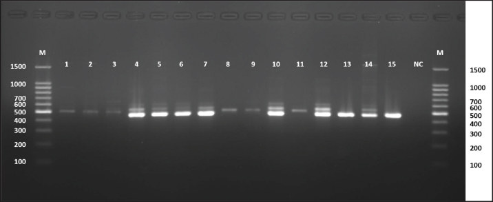
ABSTRACTBackground: Goat theileriosis is a disease caused by a protozoan parasite Theileria ovis transmitted by blood-feeding ticks. It is an emerging disease that is of concern because of its high incidence and impact on animal health and productivity. In contrast to goats, goats are often considered more tolerant of hemoparasitic infections. However, a growing body of evidence indicates that goats can also be significant reservoirs of T. ovis, where the parasite can persist and be transmitted to other hosts in endemic regions. However, in Iraq, research describing the molecular characteristics of T. ovis infections in goats is almost nonexistent. Aim: This study aimed to provide the first molecular characterization of T. ovis parasite infections in goats in the central region of Iraq and to characterize T. ovis local isolates using 18S ribosomal RNA (rRNA) gene partial sequencing and phylogenetic analysis. Methods: Genomic Deoxyribonucleic acid (DNA) was extracted from whole blood samples collected from goats using a commercial kit. The molecular detection of T. ovis was performed using a nested conventional polymerase chain reaction (PCR) that amplifies the 18S rRNA gene. Positive PCR products were purified, and Sanger sequencing was performed. Sequences were edited and deposited in the GenBank database. The sequences were then analyzed and compared using the National Center for Biotechnology Information Basic Local Alignment Search Tool database for species confirmation. Phylogenetic analysis was conducted using the maximum likelihood method based on the Tamura–Nei model. The nucleotide similarity between local and global T. ovis isolates was determined using multiple sequence alignment. Results: The DNA of T. ovis was successfully amplified by nested PCR in the blood samples of goats with the predicted 520 bp amplicon related to the 18S rRNA gene. Sequencing of 10 representative goat-derived blood samples was performed, and the results were deposited in the GenBank database with the following accession numbers: PX755,072 to PX755,081. All isolates were confirmed as T. ovis from the Basic Local Alignment Search Tool analysis, which showed a cross-nucleotide identity of 98.75%–98.81% from the global references of Brazilian, Egyptian, Turkish, Pakistani, Chinese, Japanese, and Iraqi T. ovis. Phylogenetic analysis showed that all goat isolates clustered together in the T. ovis clade, and the isolates had robust, close evolutionary relationships with T. ovis strains from other countries. Little to no sequence divergence was observed in the goat isolates, as seen in the other consensus sequence of most of the constituents in the multiple sequence alignment. Conclusion: Theileria ovis infection in goats from Central Iraq and the genetic constituents of T. ovis caprine isolates are closely related to other global T. ovis. This study emphasizes the need to consider goats as a significant circulating host of T. ovis and the need to integrate goats into molecular surveillance programs to identify tick-borne hemoparasites. Keywords: 18S rRNA gene, Goats, Nested PCR, Phylogenetic analysis, Theileria ovis. IntroductionCaprine theileriosis infection measurement entails challenging processes involving the use of the blood-parasitic tick-borne disease Theileria ovis is one of the common parasites that infects goats in epidemic regions. Although T. ovis is typically viewed as a low-pathogenic disease, epidemiologic data pose concern about T. ovis being pathogenic to goats, primarily due to the widespread infection of small ruminants (Arif et al., 2023; Almahallawi et al., 2024). The disease is exacerbated when goats are put in pasture systems where they share grazing areas with goats, as both species are highly susceptible to infected tick vectors, leading to cross-species disease transmission (Prajapati et al., 2023). The sheer number of T. ovis in goats, especially when exposed to other goats and small ruminants, poses challenges to pinpointing specific areas of infection without using advanced diagnostic molecular analysis methods. Infection with T. ovis remains undetectable even with the use of advanced diagnostic molecular analysis and fails to raise other disease symptoms that are detectable with conventional microscopic methods. Nested polymerase chain reaction (PCR) using small fragments of ribosomal 18S ribosomal RNA (rRNA) creates highly sensitive and specific strains with the intention of identifying varying strains within Theileria species (Nangru et al., 2022). The rRNA 18S genes are frequently employed in the field of molecular epidemiology because they remain highly conserved within distinct geographic regions and can show more subtle variations. Recent molecular studies have shown that T. ovis shows high genetic relatedness across different areas, such as Pakistan, Saudi Arabia, India, and China (Tanveer et al., 2022; Irfan et al., 2023). Thus, T. ovis populations are genetically stable and have strong adaptation to the hosts. Different phylogenetic analyses based on 18S rRNA sequences showed the same results, as they found that the isolates from goats are all found in the T. ovis clade and are separated from all of the other pathogenic Theileria species like T. lestoquardi (Arif et al., 2023; Prajapati et al., 2023). In the Middle East, molecular research on T. ovis infections in goats has increased, signifying the increasing attention to the epidemiological significance of caprine hosts. Research from Saudi Arabia and adjacent regions has reported an increasing prevalence of T. ovis in goats, suggesting the need for molecular surveillance in high tick regions (Almahallawi et al., 2024; AlFaleh, 2025). These studies show that goats can be important hosts and perpetuators of the parasite, even in the absence of clinical symptoms. In Iraq, goats are essential for rural economies and smallholder production systems. Nevertheless, there is a lack of molecular data describing T. ovis infections in goats compared with studies focused on goats. Few regional studies of small ruminants have focused on Theileria, and even fewer genetically characterized T. ovis isolates from goats have been identified through sequencing (Aziz and Hamadamin, 2025). This lack of data on T. ovis isolates from goats results in a deficient comprehension of the parasite’s distribution, genetic diversity, and role in the epidemiology of ovine and caprine theileriosis in the country. It is necessary to integrate molecular identification, sequencing, and phylogenetic analysis to understand the genetic diversity of T. ovis in goats and how local isolates differ from those found in other parts of the world. This will help to understand the T. ovis epidemiology in Iraq and help in the efforts to control small ruminant tick-borne hemoparasitic infections (Tanveer et al., 2022; Arif et al., 2023). Therefore, this study aimed to detect T. ovis infection in goats from Central Iraq using molecular methods and to genetically characterize the obtained isolates. Materials and MethodsStudy area and collection of samplesBetween January and November 2025, 146 goat blood samples were collected using aseptic procedures. Goats aged 6 months or older were included. Many goats on the research sites shared pastures with sheep and other small ruminants. During blood sample collection, the animals were checked for ticks and other ectoparasites. The owners were asked about the presence of ectoparasite control, but this was often not the case because many of the animals did not have complete medical records. Blood samples were collected from the animals before being sent to the Laboratory of Parasitology, College of Veterinary Medicine, University of Al-Qadisiyah, for further research. Blood samples were processed immediately, then stored at 4°C, and later at −20°C until genomic and other microbial analysis. Extraction of genomic Deoxyribonucleic acid (DNA) from blood samplesDNA was extracted from the blood samples using an AddPrep Genomic DNA Extraction Kit (AddBio Inc, South Korea). In brief, 200 µl of blood was placed into sterile 1.5 ml microcentrifuge tubes and processed according to the kit’s standard protocol. DNA was extracted using the provided elution buffer and stored at −20°C until further analysis. DNA concentration and purity were measured using the manufacturer’s protocol using a Quantus™ Fluorometer (Promega, USA). Nested polymerase chain reaction amplificationNested conventional PCR was used to detect T. ovis, targeting the 18S rRNA small subunit of the ribosomal RNA gene, as described in previous studies. For sensitivity and specificity, two rounds of amplification were performed. First round of PCRDuring the first round of PCR amplification, the following primers were used: 5′–AAGCCATGCATGTCTAAGTATAAGCTTTT–3′ (forwards) and 5′– CTTCTCCTTCCTTTAAGTGATA AGGTTCAC–3′ (reverse) to amplify a 1,600-bp fragment of the 18S rRNA gene (GenBank reference accession AY533144). Each PCR reaction was performed in 20 µl reaction volumes, consisting of 10 µl PCR Master Mix (AddBio Inc., South Korea), 1 µl of the forward primer, 1 µl of the reverse primer, 6 µl PCR-grade water, and 2 µl of the genomic DNA template (approximately 100 ng). Second round (nested PCR)During the second round of amplification, the nested primers used were: 5′–TCGAGACCTTCGGGT–3′ (forwards) and 5′–AAAGACTCGTAAAGGAG CAA–3′ (reverse), targeting an internal fragment of the 18S rRNA gene, with an expected amplicon size of 520 bp. Each nested PCR reaction consisted of 10 µl of PCR Master Mix, 1 µl of the forward primer, 1 µl of the reverse primer, 7 µl of PCR-grade water, and 1 µl of the first round PCR product. Two rounds of PCR amplification were conducted using a T100™ Thermal Cycler (Bio-Rad, USA). The PCR thermal cycling program included 1 initial denaturation step at 95°C for 3 minutes, followed by 39 cycles of denaturation at 95°C for 35 seconds, annealing at 60°C for 35 seconds, and extension at 72°C for 35 seconds, and then 1 final extension step at 72°C for 5 minutes. Agarose gel electrophoresis was used to analyze the PCR products. A 1.5% agarose gel was prepared by dissolving 1.5-g Agarose into 100 ml of 1 × TBE buffer and heating until all the agarose was dissolved. Then, the solution was cooled to approximately 60°C, and 30 µl of Ethidium Bromide was added for DNA staining. The gel was then placed into the gel electrophoresis. The gel was electrophoresed at 100 V and 80 mA for approximately 60 minutes. After the run, the gel could be viewed with the gel doc system. DNA sequencing and phylogenetic analysisOnly 10 of 15 positive PCR samples were selected and sent for sequencing using Sanger sequencing at Macrogen Inc. (South Korea). Sequences were processed to remove low-quality areas and then assembled into consensus sequences using BioEdit (version 7.2.5). GenBank assigned accession numbers to the edited sequences submitted. The species were confirmed by Basic Local Alignment Search Tool (BLAST) of the sequences obtained and the reference sequences in the National Center for Biotechnology Information database. MEGA version 11 was used to analyze the phylogenetic relationships between the obtained sequences and the published T. ovis sequences. For the published sequences, T. ovis phylogenetic trees were made with the Maximum Likelihood method based on the Tamura-Nei substitution model with 1,000 bootstrap replicates. The ClustalW algorithm was used for multiple sequence alignment to analyze the nucleotide similarity and divergence of local Iraqi isolates and the global T. ovis sequences. Ethical approvalNot needed for this study. ResultsMolecular detection of Theileria ovis by Nested PCRNested PCR for T. ovis DNA targeting the 18S rRNA gene was successful in the blood samples. After the first amplification step, a detectable PCR product was detected for at least one outer primer in the positive samples. The second (nested) round of PCR amplification resulted in a 520-bp DNA fragment unique to T. ovis. The PCR-positive goat samples showed distinct gel bands for the amplified products as seen in agarose gel electrophoresis, whereas the negative control showed no amplification. This confirms the absence of contamination and the specificity of the assay (Fig. 1).
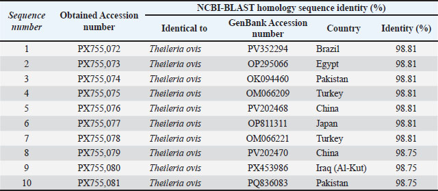
Fig. 1. Agarose gel electrophoresis (1.5%) shows the positive amplicons (1–15) of Theileria ovis in goats using the inner primers of the nested PCR technique (size=520 bp). NC is the negative control in which H2O was added instead of the template DNA. M is a molecular marker (Science Direx, South Korea). Sequencing and GenBank accessions resultsHigh-quality, clearly resolved sequenced bands were acquired from 10 nested PCR-positive goat samples. Partial 18S rRNA gene sequences were successfully acquired from every selected sample. The sequences were then subjected to trimming and quality control. We obtained products at 419 bp, and the sequences are now assigned GenBank accession numbers PX755072 to PX755081 (Table 1). Table 1. NCBI-BLAST homology sequence identity (%) in local
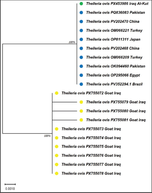
All GenBank submissions included metadata documenting the host species, sample source (whole blood), country (Iraq), and year the sample was collected. Homology BLAST and sequence identity examinationThe sequences obtained were evaluated using the NCBI BLAST to establish the degree of similarity among previously reported T. ovis isolates. BLAST analysis revealed that all sequences acquired from the goat samples matched T. ovis. The nucleotide identity was between 98.75% and 98.81% with isolates previously reported from Iraq, Brazil, Egypt, Türkiye, Pakistan, China, and Japan (Table 1). Although the identity of the sequences obtained from goat samples was somewhat lower than that reported in studies involving isolates from sheep, the results from this study confirmed the identification of T. ovis at the species level. The nucleotide differences among the goat isolates were very few, indicating that the genetic variability was low in the 18S rRNA gene analyzed region, and no particular lineage patterns were found in the obtained sequences. Goat-derived T. ovis isolates in phylogeneticsUsing the maximum likelihood method and the Tamura-Nei substitution model, we examined the phylogenetics of the partial 18S rRNA, which revolves around the Iraqi goat isolates T. ovis and other T. ovis genomes collected from GenBank, for comparative analysis. The phylogenetic tree was constructed using 419 nucleotides. The Iraqi goat-derived isolates uniformly fell under the Theileria ovis clade and were distinctly removed from the other Theileria species. The isolates exhibited phylogenetic proximity to reference strains from Turkey, Pakistan, Egypt, China, Japan, and Brazil, indicating that T. ovis is of close evolutionary origin. Bootstrap analysis was used to establish the phylogenetics of the Iraqi goat-derived isolates for inclusion within the T. ovis lineage, thereby solidifying the phylogenetic tree (Fig. 2).
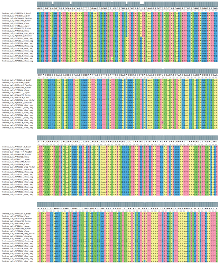
Fig. 2. Evolutionary tree analysis using the maximum likelihood method. This was inferred by using the maximum likelihood method and Tamura-Nei model. The tree is drawn to scale, with branch lengths measured in the number of substitutions per site. Green circles: Goat isolate from Iraq, blue circles: Reference isolates from sheep (different countries). Yellow circles: Current study isolates from goats (10 isolates). Multiple sequence alignment analysisPartial 18S rRNA sequences from goat isolates and T. ovis reference sequences from the National Center for Biotechnology Information were aligned. Comparative sequences from various locations/hosts showed more than 90% nucleotide identity (Fig. 3). Therefore, the aligned region of the 18S rRNA gene was considered highly conserved.
Fig. 3. Multiple sequence alignment of a partial genetic region of T. ovis in goats compared with other global sequences This shows the similarities and differences between the sequences. Regarding the reference sequences, some isolates (especially PX755079 and PX755081) had a few single-nucleotide differences that were trivial and random. These differences did not result in a recognizable pattern and did not appear to be related to the host or isolate location. The analyzed sequences did not contain insertions, deletions, or frameshift modifications. The 18S rRNA gene region revealed the genetic uniformity of the aligned sequences and demonstrated its potential to be used as a reliable marker for the detection and phylogenetic assessment of T. ovis in goats. DiscussionThe current investigation illustrates the molecular validation and genetic characterization of T. ovis infesting Central Iraq. The successful amplification of the 520 bp fragment and the sequence identity of the current goat isolates correlate with the increasing literature supporting the global distribution of T. ovis. The same molecular approaches targeting the 18S rRNA gene have been used in previous studies and have confirmed the reliability of this gene for species identification and molecular characterization of the same organism (Celik et al. 2025; Na et al. 2025). In this study, the goat-derived Iraqi isolates, when compared with global T. ovis reference sequences, showed sequence identity greater than or equal to 98.75% and less than or equal to 98.81% for all Iraqi isolates. These results are similar to the results reported by Na (2025) with sheep and goats in the Xinjiang region of China, where T. ovis had been reported to be genetically homogeneous with low intrapopulation variation. All phylogenetic studies demonstrated that the isolates from Iraqi goats were in the T. ovis clade and had close affinities with isolates from Türkiye, Pakistan, Egypt, China, Japan, and Brazil. In studies in Pakistan and Türkiye, T. ovis isolates from goats were related to sheep isolates, thus supporting the idea of host transmission among small ruminants (Riaz et al., 2024; Celik et al., 2025). The lack of specific host subclades in the phylogenetic tree also implies that the T. ovis populations in sheep and goats are genetically close and that there is considerable genetic connectivity in T. ovis populations across host species. Research from Egypt has described T. ovis infecting goats with a congruent molecular description, including high sequence similarity and low nucleotide divergence within the 18S rRNA gene (Mahmoud et al., 2024). The molecular similarity between the Iraqi and Egyptian isolates, coupled with the proposed Middle Eastern geographic region, infers that the T. ovis population exhibits genetic homogeneity, which is likely the result of prolonged endemicity and comparable ecological settings. These conditions may partially explain the genetic stasis of T. ovis populations and the subclinical infections described in goats. The involvement of ticks in the upkeep and spread of T. ovis is abundantly clear. Some studies show the molecular detection of T. ovis DNA in both hard and soft ticks associated with small ruminants in countries such as Iran, China, and Türkiye, which emphasizes the significance of ticks as biological vectors in the parasite life cycle (Ma et al., 2023; Norouzi et al., 2023; Liu et al., 2024). Although no tick samples were analyzed in this study, the presence of T. ovis in goats from Central Iraq indicates possible tick-active transmission cycles in the region. This finding is in agreement with previous studies, which state that small ruminants in endemic regions are often at high risk of being bitten by infected tick vectors. Numerous studies have pointed out the potential of goats to serve as silent reservoirs for T. ovis, perpetuating parasite transmission within mixed small ruminant systems, even in the absence of any clinical signs (Abdelsalam et al., 2023, Riaz et al., 2024). The molecular confirmation of T. ovis infection in goats in the current study supports this and indicates the epidemiological significance of considering goats as an integral part of the surveillance and control efforts of tick-borne hemoparasites. Recent studies have begun to examine the far-reaching effects of hemotropic infections in small ruminants. Jurković Žilić et al. (2025) found hemotropic pathogens in aborted ruminant fetuses, which may be suggestive of some of the effects of chronic coinfection with other pathogens on the reproductive system. Although the current study did not include an evaluation of reproductive parameters, the confirmed circulation of T. ovis in goats warrants further studies on the possible effects of T. ovis on reproduction and the overall productivity of the goats in the field. The distribution of tick-borne pathogens is influenced by a particular ecosystem’s environmental and ecological parameters. Research conducted in the Mediterranean ecosystem showed the presence of diverse tick-borne agents associated with ruminants and further described the interplay between the climate, habitat, and pathogens (Masià-Castillo et al., 2025). In this regard, the current findings provide important molecular evidence from Iraq, a country with similar ecological features, which helps to further delineate the epidemiological landscape of T. ovis in goats. The findings from this study correlate with recent molecular research from Europe, Asia, and the Middle East, confirming that T. ovis, which infects goats, shows a high level of genetic conservation. The inclusion of the newly generated goat-derived Iraqi sequences into worldwide phylogenetic analyses further substantiates the significance of goats as hosts in the epizootiology of T. ovis and emphasizes the need to integrate such caprine populations in tick-borne disease monitoring and control programs. The 6.8% (10/146) prevalence of T. ovis infection in this study is comparable to the documented figures for analogous endemic areas, being 5%–15% (Arif et al., 2023; Mahmoud et al., 2024). ConclusionThe present study proves the molecular confirmation of T. ovis infection in goats in Central Iraq by means of ‘nested PCR’ amplification, and partial sequencing of the 18S rRNA gene. The overall closeness of phylogenetic ties and Iraq isolate goat nucleotide, and high phylogenetic relationship with T. ovis of goats worldwide, suggests the presence of a genetically and conservatively populated parasite with constricted region variation. This study illustrates goats as competent hosts and even possible reservoirs for T. ovis. AcknowledgmentThe authors would like to express their sincere appreciation to the College of Veterinary Medicine, University of Al-Qadisiyah, for providing laboratory support and guidance throughout this study. Conflict of interestThe authors have no conflicts of interest to declare. FundingThe authors have self-funded the study. No external funding source is available. Authors’ contributionsAll authors have participated in the study. Data availabilityData are available when requested by the corresponding author. ReferencesAbdelsalam, M.A., Felefel, W., Fadl, S. and Bessat, M. 2023. Molecular prevalence and associated infection risk factors of tick-borne protozoan and rickettsial blood pathogens in small ruminants. BMC. Vet. Res. 19(1), 138; doi:10.1186/s12917-023-03702-4 Alfaleh, F.A. 2025. Molecular prevalence, associated risk factors, and genetic characterization of Theileria ovis in sheep and goats. Acta Parasitologica 70(4), 178; doi:10.1007/s11686-025-01110-2 Almahallawi, R.S., Omer, S.A., Al-Shaebi, E.M., Al-Hoshani, N., Al-Malki, E.S., Abdel-Gaber, R. and Mohammed, O.B. 2024. Prevalence of Theileria ovis in sheep and goats in northwestern Saudi Arabia with notes on potential vectors. PeerJ 12; doi:10.7717/peerj.18687 Arif, M., Saeed, S., Bashir, A., Farooq, M., Nasreen, N., Khan, A., Asif, M., Khalil, M.A., Ijaz, M., Muqaddas, H., Mehmood, N., Iqbal, F. and Chen, C.C. 2023. Molecular prevalence and phylogeny of Anaplasma marginale, Anaplasma ovis and Theileria ovis in goats and sheep enrolled from a hill station in Punjab, Pakistan. PLos One. 18(11), 291302; doi:10.1371/journal.pone.0291302 Aziz, K.J. and Hamadamin, B.Q. 2025. Epidemiological and molecular study of Theileria spp. in sheep and goats in Erbil, Iraq. Trop. Anim. Health Prod. 57, 80; doi: 10.1007/s11250-025-04330-w Celik, B.A., Kara, M., Ayan, A., Selcuk, M.A. and Celik, O.Y. 2025. Molecular prevalence and phylogenetic analysis of Theileria ovis and Theileria lestoquardi in small ruminants in Siirt Province, Türkiye. Vet. Med. Sci. 11(5), e70522; doi:10.1002/vms3.70522 Irfan, M., Chang, S.C., Iqbal, R.K., Tanveer, M., Asif, M., Khan, A., Nasreen, N., Atif, F.A., Shaikh, R.S., Aktas, M., Ben Said, M., Iqbal, F. and Chen, C.C. 2023. Seasonality, epidemiology and phylogeny of Theileria ovis with a note on hematological and biochemical changes in asymptomatic infected goats from Pakistan. PLos One 18(8), 290620; doi:10.1371/journal.pone.0290620 Liu, D., Wang, J., Liu, Y., Wang, S., Zhu, H., Jiang, B., Li, Y., Zhang, Y., Chahan, B. and Zhang, W. 2024. Molecular analysis of Anaplasma ovis, Theileria ovis and Brucella abortus in adult Ornithodoros lahorensis soft ticks (Acari: ixodida: Argasidae) isolated from the Xinjiang Uygur Autonomous Region, China. J. Vet. Res. 68(3), 355–361; doi:10.2478/jvetres-2024-0049 Ma, Z., Ceylan, O., Galon, E.M., Mohanta, U.K., Ji, S., Li, H., Do, T.T., Umemiya-Shirafuji, R., El-Sayed, S.A.E.S., Zafar, I., Liu, M., Sevinc, F. and Xuan, X. 2023. Molecular identification of piroplasmids in ticks from infested small ruminants in Konya Province, Turkey. Pathogens 12(9), 1123; doi:10.3390/pathogens12091123 Mahmoud, H.Y.A.H., Tanaka, T., Ali, A.O. and Emeish, W.F.A. 2024. Molecular detection and characterization of Anaplasma ovis, Theileria ovis, and Theileria lestoquardi in sheep and goats in Luxor, Egypt. BMC. Vet. Res. 20(1), 260; doi: 10.1186/s12917-024-04109-5 Masià-Castillo, M.T., Maiques-Rodriguez, E., Martínez-Alfaro, A., García-Bacete, I., Ingresa-Capaccioni, S., Ahuir-Baraja, A.E., Alarcón-Elbal, P.M., Garijo-Toledo, M.M. and Sansano-Maestre, J. 2025. Tick-borne pathogens from ruminant ticks in a Mediterranean ecosystem from Eastern Spain. Med. Vet. Entomol. 39(2), 301–314; doi:10.1111/mve.12781 Nangru, A., Maharana, B.R., Vohra, S. and Kumar, B. 2022. Molecular identification of Theileria species in naturally infected sheep using nested PCR-RFLP. Parasitol. Res. 121(5), 1487–1497; doi:10.1007/s00436-022-07489-5 Norouzi, M., Dayer, M.S. and Ghaffarifar, F. 2023. Molecular detection and characterisation of Theileria in hard ticks of small ruminants in Zarrin Dasht County, Southern Iran. Vet. Med. Sci. 9(1), 372–379; doi: 10.1002/vms3.1027 Prajapati, A., Prajapati, B., Patel, A., Chauhan, P., Das, B., Raval, S., Suthar, A., Sutaria, T., Chaudhari, R.K., Patel, P., Chauhan, V. and Patel, R. 2023. Molecular identification and genetic characterization of Theileria and Anaplasma infection in sheep and goat of North Gujarat, India. Parasitology. Res. 122(6), 1427–1433; doi:10.1007/s00436-023-07848-w Riaz, M., Chang, S.C., Tasawar, Z., Sajid, M., Nasreen, N., Amaro-Estrada, I., Cossío-Bayúgar, R., De La Fuente, J., Alanazi, A.D., Chen, C.C., Khan, A. and Ben Said, M. 2024. Molecular epidemiology and phylogeny of Theileria ovis and Theileria lestoquardi in sheep and goats from Southern Punjab, Pakistan. Vector-Borne. Zoonotic. Dis. 24(10), 656–665; doi:10.1089/vbz.2023.0118 Tanveer, M., Farooq, M., Amjad, M., Asif, M., Kashif, M., Latif, M., Khan, A., Aktas, M., Ben Said, M. and Iqbal, F. 2022. Molecular prevalence, associated risk factors and phylogeny of Anaplasma marginale, Theileria ovis and T. lestoquardi in sheep from Pakistan. Comparative Immunol. Microbiol. Infect. Dis. 86, 101822; doi:10.1016/j.cimid.2022.101822 Zhou, N., Wang, M., Zhao, X., Mijiti, A., Dang, W., Wen, L., Shi, W., Gan, L., Li, C. and Gailike, B. 2025. Molecular detection and population genetic diversity analysis of Theileria ovis in partial regions of Xinjiang, China. Mol. Biochem. Parasitol. 263, 111689; doi:10.1016/j.molbiopara.2025.111689 Žilić, D.J., Naletilić, S., Mihaljević, Z., Gagović, E., Špičić, S., Reil, I., Duvnjak, S., Tuk, M.Z., Hodžić, A. and Beck, R. 2025. Hemotropic pathogens in aborted fetuses of domestic ruminants: transplacental transmission and implications for reproductive loss. Front. Microbiol. 16, 1632135; doi:10.3389/fmicb.2025.1632135 | ||
| How to Cite this Article |
| Pubmed Style Hadi MQ, Alfatlawi MAA. Genetic evidence of Theileria ovis infection in goats based on 18S rRNA gene sequencing. Open Vet. J.. 2026; 16(6): 3376-3383. doi:10.5455/OVJ.2026.v16.i6.9 Web Style Hadi MQ, Alfatlawi MAA. Genetic evidence of Theileria ovis infection in goats based on 18S rRNA gene sequencing. https://www.openveterinaryjournal.com/?mno=307169 [Access: June 26, 2026]. doi:10.5455/OVJ.2026.v16.i6.9 AMA (American Medical Association) Style Hadi MQ, Alfatlawi MAA. Genetic evidence of Theileria ovis infection in goats based on 18S rRNA gene sequencing. Open Vet. J.. 2026; 16(6): 3376-3383. doi:10.5455/OVJ.2026.v16.i6.9 Vancouver/ICMJE Style Hadi MQ, Alfatlawi MAA. Genetic evidence of Theileria ovis infection in goats based on 18S rRNA gene sequencing. Open Vet. J.. (2026), [cited June 26, 2026]; 16(6): 3376-3383. doi:10.5455/OVJ.2026.v16.i6.9 Harvard Style Hadi, M. Q. & Alfatlawi, . M. A. A. (2026) Genetic evidence of Theileria ovis infection in goats based on 18S rRNA gene sequencing. Open Vet. J., 16 (6), 3376-3383. doi:10.5455/OVJ.2026.v16.i6.9 Turabian Style Hadi, Mohammed Qasim, and Monyer Abdulameir Abd Alfatlawi. 2026. Genetic evidence of Theileria ovis infection in goats based on 18S rRNA gene sequencing. Open Veterinary Journal, 16 (6), 3376-3383. doi:10.5455/OVJ.2026.v16.i6.9 Chicago Style Hadi, Mohammed Qasim, and Monyer Abdulameir Abd Alfatlawi. "Genetic evidence of Theileria ovis infection in goats based on 18S rRNA gene sequencing." Open Veterinary Journal 16 (2026), 3376-3383. doi:10.5455/OVJ.2026.v16.i6.9 MLA (The Modern Language Association) Style Hadi, Mohammed Qasim, and Monyer Abdulameir Abd Alfatlawi. "Genetic evidence of Theileria ovis infection in goats based on 18S rRNA gene sequencing." Open Veterinary Journal 16.6 (2026), 3376-3383. Print. doi:10.5455/OVJ.2026.v16.i6.9 APA (American Psychological Association) Style Hadi, M. Q. & Alfatlawi, . M. A. A. (2026) Genetic evidence of Theileria ovis infection in goats based on 18S rRNA gene sequencing. Open Veterinary Journal, 16 (6), 3376-3383. doi:10.5455/OVJ.2026.v16.i6.9 |


