





| Research Article | ||
Open Vet. J.. 2026; 16(6): 3912-3924 Open Veterinary Journal, (2026), Vol. 16(6): 3912-3924 Research Article Effect of bitter melon (Momordica charantia) extract on oxidative stress markers and antioxidant enzyme activities in rats with streptozotocin-induced diabetesI Nyoman Suarsana1*, I Made Kardena2, Ni Luh Suriani3 and Palagan Senopati Sewoyo21Department of Biochemistry, Faculty of Veterinary Medicine, Udayana University, Bali, Indonesia 2Department of Pathobiology, Faculty of Veterinary Medicine, Udayana University, Bali, Indonesia 3Biology Study Program, Faculty of Mathematics and Natural Sciences, Udayana University, Bali, Indonesia *Corresponding Author: I Nyoman Suarsana. Department of Biochemistry, Faculty of Veterinary Medicine, Udayana University, Bali, Indonesia. Email: suarsana65 [at] unud.ac.id Submitted: 12/01/2026 Revised: 15/05/2026 Accepted: 25/05/2026 Published: 20/06/2026 © 2025 Open Veterinary Journal
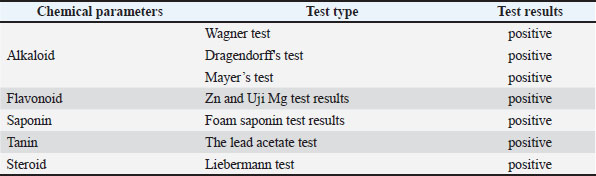
AbstractBackground: Diabetes mellitus is a major global health problem characterized by chronic hyperglycemia-induced oxidative stress and impaired antioxidant defense mechanisms. Bitter melon (M. charantia), a plant rich in antioxidant phytochemicals, has been proposed as an antihyperglycemic agent. Aim: This study aimed to investigate the effects of bitter melon extract on serum 8-Iso-PGF₂, 8-Hydroxy-2- Deoxiguanosine (8-OHdG), and antioxidant enzyme levels, as well as pancreatic histopathological changes and the expression of pancreatic superoxide dismutase (SOD) and insulin in Streptozotocin (STZ)-induced diabetic rats. Methods: A total of 25 male Sprague–Dawley rats were randomly assigned to five groups: a normal control group (P0), an untreated diabetic group (P1), and three diabetic groups (P2–P4). Oral administration of bitter melon extract at doses of 25, 50, or 100 mg/kg body weight was performed for 21 days. At the end of the treatment period, serum and pancreatic tissues were collected to evaluate the levels of 8-isoprostaglandin (8-Iso-PGF₂) and 8-OHdG, antioxidant enzyme levels [SOD, catalase (CAT), and glutathione peroxidase (GPx)], pancreatic histopathological changes, and SOD and insulin immunohistochemical expression. Results: The results showed that treatment with the ethanolic extract of bitter melon (M. charantia) in diabetic rats was able to reduce blood glucose levels, exert protective effects against pancreatic β-cell damage and preservation of the pancreatic islets of Langerhans area, and decrease antioxidant enzyme levels, as well as serum 8-Iso-PGF₂ and 8-OHdG levels. However, these parameters did not fully return to normal control levels. In addition, increased expression intensity of SOD and insulin was observed in the pancreatic islets of Langerhans. Conclusion: Treatment with ethanolic extract of Momordica charantia in STZ-induced diabetic rats showed potential antihyperglycemic effects, protection against pancreatic β-cell damage, and the ability to reduce 8-OHdG, 8-Iso-PGF2α, and serum antioxidant enzyme levels. Keywords: Antioxidant enzyme, Bitter melon, Diabetic, Hyperglycemia, Oxidative stress. IntroductionDiabetes is a chronic metabolic disease characterized by elevated blood glucose levels (hyperglycemia) due to impaired insulin secretion, insulin action, or a combination of both (ElSayed et al., 2023). Prolonged and uncontrolled hyperglycemia can trigger oxidative stress, a condition in which the production of free radicals, reactive oxygen species (ROS), exceeds the capacity of the body’s antioxidant defense mechanisms (Newsholme et al., 2019). Oxidative stress significantly contributes to the development of diabetic mellitus (DM) complications, including damage to pancreatic beta cells, which produce insulin (Ghasemi-Dehnoo et al., 2020). Understanding the mechanisms of oxidative damage is crucial for managing and preventing complications of diabetes. One approach that can be used is to minimize the impact of oxidative damage on diabetic-related complications. Oxidative stress biomarkers can be assessed by measuring the levels of malondialdehyde (MDA), 8-Hydroxy-2- Deoxiguanosine (8-OHdG), and 8-iso-prostaglandin (Goycheva et al., 2023; Goriuc et al., 2024), as well as the activity of antioxidant enzymes such as SOD, CAT, and GPx in plasma or serum (Tan et al., 2022). Diabetes places a substantial economic burden on countries, health systems, people with diabetes, and their families (ADA, 2018). The direct costs of healthcare expenditures due to diabetes are significant. According to the IDF (2025) the increase in global health care expenditures due to diabetes is substantial, growing from USD 232 billion in 2007 to over USD 1,015 billion in 2024 for adults aged 20–79 years. Traditional medicine has been used for a long time and remains an important public health practice component (Yakubu et al., 2020). According to the World Health Organization, approximately 75%–80% of the world’s population, particularly in developing countries with abundant biodiversity, still relies on plant-based medicine systems (Tran et al., 2020). These options are often preferred because they are more culturally relevant, considered more in tune with the physiological functions of the body, and generally have fewer adverse effects than modern therapies. Bitter melon (Momordica charantia) is a Cucurbitaceae family medicinal plant that is widely cultivated in Asia, including Indonesia (Oyelere et al., 2022). The use of medicinal plants as alternative therapeutic agents continues to be developed, one of which is in the management of DM. Bitter melon fruit is known to have antioxidant potential due to its high antioxidant activity content of bioactive compounds such as phenolics, flavonoids, saponins, phytosterols, terpenoids, and polyphenols (Salehi et al., 2021; Oyelere et al., 2022), as well as the compound P-insulin. P-insulin is a polypeptide phytoconstituent with a molecular weight of approximately 11 kDa that is suspected to have hypoglycemic effects and can be found in both bitter melon fruit and seeds (Jia et al., 2017). Several studies have reported that bitter melon fruit extract exhibits hepatoprotective, anti-inflammatory, antidiabetic, and antioxidant activities (Joseph and Jini, 2013; Muronga et al., 2021). However, information regarding the effect of bitter melon extract in evaluating oxidative stress markers in hyperglycemic conditions is still limited. The effectiveness of bitter melon extract therapy in preventing oxidative damage in DM can be assessed by measuring oxidative stress biomarkers, such as MDA, 8-OHdG, and 8-iso-prostaglandin (Yamaguchi and Yamaguchi, 2025), and the activity of antioxidant enzymes, such as SOD, CAT, and GPx (Suarsana et al., 2021). This study is significant because it provides a clearer picture of the molecular mechanisms underlying the potential protective effects of bitter melon on markers of oxidative stress and antioxidant enzyme activity in diabetes. The findings of this study are expected to strengthen the scientific basis for the use of bitter melon as a candidate for complementary therapy in diabetic management, particularly to help reduce the risk of complications. Materials and MethodsEthanol extract of bitter melon (M. charantia)Ripe bitter melons were obtained from a traditional market in Jimbaran, Bali, Indonesia. The fruits were thinly sliced and dried in an oven at 40°C for 48 hours. After drying, the simplicia was ground into powder using a blender. A total of 10 g of bitter melon powder was macerated in 500 ml of 80% ethanol for 24 hours. The mixture was then filtered using a clean, dry cotton cloth to obtain the supernatant. The supernatant was concentrated using a vacuum evaporator at 50°C until a thick extract was obtained (Oyelere et al., 2022). The thick extract was stored in a refrigerator at 4°C–5°C. Subsequently, this extract was prepared at doses of 25, 50, and 100 mg/kg body weight for administration to the experimental animals. Phytochemical screeningPreliminary qualitative phytochemical screening was conducted to determine the bioactive compounds in the bitter melon ethanol extract. The concentrated extract obtained from the sub-method Ethanol extract of bitter melon (M. charantia) was subsequently subjected to phytochemical screening to identify the presence of alkaloids, flavonoids, saponins, tannins, and steroids (Nortjie et al., 2022). Experimental treatment of animalsTwenty-five male Sprague–Dawley rats weighing 140–145 g were obtained from the Bio-Mice and Rat Breeding Centre in Denpasar, Bali, Indonesia, and acclimatized for 1 week. The animals were housed under controlled conditions at 23°C with a 12-hour light/dark cycle and provided food and water ad libitum. A single-dose injection of 1% streptozotocin in 50 mM sodium citrate buffer (potential of hydrogen 4.5) at 40 mg/kg body weight was administered to diabetic rats via intraperitoneal injection (Wu et al., 2025). Before Streptozotocin (STZ) administration, the rats were fasted overnight (12 hours) with no access to food or water. According to Furman (2021), rats were considered diabetic when their blood glucose levels exceeded 200 mg/dl. The average normal fasting blood glucose level in Wistar rats was 3.95 ± 1.31 mmol/l or equivalent to 71.1 ± 23.6 mg/dl (Wang et al., 2010). The animals were randomly assigned to five groups (n=5 per group): a normal control group (P0), a diabetic control group (P1), and three diabetic groups receiving bitter melon extract at doses of 25 mg/kg body weight (P2), 50 mg/kg body weight (P3), or 100 mg/kg body weight (P4). The extract was orally administered at 1 ml per day for 21 days. The 21-day treatment period was selected to allow sufficient time for bitter melon extract to produce measurable physiological and biochemical effects in streptozotocin-induced diabetic rats, including hyperglycemia stabilization, oxidative stress markers, and therapeutic response observation. On day 22, all rats were anesthetized with 40 mg/kg ketamine-Hydrochloric acid administered intramuscularly. Blood samples were collected from the orbital vein using a microhematocrit capillary tube and transferred into clean, anticoagulant-free tubes to obtain serum for analysis of antioxidant enzymes and oxidative stress markers. Subsequently, the rats were dissected, and the pancreas was harvested for hematoxylin staining and immunohistochemical examination. Measurement of blood glucose levelThe blood glucose levels of the experimental animals were measured using a GlucoDr auto™ AGM-4000 blood glucose meter (All Medicus Co., Ltd., Anyang-si, Republic of Korea) (Jendrike et al., 2017). Measurements were performed on day 0 (baseline) and day 22 after the treatment period. Before measurement, the rats were fasted overnight (12 hours) with no access to food or water. Analysis of 8-OHdG [8-hydroxy-dsoxyguanosine] and 8-iso-prostaglandin levelsThe quantification of 8-OHdG using an Enzyme-Linked Immunosorbent Assay (ELISA) sandwich test was performed as previously described by Korkmaz et al. (2018), and the quantification of 8-iso-prostaglandin was performed using the method by Hursitoğlu and Kurutas (2023). The analysis procedure for 8-OHdG and 8-Iso-prostaglandin levels using an ELISA test complies with the standard procedure guidelines listed on the 8-OHdG (Rat 8-Hydroxy-desoxyguanosine) ELISA Kit (Cat. No. E0032Ra), and Rat ELISA Kit 8-Iso-prostaglandin (Cat. No. E1179Ra; Bioassay Technology Laboratory products). Determination of antioxidant enzymes (SOD, CAT, and GPx)The activity of SOD, CAT, and GPx enzymes in serum was measured spectrophotometrically using an enzyme-linked immunosorbent assay kit as described by Zeni-Graiff et al. (2019). The analysis procedure for SOD, CAT, and GPx levels using the ELISA test followed the standard procedure guidelines for the product kits using the Rat Superoxidase Dismutase ELISA kit (Cat. No. E0168Ra), Rat CAT ELISA kit (Cat. No. E0869Ra), and Rat GPX ELISA kit (Cat. No. E1242Ra) products from the Bioassay Technology Laboratory. Immunohistochemical staining for insulin and SODImmunohistochemical staining was performed following the procedure described by Suarsana et al. (2021). Tissue sections were incubated with a 1:200 dilution of monoclonal antibodies against insulin or SOD. After 1 hour of incubation, Histofine Simple Stain MAX PO reagent was applied and allowed to react at room temperature for 30 minutes. Antigen-antibody complexes were visualized using diaminobenzidine, followed by hematoxylin counterstaining. The slides were examined under a light microscope at 400x magnification. Positive immunoreactivity was indicatedby the presence of brown cells. Histopathological stainingPreparation and hematoxylin–eosin staining were performed according to the procedures described by Suarsana et al. (2020). Pancreatic tissue was fixed in 10% buffered formalin for 24 hours and subsequently processed using standard paraffin-embedding techniques. Sections were cut at a thickness of 4 µm, mounted onto glass slides, and stained with hematoxylin and eosin. Histological observations were performed using a light microscope at 400× magnification. The area of the islets of Langerhans was measured using Fiji (ImageJ) software on microscope photographs at 400x magnification. Data analysisThis study used a completely randomized design. The data levels of 8-hydroxydeoxyguanosine, 8-iso-prostaglandin, SOD, CAT, and GPx were analyzed using analysis of variance. Immunohistochemical and histopathological data were descriptively analyzed. Data analysis was performed using the Statistical Package for the Social Sciences version 26 software. Ethical approvalThe ethics committee of the Faculty of Veterinary Medicine, Udayana University, Bali, Indonesia, has approved this research protocol for experimental animal research (No: B/180/UN14.2.9/PT.01.04/2025). ResultsPhytochemical compositionQualitative phytochemical analysis revealed the presence of bioactive compounds, including alkaloids, saponins, tannins, flavonoids, and steroids, in the ethanol extract of bitter melon (Table 1). Table 1. Results of phytochemical tests on bitter melon ethanol extract.
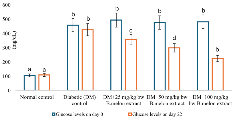
Effect of bitter melon extract on blood glucose levelsAs shown in Figure 1, the STZ-induced diabetic rats exhibited increased fasting blood glucose levels compared to the control group; this indicates the development of hyperglycemia in the STZ-induced diabetic rats. The results showed that the administration of bitter melon (M. charantia) extract (100 mg/kg body weight) to diabetic-induced rats (P4) significantly (p < 0.05) reduced glucose levels compared to diabetic rats (P1).
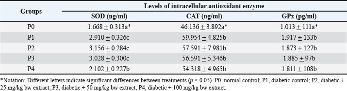
Fig. 1. Blood glucose levels in diabetic rats treated with bitter melon extract. Antioxidant enzyme analysis (SOD, CAT, and GPx)The highest SOD levels were observed in treatments P2 (3.156 ng/ml) and P3 (3.028 ng/ml), which were not significantly different (p > 0.05) compared to the DM rat group (P1), which reached 2.910 ng/ml. The 100 mg/kg body weight treatment (P4) showed a lower level of 2.102 ng/ml and was significantly different from the DM treatment (P1). The lowest SOD level was observed in the normal group (P0), reaching 1.668 ng/ml The highest CAT levels were observed in the DM rat group (P1), reaching 59.954 ng/ml; however, this value was not significantly different (p > 0.05) compared to P2 (57.591 ng/ml), P3 (56.591 ng/ml), and P4 (54.318 ng/ml). The lowest CAT level, 46.136 ng/ml, was observed in the normal group (P0). The highest GPx levels were observed in the DM rat group (P1), reaching 1.917 ng/ml; however, this value was not significantly different (p > 0.05) compared with P2 (1.873 ng/ml), P3 (1.885 ng/ml), and P4 (1.811 ng/ml). The lowest GPx level was observed in the normal group (P0), reaching 1.013 ng/ml (Table 2). Table 2. Average levels of intracellular enzymes in the serum of diabetic rats treated with bitter melon extract.
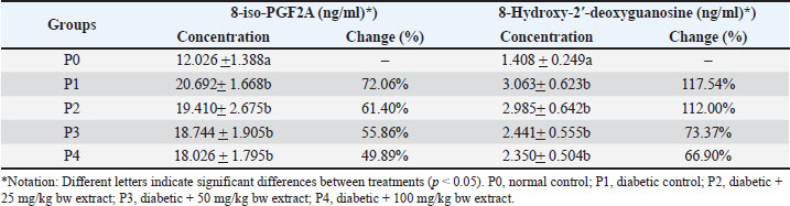
Effect of bitter melon extract on 8-iso-PGF₂α and 8-OHdG in serumThe effect of bitter melon extract treatment on the levels of 8-iso-PGF₂α and 8-OHdG in the serum of diabetic rats is presented in Table 3. Table 3. Average levels of 8-iso-PGF₂α and 8-Hydroxy-2′-deoxyguanosine in the serum of diabetic rats treated with bitter melon extract.
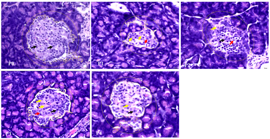
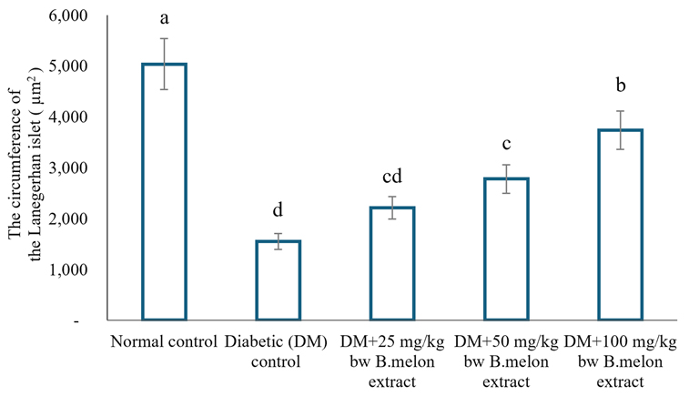
The average level of 8-iso-PGF₂α serum in the diabetic control group (P1) is higher and significantly different (p < 0.05) compared to the normal control group (P0). Treatment with bitter melon extract at doses of 25 mg/kg body weight (P2), 50 mg/kg body weight (P3), and 100 mg/kg body weight (P4) resulted in lower levels of 8-iso-PGF₂α compared to the diabetic control group (P1). However, this reduction was not statistically significant (p > 0.05). The serum level of 8-OHdG in the diabetic control group (P1) was significantly higher (p < 0.05) compared to the normal control group (P0). Treatment with bitter melon extract at doses of 25 mg/kg body weight (P2), 50 mg/kg body weight (P3), and 100 mg/kg body weight (P4) resulted in lower levels of 8-OHdG compared to the diabetic control group (P1). However, this decrease was not statistically significant (p > 0.05). Histopathological staining of the pancreas in diabetic ratsIn general, the histological appearance of the pancreatic islets of Langerhans in the diabetic positive group showed various types of damage, including changes in shape and size and a decrease in beta cell mass compared with normal controls. The area of the islets of Langerhans was smaller, and some islets were destroyed and almost shrunk (Figs. 2 and 3).
Fig. 2. Hematoxylin and eosin staining of pancreatic tissue of experimental rats. P0, normal control; P1, diabetic control; P2, diabetic + 25 mg/kg bw extract; P3, diabetic + 50 mg/kg bw extract; P4, diabetic + 100 mg/kg bw extract. Black arrows indicate beta cells, yellow arrows indicate necrosis, red arrows indicate pyknotic cell nuclei, and yellow circles indicate islets of Langerhans. Scale bar=10 μm, 400x magnification.
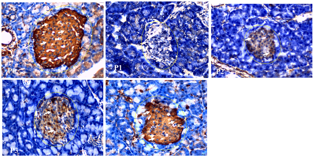
Fig. 3. The Langerhans islet area circle for all treatment groups. Different superscript letters indicate significant differences among treatments (p < 0.05). Immunohistochemical profile of pancreatic SODStrong brown staining was observed in normal pancreatic tissue (P0), spreading throughout the islets of Langerhans and some acinar cells. SOD expression was decreased in P1, with a weaker brown staining intensity than that in normal tissue (P0). Brown staining began to increase compared with the untreated diabetic group, although it remained weaker than in normal tissue (P0) The brown coloration appeared stronger and more even in group P3 than in group P2. This indicates an increase in SOD enzyme activity in response to the administration of bitter melon extract. The brown coloration was more intense and closer to the pattern in normal tissue (P0). This indicates that the administration of bitter melon extract at a dose of 100 mg/kg body weight had a better effect on increasing SOD expression than the other treatments (Fig. 4).
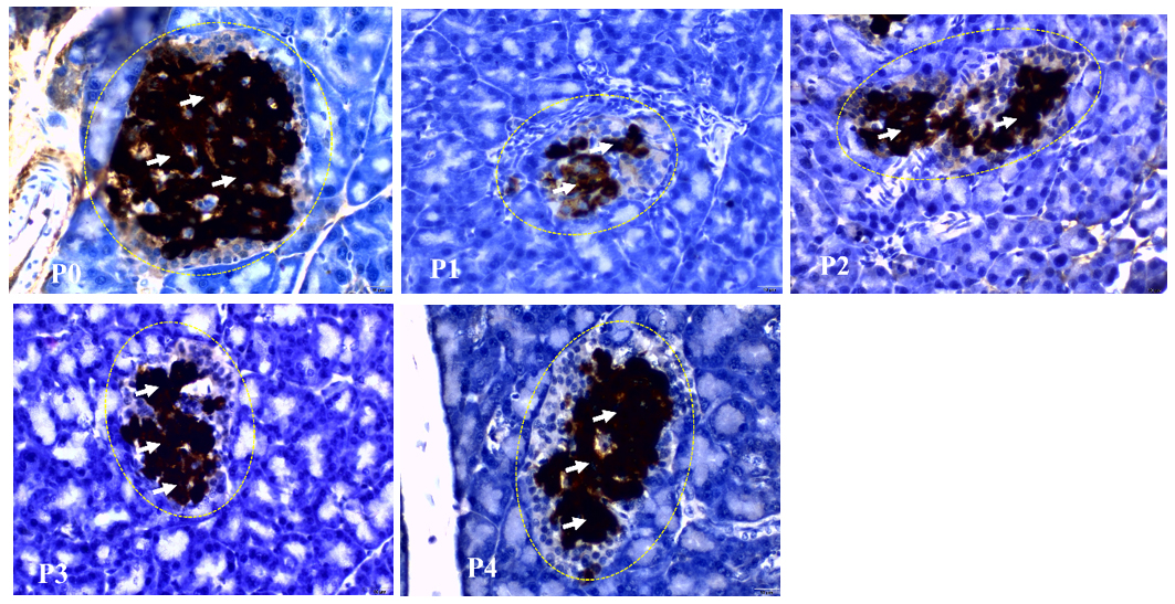
Fig. 4. SOD immunohistochemical staining of the pancreas. P0, normal control; P1, diabetic control; P2, diabetic + 25 mg/kg bw extract; P3, diabetic + 50 mg/kg bw extract; P4, diabetic + 100 mg/kg bw extract. The white arrow indicates positive SOD (brown color). Scale bar=10 µm, magnification 400x. Immunohistochemical profile of pancreatic insulin in ratsImmunohistochemical staining of insulin in pancreatic tissue showed clear differences in expression between the normal, diabetic, and bitter melon extract treatment groups. A positive immunohistochemical reaction was observed in the normal pancreas (Fig. 5, P0), characterized by intense brown coloration in the cytoplasm of cells in the islets of Langerhans. Positive staining indicates high insulin expression in pancreatic β cells. In contrast, in the diabetic pancreas (Fig. 5, P1), the intensity of the brown staining appears much weaker or significantly reduced, with some areas of the islets of Langerhans showing a weakening of the brown staining or even almost no positive staining. Bitter melon extract administration in treatments P2–P4 (Fig. 5, P2–P4) showed weak and uneven brown staining in the islets of Langerhans. The positive insulin intensity was still low compared with that of normal pancreatic tissue. Treatment P3 increased the intensity of brown staining in the islets of Langerhans. The staining distribution appeared more homogeneous and widespread. In the P4 treatment group, stronger and more widespread brown staining was observed in the islets of Langerhans, approaching the intensity seen in the normal pancreas (Fig. 5).
Fig. 5. Insulin immunohistochemical staining of the pancreas. P0, normal control; P1, diabetic control; P2, diabetic + 25 mg/kg bw extract; P3, diabetic + 50 mg/kg bw extract; P4, diabetic + 100 mg/kg bw extract. The white arrow indicates insulin (brown color). Scale bar=10 µm, magnification 400x. DiscussionThe body’s response to blood glucose levels involves a complex coordination of various physiological mechanisms. Disruption in any component involved in blood glucose regulation, insulin secretion, or glucose uptake can lead to glucose accumulation in the bloodstream. In STZ-induced diabetic conditions, damage occurs to pancreatic β-cells as insulin-producing cells, which ultimately triggers hyperglycemia (Mahmoud et al., 2017). Various approaches have been employed to reduce the incidence of DM. Insulin and oral hypoglycemic agents are commonly used in diabetic management; however, their use is associated with side effects, such as hypoglycemia, weight gain, and gastrointestinal discomfort (Philippe, 2010), and they often fail to significantly alter the development of diabetic complications, particularly macrovascular complications (Marco, 2015). M. charantia has a long history of use in traditional medicine for the treatment of diabetes and its associated complications. It is widely used in Asia, South America, India, and East Africa, where the fruit is consumed either as food or in traditional medicinal preparations (Joseph and Jini, 2013; Çiçek, 2022). Qualitative phytochemical screening of the ethanol extract of bitter melon fruit revealed the presence of alkaloids, flavonoids, saponins, tannins, and steroids (Table 1). These findings are consistent with those reported by Gayathry and John (2022) and Peeran et al. (2024), who also identified alkaloids, flavonoids, saponins, tannins, and steroids in bitter melon ethanol extract. This study demonstrated that bitter melon fruit extract reduced glucose levels. A positive dose–response relationship was observed, whereby higher doses of the extract produced a greater reduction in blood glucose levels in diabetic rats, as seen in treatments P2, P3, and P4. These findings indicate that bitter melon extract has potential as a natural antihyperglycemic agent. The results are consistent with those of several previous studies. Laczkó-Zöld et al. (2024) reported that bitter melon extract reduced blood glucose levels in diabetic rats. Çiçek (2022) found that bitter melon extract reduced lipid levels in diabetic rats in addition to lowering blood glucose levels, while Romdhoni et al. (2025) reported that bitter melon extract not only decreased blood glucose levels in diabetic rats but also increased glycogen storage. Ethanol extract of bitter melon (M. charantia var. abbreviata Ser.) is effective in lowering blood glucose and lipid levels and alleviating glucose intolerance and insulin resistance in diabetic rats. Mechanistic studies have shown that this extract can work by regulating the AMP-activated protein kinase (AMPK)/phosphoinositide 3-kinase (PI3K) signaling pathway (Sun et al., 2023). AMPK and PI3K, which are insulin signaling pathways, will trigger Glucose Transporter 4 translocation to the surface of muscle and adipose cells, thereby increasing glucose uptake and improving insulin sensitivity by reducing lipotoxicity. The low activity of these enzymes leads to the accumulation of ROS, damaging various cellular components (lipids, proteins, and DNA), thereby causing cell damage (Juan et al., 2021). Cellular damage in various tissues can disrupt the antioxidant system and affect the activity of serum antioxidant enzymes (Goycheva et al., 2023). The diabetic rat group (P1) showed increased levels of the antioxidant enzymes SOD, CAT, and GPx compared with the normal group (P0), reflecting a compensatory response to elevated oxidative stress under hyperglycemic conditions. The administration of M. charantia extract at doses of 25–50 mg/kg body weight (P2–P3) resulted in a greater increase in SOD, CAT, and GPx enzymes compared with P0, indicating a potentiating effect on the endogenous antioxidant defense system. However, at a dose of 100 mg/kg body weight (P4), enzyme levels remained higher than those in the normal group (P0) but began to decline compared with those in P2–P3. This finding demonstrates a tendency toward high-dose effects, such as saturation or a hormesis response. Overall, these results indicate that diabetic conditions trigger activation of the antioxidant system, and M. charantia administration enhances this response in a dose-dependent manner up to an optimal level. Polyphenolic phytochemical compounds follow a biphasic dose-response pattern known as hormesis, characterized by low-dose stimulation and inhibition or a decline at higher doses (Calabrese and Mattson, 2017). Natural products or phytochemicals that exhibit this hormesis dose–response relationship include curcumin, quercetin, ginseng, green tea, and sulforaphane (Calabrese et al., 2020; Calabrese and Kozumbo, 2021). Based on the above explanation, the bitter melon extract in this study may exhibit a hormesis dose-response relationship, as phytochemical screening indicated the presence of flavonoid compounds (Table 1). Several studies have reported the effects of bitter melon (M. charantia) on antioxidant enzymes such as SOD, CAT, and GPx in diabetic rat models. Bitter melon extract in nanoparticle formulations increases serum SOD and CAT activities while reducing MDA levels (Elekofehinti, 2022). Administration of bitter melon extract for 28 days under hyperglycemic conditions was also reported to enhance SOD, CAT, and GSH activity (Abas et al., 2014). Furthermore, the phytochemical constituents of bitter melon have been demonstrated to reduce oxidative stress and improve serum SOD and CAT levels in diabetic rats (Abou-El-Naga et al., 2025). Induction with STZ in rats (P1) significantly increased the levels of 8-iso-prostaglandin F2α (8-iso-PGF2α) and 8-OHdG compared to the normal control group (P0) (p < 0.05). This increase was 72.06% for 8-iso-PGF2α and 117.54% for 8-OHdG, respectively, indicating enhanced lipid peroxidation and DNA damage due to oxidative stress under diabetic conditions. Studies specifically investigating the effects of M. charantia on 8-OHdG and 8-iso-PGF₂α levels in diabetic rats as biomarkers of oxidative stress remain limited. A study involving 60 patients with type 2 diabetes mellitus (T2DM) and 30 healthy controls, 8-iso-PGF2α levels were found to be significantly elevated in diabetic patients, suggesting its potential utility as a biomarker for diabetes-associated complications (Muftin and Kashtl, 2026). Both 8-iso-PGF2α and 8-OHdG have also been reported to significantly increase in individuals with diabetes (Msigwa et al., 2026). Furthermore, 8-iso-PGF2α is recognized as a specific biomarker of lipid peroxidation, while 8-OHdG serves as an indicator of oxidative DNA damage (Di Minno et al., 2022). The increased levels of 8-iso-PGF2α and 8-OHdG in STZ-induced rats are closely associated with hyperglycemic conditions that trigger the overproduction of ROS, thereby leading to oxidative stress (Mimura and Noma, 2025). Oxidative stress causes damage to essential biomolecules such as lipids, proteins, and DNA, leading to oxidative injury in cellular structures (Martemucci et al., 2023). ROS induce the non-enzymatic peroxidation of arachidonic acid, generating F2-isoprostanes, including 8-iso-PGF2α, which is a stable biomarker of oxidative stress (Urbaniak et al., 2020). In addition, ROS oxidize guanine bases in DNA, resulting in the formation of the end product 8-OHdG (Goriuc et al., 2024). The administration of M. charantia extract in groups P2–P4 showed a decreasing trend in 8-iso-PGF2α and 8-OHdG levels compared with those in the diabetic group (P1). This effect is likely associated with its hypoglycemic activity (Fig. 3) and the presence of bioactive compounds such as flavonoids and saponins (Table 1). Bitter melon exhibits hypoglycemic effects that can reduce blood glucose levels, thereby indirectly decreasing the primary source of ROS generation under diabetic conditions (Joseph and Jini, 2013). The antioxidant activity of the bitter melon extract was not evaluated; therefore, its antioxidant capacity remains unknown. However, evidence from the international literature indicates that bitter melon extract possesses antioxidant activity. As reported by Abdullah et al. (2024), bitter melon is rich in phytochemical compounds, such as flavonoids, polyphenols, saponins, triterpenoids, and alkaloids, which exhibit antioxidant properties and inhibit lipid peroxidation reactions. The reduction in 8-iso-PGF2α levels is directly associated with a decrease in lipid peroxidation. Antioxidant activity can inhibit the oxidation of arachidonic acid in cell membranes, thereby reducing the formation of 8-iso-PGF2α (Abdullah et al., 2024). Meanwhile, the decrease in 8-OHdG levels indicates protection against oxidative DNA damage, particularly by preventing the oxidation of guanine bases in DNA, thus reducing the formation of 8-OHdG (Oyelere et al., 2022). Diabetes mellitus is a chronic metabolic disorder characterized by hyperglycemia (high blood glucose levels) (ElSayed et al., 2023). Under hyperglycemic conditions, ROS production is increased, leading to an imbalance between oxidants and antioxidants and oxidative stress (Papachristoforou et al., 2020). ROS can attack cellular components, including lipids, proteins, and DNA. The formation of 8-OHdG as a marker of DNA damage begins when ROS (particularly hydroxyl radicals, •Hydroxide) attack the guanine bases in DNA. Oxidation of guanine leads to the formation of 8-OHdG (Urbaniak et al., 2020). Both 8-OHdG and 8-Iso-PGF₂ are used as biomarkers to evaluate oxidative stress-induced damage in diabetic (Kant et al., 2016), as they represent two primary targets of oxidative injury: 8-OHdG for DNA damage and 8-iso-PGF₂α for lipid damage through the mechanism of lipid peroxidation (Liu et al., 2022) Figures 2 and 3 show that the normal control group (P0) shows clearly defined islets of Langerhans. The islets of Langerhans show an area of the circle with the mass and number of beta cells spread throughout the islets of Langerhans. The area of the islets of Langerhans was reduced, and a decrease in beta cell mass was observed in the diabetic group (P1) compared to the normal control group. Beta cells were necrotic, and pyknotic cells were found (Fig. 2, P1). Pancreatic β cell damage, in the form of degeneration and necrosis, is caused by streptozotocin, which is used to induce diabetes (Ghasemi and Jeddi, 2023), and free radicals are produced during diabetes (Juan et al., 2021). The pancreatic histopathology of the diabetic group given the extract showed improvements in the islets of Langerhans, as shown in Fig. 2 (P2), 2 (P3), and 2 (P4). Treatment with 100 mg/kg bw of extract significantly maintained the size or area of the circle of the islets of Langerhans compared to doses of 25 and 50 mg/kg bw. This reflects a dose-dependent effect, in which higher doses more effectively maintain or increase the size of the islets of Langerhans. A study examining the effects of metformin on rat islets of Langerhans showed that a higher dose (300 mg/kg) significantly increased islet volume compared with lower doses (75 and 150 mg/kg) (Khorsandi et al., 2011). A high-dose (500 mg/kg) methanol extract of A. borneensis improved the morphology of islets of Langerhans in diabetic rats and showed a more significant increase in the islet size ratio compared to a low-dose (250 mg/kg) (Matusin et al., 2021). The M. charantia polysaccharide (MCP) is one of the main bioactive components of bitter melon. MCP is a heterogeneous polysaccharide derived from bitter melon fruit that functions as a bioactive substance and is an important component of bitter melon (Alam et al., 2018).. MCP, an extract from bitter melon, exhibits various functions and effects, including blood glucose-lowering activity (Perumal et al., 2022) and antioxidant activity (Akyüz et al., 2020). The exploration of the protective effect of MCP against retinal damage in type 1 DM rats was investigated by Liu et al. (2023). The results of this study indicate that MCP can delay the development of retinal neurodegeneration, increase antiapoptotic retinal tissue, inhibit inflammation and apoptosis of retinal ganglion cells, and reduce the expression of proteins and genes involved in the NF-κB and Caspase-3 pathways. Clinical evidence supporting the benefits of commonly used antidiabetic therapies in increasing intracellular antioxidants to protect against β-cell dysfunction in type 2 diabetes is limited (Dludla et al., 2023). In this study, bitter melon extract administration was able to further protect against pancreatic β-cell necrosis, so that the concentration of SOD, Cat, and GPx antioxidants in the pancreas did not decrease drastically. ConclusionThe administration of bitter melon (M. charantia) fruit extract at doses up to 100 mg/kg body weight for 21 days in STZ-induced diabetic rats demonstrated the potential to lower blood glucose concentrations, possibly through the expansion of the islets of Langerhans and protection against pancreatic β-cell damage during the recovery period. It also reduced the formation of 8-OHdG and 8-iso-PGF2α, as well as decreased intracellular antioxidant enzyme levels in the serum. The limitations of this study include the lack of identification of active compounds using different solvents, antioxidant activity assays, and toxicity testing. Therefore, further studies are needed to elucidate the mechanisms of action of the bioactive compounds in M. charantia ethanolic extract underlying its antihyperglycemic and protective effects on pancreatic β-cells, as well as to evaluate its toxicity. AcknowledgmentsWe would like to express our sincere gratitude to the Directorate General of Research and Development, Directorate of Research and Community Service, Ministry of Higher Education, Science, and Technology of the Republic of Indonesia, for providing research funding. We would like to thank Grace Annie Patricia and Ni Nyoman Sinta Saputri, students of the Faculty of Veterinary Medicine, for their assistance in this research. Conflict of interestThe authors declare no conflict of interest. FundingThis research was funded by a Regular Fundamental Scheme grant from the Directorate General of Research and Development, Directorate of Research and Community Service, Ministry of Higher Education, Science, and Technology of the Republic of Indonesia (Grant No. 0419/C3/DT.05.00/2025). Authors' contributionsI Nyoman Suarsana: research design, methodology, statistical analysis, enzyme-linked immunosorbent assay analysis, and manuscript preparation; I Made Kardena and Palagan Senopati Sewoyo: histopathology and immunohistochemistry, figures, tables, and manuscript review; Ni Luh Suriani: extract preparation, phytochemical testing, and manuscript review. All authors have read and approved the final version of the manuscript. Data availabilityAll relevant data are included in the manuscript. ReferencesAbas, R., Othman, F. and ChiThent, Z. 2014. Protective effect of Momordica charantia fruit extract on hyperglycemia-induced cardiac fibrosis. Oxidative. Med. Cellular. Longevity. 429060, 1–8; doi:10.1155/2014/429060 Abdullah, A.T.M., Rahman, M.M., Sharif, M., Khan, T.A. and Islam, S.N. 2024. Bioactive polyphenolic compounds and antioxidant potentials of two leafy vegetables in Bangladesh: the Momordica charantia and the Ipomoea aquatica. Food. Prod. Process. Nutr. 6(35), 1–15; doi:10.1186/s43014-023-00173-w Abou-El-Naga, A.M., Mansour, H.A.E.H., El-Sawi, M.R., El-Dein, M.A., Tag, Y.M., Ghanem, R.A. and Shawki, M.A. 2025. Restorative effects of Momordica charantia extract on cerebellar GFAP and NGF expression in pregnant diabetic rats and their offspring. PLos One 20(4), 1–25; doi:10.1371/journal.pone.0321022 Akyüz, E., Türkoğlu, S., Sözgen Başkan, K., Tütem, E. and Apak, M.R. 2020. Comparison of antioxidant capacities and antioxidant components of commercial bitter melon (Momordica charantia L.) products. Turk. J. Chem. 44(6), 1663–1673; doi:10.3906/kim-2007-67 Alam, F., Shafique, Z., Amjad, S.T. and Bin Asad, M.H.H. 2018. Enzymes inhibitors from natural sources with antidiabetic activity: a review. Phytother. Res. 33(1), 41–54; doi:10.1002/ptr.6211 ADA [American Diabetes Association]. 2018. Economic costs of diabetic in the U.S. in 2017. Diabetic. Care. 41(5), 917–928; doi: 10.2337/dci18-0007 Calabrese, E.J. and Kozumbo, W.E.J. 2021. The protective agent sulforaphane prevents inflammatory degenerative diseases and age-related pathologies via Nrf2-mediated hormesis. Pharmaco. Research. 163, 1–13; doi:10.1016/j.phrs.2020.105283 Calabrese, E.J. and Mattson, M.P. 2017. How does hormesis impact biology, toxicology and medicine? Aging Mech. Dis. 3(13), 1–8; doi:10.1038/s41514-017-0013-z Calabrese, E.J., Tsatsakis, A., Agathokleous, E., Giordano, J. and Calabrese, V. 2020. Does green tea induce hormesis? Dose-Response. Int. J. 18(3), 1–13; doi:10.1177/1559325820936170 Çiçek, S.S. 2022. Momordica charantia L. diabetes related bioactivities, quality control, and safety considerations. Front. Pharmacol. 13, 1–9; doi:10.3389/fphar.2022.904643 Di Minno, A., Aveta, A., Gelzo, M., Tripodi, L., Pandolfo, S.D., Crocetto, F., Imbimbo, C. and Castaldo, G. 2022. 8-hydroxy-2-deoxyguanosine and 8-iso-prostaglandin F2: putative biomarkers to assess oxidative stress damage following robot-assisted radical prostatectomy (RARP). J. Clin. Med. 11, 6102–6111; doi:10.3390/jcm11206102 Dludla, P.V., Mabhida, S.E., Ziqubu, K., Nkambule, B.B., Mazibuko-Mbeje, S.E., Hanser, S., Basson, A.K., Pheiffer, C. and Kengne, A.P. 2023. Pancreatic β-cell dysfunction in type 2 diabetes: implications of inflammation and oxidative stress. World. J. Diabetes. 14(3),130–146; doi 10.4239/wjd.v14.i3.130 Elekofehinti, O.O. 2022. Momordica charantia nanoparticles potentiate insulin release and modulate antioxidant gene expression in pancreas of diabetic rats. Egypt. J. Med. Hum. Genet. 23(63), 1–10; doi:10.1186/s43042-022-00282-0 Elsayed, N.A., Aleppo, G., Aroda, V.R., Raveendhara, R., Bannuru, R.R., Brown, F.M., Bruemmer, D., Collins, B.S., Hilliard, M.E., Isaacs, D., Johnson, E.L., Kahan, S., Khunti, K., Leon, J., Lyons, S.K., Perry, M.L., Prahalad, P., Pratley, R.E., Seley, J.J., Stanton, R.C., and Gabbay, R.A. 2023. 2.Classification and diagnosis of diabetes: Standards of Care in Diabetes. Diabetes Care. 46(Suppl.1), S19–S40; doi: 10.2337/dc23-er05 Furman, B.L. 2021. Streptozotocin-induced diabetic models in mice and rats. Curr. Protocols. 1(4), 1–21; doi:10.1002/cpz1.78 Gayathry, K.S. and John, J.A. 2022. A comprehensive review on bitter gourd (Momordica charantia L.) as a gold mine of functional bioactive components for therapeutic foods. Food. Prod. Process. Nutr. 4(10), 1–14; doi:10.1186/s43014-022-00089-x Ghasemi, A. and Jeddi, S. 2023 streptozotocin as a tool for induction of rat models of diabetes: a practical guide. EXCLI J. 22, 274–294; doi: 10.17179/excli2022-5720 Ghasemi-Dehnoo, M., Amini-Khoei, H., Lorigooini, Z. and Rafieian-Kopaei, M. 2020. Oxidative stress and antioxidants in diabetes mellitus. Asian Pac. J. Trop. Med. 13(10), 431–438; doi: 10.4103/1995-7645.291036 Goriuc, A., Cojocaru, K.A., Luchian, I., Ursu, R.G., Butnaru, O. and Foia, L. 2024. Using 8-hydroxy-2 deoxiguanosine (8-OHdG) as a reliable biomarker for assessing periodontal disease associated with diabetes. Int. J. Mol. Sci. 25, 1–18; doi:10.3390/ijms25031425 Goycheva, P., Petkova-Parlapanska, K., Georgieva, E., Karamalakova, Y. and Nikolova, G. 2023. Biomarkers of oxidative stress in diabetes mellitus with diabetic nephropathy complications. Int. J. Mol. Sci. 224, 1–16; doi: 10.3390/ijms241713541 Hursitoğlu, O. and Kurutas, E.B. 2023. Serum levels of 8-Iso-Prostaglandin F2α and raftlin in patients with generalized anxiety disorder. CNS. & Neurological. Disord. - Drug. Targets. 21(2), 370–376; doi:10.9758/cpn.2023.21.2.370 IDF [International Diabetes Federation]. 2025. Diabetes Atlas 11th edition. Available via https://diabetesatlas.org/resources/idf-diabetes-atlas-2025/ Jendrike, N., Baumstark, A., Kamecke, U., Haug, C. and Freckmann, G. 2017. ISO 15197: 2013 evaluation of a blood glucose monitoring system’s measurement accuracy. J. Diabetes Sci. Tech. 11(6), 1275–1276; doi:10.1177/1932296817727550 Jia, S., Shen, M., Zhang, F. and Xie, J. 2017. Recent advances in Momordica charantia: functional components and biological activities. Int. J. Mol. Sci. 18(12), 2555–2570; doi:10.3390/ijms18122555 Joseph, B. and Jini, D. 2013. Antidiabetic effects of Momordica charantia (bitter melon) and its medicinal potency. Asian. Pac. J. Trop. Dis. 3(2), 93–102; doi:10.1016/S2222-1808(13)60052-3 Juan, C.A., Pérez De La Lastra, J.M., Plou, F.J. and Pérez-Lebeña, E. 2021. The chemistry of reactive oxygen species (ROS) revisited: outlining their role in biological macromolecules (DNA, Lipids and Proteins) and induced pathologies. Int. J. Mol. Sci. 22, 1–21; doi:10.3390/ijms22094642 Kant, M., Akıs, M., Calan, M.¸ Arkan, T., Bayraktar, F., Dizdaroglue, M. and Islekel, H. 2016. Elevated urinary levels of 8-oxo-2-deoxyguanosine, (5 R) and (5 S)-8,5 -cyclo-2 -deoxyadenosines, and 8-iso-prostaglandin F2 as potential biomarkers of oxidative stress in patients with prediabetes. DNA Repair 48, 1–7; doi: 10.1016/j.dnarep.2016.09.004 1568-7864 Khorsandi, L.S., Bahramzadeh, S., Hashemitabar, M. and Mahdavi, K.S.R. 2011. Metformin effect on the mouse pancreatic Langerhans islets volume. J. Zanjan Univ. Med. Sci. Health Serv. 19(74), 1–2. Korkmaz, K.S., Butuner, B.D. and Roggenbuck, D. 2018. Detection of 8-OHdG as a diagnostic biomarker. J. Lab. Precis. Med. 3(95), 1–8; doi:10.21037/jlpm.2018.11.01 Laczkó-Zöld, E., Csupor-Löffler, B., Kolcsár, E.B., Ferenci, T., Nan, M., Tóth, B. and Csupor, D. 2024. The metabolic effect of Momordica charantia cannot be determined based on the available clinical evidence: a systematic review and meta-analysis of randomized clinical trials. Front. Nutr. 10, 1200801; doi:10.3389/fnut.2023.1200801 Liu, J., Liu, Y., Sun, J., Guo, Y., Lei, Y., Guo, M. and Wang, L. 2023. Protective effects and mechanisms of Momordica charantia polysaccharide on early-stage diabetic retinopathy in type 1 diabetes. Biomed. Pharmacother. 168, 1–14; doi:10.1016/j.biopha.2023.115726 Liu, W., Wang, B., Yang, S., Xu, T., Yu, L., Wang, X., Cheng, M., Zhou, M. and Chen, W. 2022. Associations of propylene oxide exposure with fasting plasma glucose and diabetes: roles ofoxidative DNA damage and lipid peroxidation. Environ. Pollut. 292, 1–7; doi:10.1016/j. envpol.2021.118453 Mahmoud, M.F., El Ashry, F.E.Z.Z., El Maraghy, N.N. and Fahmy, A. 2017. Studies on the antidiabetic activities of Momordica charantia fruit juice in streptozotocin-induced diabetic rats. Phar. Bio. 55(1), 758–765; doi:10.1080/13880209.2016.1275026 Marco, A. 2015. Risk factors for hypoglycemia (drugs, lifestyle, and unawareness of hypoglycemia). G. Ital. Diabetol. Metab. 35(3), 188–195. Martemucci, G., Portincasa, P., Centonze, V., Mariano, M., Khalil, M. and D'Alessandro, A.G. 2023. Prevention of oxidative stress and diseases by antioxidant supplementation. Medic. Chem. 19(6), 509–537; doi:10.2174/1573406419666221130162512 Mimura, T. and Noma, H. 2025. Oxidative stress in diabetic retinopathy: a comprehensive review of mechanisms, biomarkers, and therapeutic perspectives. Antioxidants 14(10), 1–14; doi:10.3390/antiox14101204 Matusin, A.H., Goh, M.P.Y and Ahmad, N. 2021. The antidiabetic and protective effects of the methanolic leaf extract of A. borneensis in alloxan-induced diabetic rats. Pharm. Sci. Asia. 48(6), 506–515; doi:10.29090/psa.2021.06.21.047 Muftin, N.Q. and Kashtl, G.J. 2026. Estimation of 8-iso-PGF2α as a marker of oxidative stress in T2DM patients with and without dyslipidemia. Curr. Issues Pharm. Med. Sci. 39(1), 1–6; doi: 10.12923/cipms-2026-0001 Muronga, M., Quispe, C., Tshikhudo, P.P., Msagati, T.A.M., Mudau, F.N., Martorell, M., Salehi, B., Razis, A.F., Sunusi, U., Kamal, R.M. and Sharifi-Rad, J. 2021. Three selected edible crops of the genus Momordica as potential sources of phytochemicals: biochemical, nutritional, and medicinal values. Front. Pharmacol. 12, 1–19; doi:10.3389/fphar.2021.625546 Msigwa, S.S., Mkwambec, M.C., Yud, Q.Y., Zhua, S.G., Zhuf, J.H., Zhanga, X. and Wanga, J.Y. 2026. Comparative evaluation of oxidative stress biomarkers F2-isoprostanes and 8-OHdG in Parkinson’s disease and type 2 diabetes mellitus: a systematic review and meta-analysis of human studies. Ann. Med. 58(1), 1–19; doi:10.1080/07853890.2026.2654251 Newsholme, P., Cruzat, V.F., Keane, K.N., Carlessi, R. and De Bittencourt, J.P.I.H. 2019. Molecular mechanisms of ROS production and oxidative stress in diabetes. Bioch. J. 473(24), 4527–4550; doi:10.1042/BCJ20160503C Nortjie, E., Basitere, M., Moyo, D. and Nyamukamba, P. 2022. Extraction methods, quantitative and qualitative phytochemical screening of medicinal plants for antimicrobial textiles: a review. Plants 11(15), 1–17; doi:10.3390/plants11152011 Oyelere, S.F., Ajayi, O.H., Ayoade, T.E., Santana Pereira, G.B., Dayo Owoyemi, B.C., Ilesanmi, A.O. and Akinyemi, O.A. 2022. A detailed review on the phytochemical profiles and anti-diabetic mechanisms of Momordica charantia. Heliyon 8, 1–11; doi:10.1016/j.heliyon.2022.e09253 Papachristoforou, E., Lambadiari, V., Maratou, E. and Makrilakis, K. 2020. Association of glycemic indices (hyperglycemia, glucose variability, and hypoglycemia) with oxidative stress and diabetic complications. J. Diabetes Res. 2020, 1–7; doi:10.1155/2020/7489795 Peeran, S.W., Murugan, M., Fageeh, H., Ibrahim, W., Al-Ak’hali, M.S., Basheer, S.N. and Mohamed, A.M.B. 2024. Invitro evaluation of wild bitter melon (Momordica charantia) extracts against periodontopathic bacteria (Prevotella intermedia and Porphyromonas gingivalis) using antibacterial, antiinflammatory and molecular docking analysis. J. Pharm. Bioall. Sci. 16, S1554–S1565; doi:10.4103/jpbs.jpbs_512_23 Perumal, N., Nallappan, M., Shohaimi, S., Kassim, N.K., Tee, T.T. and Cheah, Y.H. 2022. Synergistic antidiabetic activity of Taraxacum officinale (L.) Weber ex F. H. Wigg and Momordica charantia L. polyherbal combination. Biomed. Pharmacother. 145, 1–13; doi:10.1016/j.biopha.2021.112401 Philippe, J. 2010. How to minimize weight gain associated with insulin treatment. Reviu. Medicale. Suisse. 6(252), 1199–1204. Available via https://pubmed.ncbi.nlm.nih.gov/20614755/ Romdhoni, M.F., Doewes, M., Soetrisno, S. and Febrinasari, R.P. 2025. Antidiabetic activity of Momordica charantia extracts through incretin pathway in streptozotocin-nicotinamide induced diabetic rat depends on dose differences. Avicenna. J. Med. Biotech. 17(1), 47–55; doi:10.18502/ajmb.v17i1.17677 Salehi, B., Quispe, C., Sharifi-Rad, J., Cruz-Martins, N., Nigam, M., Mishra, A.P., Konovalov, D.A., Orobinskaya, V., Abu-Reidah, I.M., Zam, W., Sharopov, F., Venneri, T., Capasso, R., Kukula-Koch, W., Wawruszak, A. and Koch, W. 2021. Phytosterols: from preclinical evidence to potential clinical applications. Front. Pharmacol. 11, 1–18; doi:10.3389/fphar.2020.599959 Suarsana, I.N., Harjono Utama, I., Kardena, I.M. and Mantik Astawa, I.N. 2021. Blood biochemical parameters and immunohistochemistry of superoxide dismutase in rats with exercise and given tempe extract. Adv. Anim. Vet. Sci. 9(9), 1324–1330; doi:10.17582/journal.aavs/2021/9.9.1324.1330 Suarsana, I.N., Utama, I.H. and Kardena, I.M. 2020. Tempe extract reduces cell damage in the liver and kidneys after intensive physical exercise in rats. Vet. World 13(8), 1510–1516; doi:10.14202/vetworld.2020.1510-1516 Sun, K., Ding, M., Fu, C., Li, P., Li, T., Fang, L., Xu, J. and Zhao, Y. 2023. Effects of dietary wild bitter melon (Momordica charantia var. abbreviate Ser.) extract on glucose and lipid metabolism in HFD/STZ-induced type 2 diabetic rats. J. Ethnopharmacol. 306, 16154; doi:10.1016/j.jep.2023.116154 Tan, Y., Cheong, M.S. and Cheang, W.S. 2022. Roles of reactive oxygen species in vascular complications of diabetes: therapeutic properties of medicinal plants and food. Oxygen 2, 246–268; doi:10.3390/oxygen2030018 Tran, N., Pham, B. and Le, L. 2020. Bioactive compounds in anti-diabetic plants: from herbal medicine to modern drug discovery. Biol 9(252), 1–31; doi: 10.3390/biology9090252. Urbaniak, S.K., Boguszewska, K., Szewczuk, M., Kaźmierczak-Barańska, J. and Karwowski, B.T. 2020. 8-Oxo-7,8-Dihydro-2′-Deoxyguanosine (8-oxodG) and 8-Hydroxy-2′-Deoxyguanosine (8OHdG) as a potential biomarker for gestational diabetes mellitus (GDM) development. Molecules 25(1), 1–13; doi:10.3390/molecules25010202 Wang, Z., Yang, Y., Xiang, X., Zhu, Y., Men, J. and He, M. 2010. Estimation of the normal range of blood glucose in rats. Wei Sheng Yan Jiu. 39(2), 133–137; Available via https://pubmed.ncbi.nlm.nih.gov/20459020/ Wu, L., Deng, S., Tang, C., Chang, H., You, H., Zhang, S., Gao, R. and Liu, C. 2025. Blood glucose reduction associated with wholewheat noodle diet in rats with type 2 diabetes mellitus. Sci. Rep. 15, 1–10; doi:10.1038/s41598-025-99446-2 Yakubu, O.E., Imo, C., Shaibu, C., Akighir, J. and Ameh, D.S. 2020. Effects of ethanolic leaf and stem-bark extracts of adansonia digitate in alloxan-induced diabetic Wistar rats. J. Pharmacol. Toxicol. 15, 1–7; doi:10.3923/jpt.2020.1.7 Yamaguchi, H. and Yamaguchi, H. 2025. Oxidative and glycation stress biomarkers: advances in detection technologies and point-of-care clinical applications. Molecules 30, 1–15; doi:10.3390/molecules30214286 Zeni-Graiff, M., Rios, A.C., Maurya, P.K., Rizzo, L.B., Sethi, S., Yamagata, A.S., Mansur, R.B., Pan, P.M., Asevedo, E., Cunha, G.R., Zugman, A., Bressan, R.A., Gadelha, A. and Brietzke, E. 2019. Peripheral levels of superoxide dismutase and glutathione peroxidase in youths in ultra-high risk for psychosis: a pilot study. CNS. Spectrums. 24(3), 333–337; doi:10.1017/S1092852917000803 | ||
| How to Cite this Article |
| Pubmed Style Suarsana IN, Kardena IM, Suriani NL, Sewoyo PS. Effect of bitter melon (Momordica charantia) extract on oxidative stress markers and antioxidant enzyme activities in rats with streptozotocin-induced diabetes. Open Vet. J.. 2026; 16(6): 3912-3924. doi:10.5455/OVJ.2026.v16.i6.60 Web Style Suarsana IN, Kardena IM, Suriani NL, Sewoyo PS. Effect of bitter melon (Momordica charantia) extract on oxidative stress markers and antioxidant enzyme activities in rats with streptozotocin-induced diabetes. https://www.openveterinaryjournal.com/?mno=306497 [Access: June 26, 2026]. doi:10.5455/OVJ.2026.v16.i6.60 AMA (American Medical Association) Style Suarsana IN, Kardena IM, Suriani NL, Sewoyo PS. Effect of bitter melon (Momordica charantia) extract on oxidative stress markers and antioxidant enzyme activities in rats with streptozotocin-induced diabetes. Open Vet. J.. 2026; 16(6): 3912-3924. doi:10.5455/OVJ.2026.v16.i6.60 Vancouver/ICMJE Style Suarsana IN, Kardena IM, Suriani NL, Sewoyo PS. Effect of bitter melon (Momordica charantia) extract on oxidative stress markers and antioxidant enzyme activities in rats with streptozotocin-induced diabetes. Open Vet. J.. (2026), [cited June 26, 2026]; 16(6): 3912-3924. doi:10.5455/OVJ.2026.v16.i6.60 Harvard Style Suarsana, I. N., Kardena, . I. M., Suriani, . N. L. & Sewoyo, . P. S. (2026) Effect of bitter melon (Momordica charantia) extract on oxidative stress markers and antioxidant enzyme activities in rats with streptozotocin-induced diabetes. Open Vet. J., 16 (6), 3912-3924. doi:10.5455/OVJ.2026.v16.i6.60 Turabian Style Suarsana, I Nyoman, I Made Kardena, Ni Luh Suriani, and Palagan Senopati Sewoyo. 2026. Effect of bitter melon (Momordica charantia) extract on oxidative stress markers and antioxidant enzyme activities in rats with streptozotocin-induced diabetes. Open Veterinary Journal, 16 (6), 3912-3924. doi:10.5455/OVJ.2026.v16.i6.60 Chicago Style Suarsana, I Nyoman, I Made Kardena, Ni Luh Suriani, and Palagan Senopati Sewoyo. "Effect of bitter melon (Momordica charantia) extract on oxidative stress markers and antioxidant enzyme activities in rats with streptozotocin-induced diabetes." Open Veterinary Journal 16 (2026), 3912-3924. doi:10.5455/OVJ.2026.v16.i6.60 MLA (The Modern Language Association) Style Suarsana, I Nyoman, I Made Kardena, Ni Luh Suriani, and Palagan Senopati Sewoyo. "Effect of bitter melon (Momordica charantia) extract on oxidative stress markers and antioxidant enzyme activities in rats with streptozotocin-induced diabetes." Open Veterinary Journal 16.6 (2026), 3912-3924. Print. doi:10.5455/OVJ.2026.v16.i6.60 APA (American Psychological Association) Style Suarsana, I. N., Kardena, . I. M., Suriani, . N. L. & Sewoyo, . P. S. (2026) Effect of bitter melon (Momordica charantia) extract on oxidative stress markers and antioxidant enzyme activities in rats with streptozotocin-induced diabetes. Open Veterinary Journal, 16 (6), 3912-3924. doi:10.5455/OVJ.2026.v16.i6.60 |


