





| Research Article | ||
Open Vet. J.. 2026; 16(6): 3832-3842 Open Veterinary Journal, (2026), Vol. 16(6): 3832-3842 Research Article Histochemical analysis of the proventriculus and the pancreatic endocrine portion of the Timneh African grey parrot (Psittacus timneh)Ramzi Abdulghafoor Al-Agele*, and Raad Shaalan Ibrahim,Department of Anatomy and Histology, College of Veterinary Medicine, University of Diyala, Diyala, Iraq *Corresponding Author: Ramzi A. Al-Agele. Department of Anatomy and Histology, College of Veterinary Medicine, University of Diyala, Diyala, Iraq. Email:al-agele [at] uodiyala.edu.iq Submitted: 09/01/2026 Revised: 05/05/2026 Accepted: 05/04/2026 Published: 18/06/2026 © 2025 Open Veterinary Journal
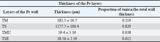
ABSTRACTBackground: The diversity of architecture and histomorphology of the avian alimentary canal significantly influences a wide range of feeding behaviors. Aim: This study aimed to examine the histological and histochemical features of the proventriculus and pancreas in adult Timneh African grey Parrot (TAGP). Methods: Eight TAGPs were utilized. Subsequently, tissue samples from the Pr and Pa were collected and prepared for histochemical analysis and stained with H&E, Periodic Acid-Schiff (PAS), and Masson’s. Results: Histological examination revealed that the wall of the proventriculus consisted of four tunicas: tunica mucosa (TM), tunica submucosa (TS), tunica muscularis, and tunica serosa. The TM exhibited numerous plicae and sulci lined with simple columnar cells that demonstrated a positive reaction to PAS and Masson’s staining. The TS contained compound tubular glands, which were significantly different from other tunicas. The openings of the proventricular glands were seen through the primary and secondary ducts as raised tubes with consistent tubular outlines. Mucosal folds that reacted positively to PAS and Masson’s staining surrounded the openings. The pancreas showed that the alpha (Ai) islets were characterized by their significantly large size, reaching 2,279.83 ± 140.7 µm² with an average diameter of 54.4 ± 13.09 µm, whereas the beta (Bi) islets were characterized by an area of 130.11 ± 8.4 µm² and an average diameter of 13.09 ± 1.08 µm. However, the intermediate mixed (M) islets had an average area of 773.6 ± 71.3 µm² and an average diameter of 26.55 ± 2.79 µm. Conclusion: The results contribute to a more thorough understanding of Pr/Pa histomorphology in TAGP and offer insights into potential functional dietary adaptations or behaviors. Keywords: Alpha cell, Beta cell, Islets, Proventriculus, Timneh African grey parrots, Tunica. IntroductionThe TAGP, referred to as the West African parrot, is classified within the order Psittaciformes, family Psittacidae, genus Psittacus, and species Psittacus timneh (Martin, 2018). They feed on a wide variety of plant species, including tall trees, shrubs, and vines, and ingest many plant components, including fruits, seeds, flowers, leaf buds, and sap, occasionally taking numerous portions from the same species (Lopes et al., 2018). The avian stomach comprises the proventriculus (Pr) (glandular stomach) and the gizzard (muscular stomach), also referred to as the ventriculus. Pr is the location where digestion starts. The epithelium comprises two primary glandular types: tubular glands that secrete mucus and gastric glands that secrete hydrochloric acid (HCl) and pepsin. The Pr glands constitute the mainstream of the Pr wall thickness. The Pr consisted of TM, TS, tunica muscularis (TMU), and tunica serosa (TSE) (Madkour and Mohamed, 2019; Abdel Maksoud et al., 2022; Nasser et al., 2024; Madkour et al., 2025). The Pr wall is considered the thickest in birds classified as omnivores, while it is the thinnest in granivores and moderate in thickness in nectarivores (Madkour et al., 2025). Understanding the mechanism behind the adaptation of the avian stomach to various feeding patterns enables us to improve our understanding of classifying different diseases (Kadhim et al., 2023) and to care for birds in captivity; therefore, it is important to pay attention to the manner in which the glandular stomach is constituted by histomorphology. The pancreas (Pa) is a crucial component of the digestive system, as it secretes enzymes that facilitate food breakdown (Beheiry et al., 2018; Deutschlander, 2024). The pancreas must be both healthy and functionally active for avian health, efficiency, and physical fitness. Several studies have confirmed the distinctions observed between the pancreas of avians and mammals, attributing these variations to various feeding strategies, which result in the avian pancreas possessing more than two lobes and a diversity of islet types (Beheiry et al., 2018; Al-Agele et al., 2021; Deutschlander, 2024). Most birds have a pancreas in the duodenal loop with dorsal and ventral lobes. The splenic lobe may be connected or detachable. The ventral lobe of galliform birds exhibits a unique third lobe expansion (Beheiry et al., 2018; Deutschlander, 2024). Dorsal, ventral, splenic, and third pancreatic lobes have been found in common wood pigeons (Al-Agele et al., 2021). The pancreas executes both endocrine and exocrine functions (Al-Agele et al., 2021; Goodarzi and Bashiri, 2024). The exocrine component of the pancreas comprises intricate tubular-acinar glands organized into lobules (Deutschlander, 2024). The contents of the pancreatic ducts are sequentially discharged from the acinus into the duodenum by intercalated ducts, intralobular ducts, interlobular ducts, and pancreatic ducts (Shaik et al., 2025). The endocrine component consists of pancreatic islets interspersed within the exocrine area as islets of Langerhans. The islets are classified into three categories: alpha, beta, and mixed islets (Goodarzi and Bashiri, 2024). Circular, oval, or elliptical islets are observed (Al-Agele et al., 2021; Mahmood et al., 2022). Currently, limited data exist regarding TAGP, and the histological and histochemical details of stomach/pancreatic islets have received insufficient attention. The aim of this study was to enhance our understanding of the proventriculus’s histological and histochemical analysis and to provide an overview of the morphometric data and the types of islets of Langerhans in TAGP that have a varied and diverse diet consisting of seeds and fruits from plants often overlooked by many other seed-eating birds. Materials and MethodsExperimental designEight adult TAGP aged 13–16 months and weighing 285–312 gm of both sexes (noting that the sex was not a variable) were obtained for histological examination and histochemical study. All birds were clinically healthy and were maintained under standardized housing and feeding conditions. The food included sunflower seeds and pellets, but they also had fresh vegetables such as leafy greens, carrots, bell peppers, and broccoli. They also received fruits such as apples, berries, mango, and papaya, along with sources of calcium and access to clean water every day whenever they wanted, at the animal house of the College of Veterinary Medicine to decrease external physiological variability. The birds were euthanized with an injection of an overdose of ketamine (25 mg/kg B.W.) and xylazine (5 mg/kg B.W.) as outlined by Baldrey et al. (2021) and Hatt et al. (2023), as it is a technique used for euthanasia in birds. Thereafter, tissue samples from the Pr and Pa were dissected from the abdominal viscera and immediately placed into the 10% neutral buffered formalin. Subsequently, all tissue samples were processed using routine methods to create paraffin blocks, which were then cut into 5 μm thick serial sections for the following stains: H&E stain (for routine examination), Masson Trichrome stain (to assess the collagen content of the tissue), and Periodic Acid-Schiff (PAS) stain (to demonstrate carbohydrates such as glycogen, glycoproteins, and mucins, appearing in bright magenta or purple) (Kashef et al., 2025). The stained sections were obtained from all tissues, and two sections were photomicrographed using systematic random sampling. Images were captured using an Omax electron digital camera. Five photomicrographs were taken from each section. The stains enabled the measurement of the boundaries of the islets of Langerhans, along with the diameter of each islet from each tissue section. Measurements were performed using Fiji image software as outlined by Al-Agele (2020) and Kamil and Al-Agele (2025). These measurements were performed first by calibration with a micrometer slide image on the same power that was captured for the tissue sections, thereafter drawing around the entire islet surface area and the two dimensions of the islets in their long and short axes (Karim and Al-Agele, 2025). The morphometric data were recorded and calculated for each sample as mean ± SEM using Excel. Data analysisA one-way analysis of variance with Tukey’s post hoc test was used to analyze the thickness of the layers of Pr and the area and diameter of the islets of Langerhans. The software used was GraphPad Prism 10. Statistical analysis was employed to identify significant differences among the various types of islets of Langerhans, with significance set at p < 0.05 (Dawson and Trapp, 2020). Ethical approvalThis study was conducted following and according to the guidelines of the Animal Experimentation Ethics Committee of the College of Veterinary Medicine, University of Diyala, Iraq (Approval No.: Vet. Med. (223); February 2025 (R, A, and R). ResultsHistological analysis of the proventriculus specimensH&EHistological analysis indicated that the wall of the Pr comprised four layers: TM, TS, TMU, and TSE (Fig. 1). The TM exhibited mucosal folds (plicae proventriculares) and sulci on its luminal surface. The luminal epithelium observed was lined by simple columnar epithelium (Fig. 1). Fig. 1. Photomicrographs of hematoxylin and eosin (H&E)-stained sections of the Pr in TAGP, showing (A) four tunics: mucosa (mu), submucosa (sm), muscularis (m), and serosa (s). Note: Proventricular submucosal glands (sg) and the primary duct (pd). Higher magnification of Figs. B, C, and D shows an opening of the primary duct (pd) and the secondary ducts (sd) of the deep submucosal glands (sg). (H&E stain image (A), the scale bar 500 µm, and images (B–D) 50 µm). PAS stainPAS staining showed a positive reaction in the TM surface epithelium located in the proventriculus (Fig. 2). The tubular glands had a simple columnar epithelium and showed a positive reaction to PAS (Fig. 2). The epithelium invaginated the lamina propria, creating branched tubular glands that tested positive for PAS (Fig. 2). Table 1 shows the morphometric data about the tunic thickness and proportions. This shows that TS plays a significant role in the overall thickness of the Pr wall. Deep proventricular (tubuloalveolar) glands originating from the branched tubular glands were identified. The deepest region of the proventricular glands had composite tubuloalveolar lobules. Lobules were encased in a connective tissue capsule (Fig. 2). Tubuloalveolar units flowed into the secondary ducts in each lobule, while the primary duct connected them to the Pr lumen (Fig. 2). Columnar to cuboidal cells in the basal parts gave the secretory units a dentate appearance (Fig. 2). The primary and secondary ducts have a tall, simple columnar epithelium. PAS-stained sections showed considerable reactivity, particularly in the primary and secondary duct epithelium (Fig. 2). Table 1. Morphometric data for the tunic thickness and proportion of each tunic.
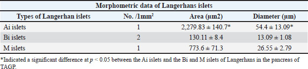
Fig. 2. Photomicrographs of PAS-stained sections of the Pr in TAGP showing (A) proventricular glands (g), primary duct (pd), and surface epithelium showing mucous secretion (se); (B) strong positive PAS-reactions in the surface epithelial cells (black arrows) and the underlined simple glands and tunica mucosa lined by simple columnar epithelium (arrowheads); (C) enteroendocrine cells (et) dispersed in the deep submucosal glands. (D) Proventricular glandular lobule (gl) contains the secretory portion (sp) lined with simple cuboidal epithelium (arrows). (PAS stain image (A), the scale bar represents 500 µm, and in images (B) and (C), 200 µm, and (D), 50 µm). Masson’s trichrome stainingThe tubular/branched tubular gland epithelial cells stained positive with Masson’s stain (Fig. 3). Conversely, the secretory cells of the tubuloalveolar glands had mild positive reactivity near their apexes. The primary and secondary duct epithelium reacted positively with Masson’s stain (Fig. 3). The inner longitudinal and outer circular smooth muscle fiber layers formed the TMU. Mesothelium-enclosed loose connective tissue was abundant in blood vessels in the TSE (Fig. 3). Fig. 3. Photomicrographs of Masson’s trichrome sections of the Pr of TAGP showing the following: (A) Four tunics: mucosa (mu), submucosa (sm), muscularis (m), and serosa (s). Note: Proventricular submucosal glands (sg) and primary duct (pd). (B) Lamina epithelialis (le) composed of simple columnar epithelium, lamina propria (lp), submucosal glands (sg), and extensive collagen fibers (co) occupying the spaces between the simple tubular glands in the lamina propria (lp). (C) Primary duct (pd) and secondary ducts (sd) of the deep submucosal glands (sg). (D) Lamina epithelial cells (le) and the underlined (lp) and secretory portion (sp) of the deep glandular lobules (gl) in the tunica submucosa. (Masson’s trichrome staine X (A), scale bar 500 µm, (B) and (C) 200 µm, (D) 50 µm). Histological analysis of the pancreas specimenH&EThe excretory ducts were characterized by flattened/low cuboidal epithelium, and the intralobular ducts were subsequently lined by simple cuboidal epithelium. These with progress showed changeable epithelium lined with simple columnar epithelium that ended in stratified columnar in the main excretory ducts (Fig. 4). The other spread part of the pancreas was identified as the endocrine portion containing islets of varying forms and sizes, which were classified as alpha (Ai), beta (Bi), and mixed (M) (Fig. 4). Fig. 4. Photomicrographs of H&E-stained sections of the pancreas in TAGP, showing two islets (Ai and Bi) demarcated by acinar glands (ac) in higher magnification in Figure A, B, C, and B; in (A), round and/or oval forms of Bi islets (Bi) and irregular shapes of M islets are shown, while in (B), large irregular Ai islets demarcated by acinar glands (ac) and intercalated duct (id) are shown. (C) An interlobar duct (il) lined by stratified cuboidal cells (arrowhead) and connective tissue (co). (D) Interlobular (inl) and intercalated ducts (id). (H&E stain A–D , scale bar 50 µm). PAS stainHistological analysis revealed two distinct areas; one formed the substantial part of the pancreatic lobules and was represented by the exocrine portion of the secretory acini. These cells comprised many triangular/columnar-like-shaped cells (Fig. 5). The ducts started with intercalated ducts characterized by flattened and low cuboidal epithelium, progressed to further intercalation, and the intralobular ducts were subsequently lined by cuboidal epithelium. The interlobular ducts were coated with tall columnar cells in the main/primary excretory ducts that transitioned to stratified columnar cells (Fig. 5). The second one formed the endocrine region, which had islets of varying forms and sizes, specifically alpha (Ai), beta (Bi), and mixed (M) (Fig. 6). Disseminated within the parenchymal tissue of the exocrine area (Fig. 6). Fig. 5. Photomicrographs of PAS-stained sections of the pancreas in TAGP, demonstrating (A) the pancreatic lobe containing interlobar and interlobular ducts; higher magnification of Figure B, C, and D, showing the main excretory interlobar duct lined by stratified cuboidal epithelium and neighbored by interlobular ducts, which seemed to be clustered around interlobar ducts, and adjacent to it, the acinar gland (ac); higher magnification of Figure. E, F, and G, showing three islets (Ai, Bi, and M) demarcated by acinar glands, in (E) showing round and/or oval forms of the Bi islet containing β-cells, while in (F) showing the irregular shape of the Ai islet containing α and β cells; (G) showing mixed islets containing (Bi and M islets). (PAS stain X (A), scale bar 500 µm, and (B–G), 50 µm). Fig. 6. Photomicrographs of PAS-stained sections of the pancreas in TAGP , showing three islets (Ai, Bi, and M) demarcated by acinar glands (ac) at higher magnification (Figs. A and B). In (A), round and/or oval forms of Bi islets contained β-cells, and irregular-shaped M islets contained α, β, and M-cells and blood vessels (bv), while in (B), Ai islets contained α-cells demarcated by acinar glands (ac) and intercalated duct (id). (PAS stain X (A and B), scale bar 50 µm). Masson’s trichrome stainingHistological analysis revealed that the Pa was encased in a thin capsule. A mesh network of collagen and reticular fibers, together with elastic fibers present in the interlobular tissues, was identified in the interacinar and/or interlobular regions (Fig. 7). The Ai islets were larger than the Bi islets, lacking a distinct boundary with the exocrine region. Bi islets were delineated from secretory acini by a delicate layer of collagen fibers, appearing distinct from the exocrine region by a thin layer of connective tissue (Fig. 7). They manifested as circular or elliptical shapes in the cross sections and predominantly included β cells. β cells exhibited a circular arrangement surrounding the tiny capillaries (Fig. 7). A few cells were also present adjacent to the Bi islets, either individually or in tiny clusters inside the parenchymal tissue of the exocrine area. Moreover, several islets had M cells, whereas an α cell was observed at the periphery of these islets, referred to as mixed islets. These islets consisted primarily of α cells, encased by one or more clusters of β cells (Fig. 7). The histomorphometric analysis of the islets of Langerhans (Table 2) revealed that the average area of the Ai islets was 2,279.83 ± 140.7 µm², with an average diameter of 54.4 ± 13.09 µm. In contrast, the Bi islets exhibited an area of 130.11 ± 8.4 µm² and an average diameter of 13.09 ± 1.08 µm. The M islets displayed intermediate values, with an area of 773.6 ± 71.3 µm² and a diameter of 26.55 ± 2.79 µm. Table 2. The morphometric data relate to the islets of Langerhans located in the pancreas of TAGP.
Fig. 7. Photomicrographs of Masson trichrome-stained sections of the pancreas in TAGP, showing (A) the pancreatic lobe (pl) and the main secretory duct (msd) surrounded by extensive collagen fibers (co) and neighboring interlobular ducts (ac). Higher magnification of Figure B shows the main excretory duct (msd) lined by stratified cuboidal epithelium (st) and surrounded by extensive collagen fibers (co). Higher magnification of Figure C shows the round and/or oval forms of Bi islet containing β-cells and Mi islet containing α- and β-cells, the islet demarcated by acinar glands. (D) A islet (Ai) containing α-cells. (Masson’s trichrome stain X (A), scale bar 200 µm, and (B–D), 50 µm). DiscussionThe histological examination of the present data indicated that the proventriculus wall in TAGP had four layers: TM, TS, TMU, and TSE. Analogous studies have delineated the proventriculus structure in various avian species, including the common starling (Sayrafi and Aghagolzadeh, 2020), bobwhite quail (Al-Kafagy et al., 2022), hooded crow (Abdel Maksoud et al., 2022), breasted white turkey (Madkour and Kandyel, 2022), barn owl (Maher and Hussein, 2023), male duck (Sultan et al., 2023), and cockatiels (Marques et al., 2025). The TM epithelium was identified as simple columnar, as has been stated in other studies on different birds (Sayrafi and Aghagolzadeh, 2020; Abdel Maksoud et al., 2022; Madkour and Kandyel, 2022; Sultan et al., 2023; Marques et al., 2025). The proventricular glands of the TS were significantly indicated, which constitute a substantial thickness of the proventricular wall, in accordance with previous studies (Al-Kafagy et al., 2022; Sultan et al., 2023; Marques et al., 2025). The existence of diverse and extensive glands in the Pr wall may be attributed to the varied diet of TAGP. The deep proventricular glands in the TAGP were observed as compound tubuloalveolar glands, which is in agreement with the findings of Saran and Meshram (2021) in guinea fowl and Abdel Maksoud et al. (2022) and Madkour et al. (2025) in hooded crows. A recent investigation of the TAGP proventriculus identified two types of glands: simple tubular branched glands in the lamina propria and deep compound tubuloalveolar glands in the submucosa. The findings concurred with those of Sayrafi and Aghagolzadeh (2020), while Abdel Maksoud et al. (2022) indicated that the simple tubular gland was located in the cranial region and the branched tubular gland in the other areas, whereas the deep proventricular glands were classified as compound tubuloalveolar. The findings of the current study contradict those of a previous study, which asserted that only compound tubular glands exist in the proventriculus of each pied crow (Udoumoh and Ikejiobi, 2017). The results demonstrated that the secondary ducts connect each tubulo-alveolus to the common cavity, which all secondary ducts converge to form, whereas the primary duct connects this cavity to the proventriculus lumen. These results are consistent with those of Abdel Maksoud et al. (2022) in hooded crows and with the findings related to the proventriculus in white-breasted turkeys (Madkour and Kandyel, 2022) and male ducks (Sultan et al., 2023). The mucosal epithelium of the TAGP exhibited a favorable response to PAS staining. The presence of PAS-positive mucin in TAGP is consistent with the observations of Beheiry (2018) in Turkey, Sayrafi and Aghagolzadeh (2020) in common starlings, and Al-Kafagy et al. (2022) in bobwhite quail. Furthermore, the PAS-positive reaction was demonstrated to identify the secretion of the tubular glands and the ductular epithelium of the tubuloalveolar glands, corroborating the results of Abdel Maksoud et al. (2022) in hooded crows, as well as findings in the white-breasted turkey (Madkour and Kandyel, 2022), male duck (Sultan et al., 2023), and cockatiels (Marques et al., 2025). Udoumoh and Ikejiobi (2017) indicated that the compound tubular glands of the Pr in the pied crow were PAS-negative, while the current study showed a positive reaction with PAS and/or Masson’s trichrome stains, which corroborates the results of Maksoud et al. (2022) in hooded crows, as well as findings in the white-breasted turkey (Madkour and Kandyel, 2022), male duck (Sultan et al., 2023), and cockatiels (Marques et al., 2025). Secretion composition may vary depending on epithelial tissue function and ambient factors. Histochemically, mucins are neutral or acidic in birds, preserving the mucosal epithelium and forming a protective barrier (Sayrafi and Aghagolzadeh, 2020). This investigation showed that a lamina muscularis mucosa encircled the apical regions of the deep submucosal glands, delineating the lamina propria from the tunica submucosa. These findings are consistent with the results of Abdel Maksoud et al. (2022) in hooded crows. In the common starling, the lamina muscularis mucosa was identified as a thin layer within the proventricular mucosal tunic, encircling the lobules of the tubuloalveolar glands (Sayrafi and Aghagolzadeh, 2020). Histochemical analysis of proventricular submucosal glands showed that the apical surface of the epithelial cells lining the compound tubuloalveolar glands’ ducts strongly reacted to PAS and Masson’s stains, while the secretory portions, composed of oxynticopeptic cells, did not, suggesting that these cells lack mucous secretory activity, as they produce HCl and pepsin (Udoumoh and Ikejiobi, 2017; Abdel Maksoud et al., 2022). Ahmed et al. (2025) stated that the cells lining the ducts of compound tubular glands contain a blend of acidic and neutral mucopolysaccharides and suggested that the secreted ductal mucin lubricates the mucosal surface and protects it from luminal contents (Udoumoh and Ikejiobi, 2017). The TS, which comprised compound tubuloalveolar lobules, constituted a significant thickness of the TAGP proventricular wall, as noted in this study. This finding agrees with the observations of Abdel Maksoud et al. (2022), who noted that the deep proventricular glands constituted the primary structural element of the proventricular wall in hooded crows. Conversely, Sayrafi and Aghagolzadeh (2020) asserted that the TS is a small connective tissue layer between the lamina muscularis mucosae and the TMU’s inner layer. The results were compared with those reported by Abdel Maksoud et al. (2022), who observed connective tissue septa abundant in collagen and elastic fibers encasing the glandular lobules, and with the findings of Sayrafi and Aghagolzadeh (2019), who noted the presence of smooth muscle fibers originating from the lamina muscularis mucosa. Enteroendocrine cells identified in this study were dispersed individually among the surface epithelial cells and were abundant in the Submucosal Glands (SMGs). This finding aligns with the observations made by Abdel Maksoud et al. (2022), who identified endocrine cells exhibiting distinctive characteristics among the Deep Proventricular Glands (DPGs). The findings also corroborated the observations made by Abdel Maksoud et al. (2022) regarding the hooded crow and by Sayrafi and Aghagolzadeh (2020) concerning the common starling. Al-Saffar and Al-Samawy (2015) observed an inverted configuration of the tunica muscularis in mallard ducks: a slender inner longitudinal layer and a robust outer circular layer. TSE comprises loose connective tissue enveloped by a layer of mesothelial cells. This finding is consistent with those of Sayrafi and Aghagolzadeh (2019) on common starlings and Abdel Maksoud et al. (2022) on the hooded crow. This suggests that the gastrointestinal tract of birds optimizes the physical and chemical features of various diets. Dietary variables may affect the Pr structures of bird species (De Moraes et al., 2024) as the Pr wall comprises many glands, which may be an adaptation to different diets (Lopes et al., 2018). Histological examination indicated that the TAGP pancreas was enveloped by a thin connective tissue capsule. These results align with the findings of Beheiry et al. (2018) in geese, Al-Haaik (2019) in kestrels, and Mahmood et al. (2022) in native ducks. Al-Agele et al. (2021) reported the existence of subcapsular adipocytes in the pancreas of the wood pigeon; however, the TAGP findings contradict this buildup of adipocytes beneath the capsule and surrounding the interlobar duct. Subcapsular adipocytes in common wood pigeons may arise from a prevalent and varied diet mostly consisting of seeds and fruits from plants overlooked by most other seed-eating avians (Junior et al., 2025). The secretory acini comprised several pyramidal cells exhibiting either a triangular or elongated columnar morphology. These findings are consistent with other studies, including Beheiry et al. (2018) on geese and Mahmood et al. (2022) on native ducks. The endocrine area of the TAGP pancreas exhibited diverse morphologies and dimensions of Ai, Bi, and M islets dispersed within the exocrine region’s parenchymal tissue. The present findings align with those reported in earlier studies, including those by Kara et al. (2014) on sparrowhawks, Al-Haaik (2019) on adult kestrels, Al-Agele et al. (2021) on common wood pigeons, and Abdellatif (2023) on moorhens. The endocrine portion of the avian pancreas typically consists of a substantial irregular formation of alpha/Ai islets and a rounded configuration of β/Bi islets, with few or no mixed/M islets (Deutschlander, 2024), aligning with the current findings in TAGP. However, Mohammadi and Goodarzi (2024) reported that no M islets were found in the pancreas of common pheasants. In Ai islets, the main α cells were located throughout the entire islet; however, some investigations indicated that these cells are in the periphery region (Al-Khakani et al., 2019; Shaik et al., 2025). In the Bi islet, β cells exhibited a circular arrangement surrounding tiny capillaries, similar to other avian species (Al-Haaik, 2019; Al-Agele et al., 2021; Shaik et al., 2025). M cells in TAGP have also been identified in both Ai and Bi islets (Kara et al., 2014). Nonetheless, in Ai islets, a limited number of β cells were identified; these were present in certain avian pancreases (Al-Haaik, 2019; Shaik et al., 2025). Based on the study findings, the Pr of TAGP displayed significant differences between different species of birds, which may be related to the vast range of feeding behaviors (Vertiprakhov et al., 2023). ConclusionHistochemical analysis revealed distinctive characteristics in the Pr/Pa histomorphology. These included the presence of two glandular types: simple tubular branched glands in the lamina propria and deep compound tubuloalveolar glands in the Pr submucosa. The diversified diet consumed by parrots may have led to the development of various and distinct glands within the proventriculus wall of TAGP. The three types of Langerhans islets (Ai, Bi, and M islets) were shown as unique features along with their Pr morphometric data. The existence of various endocrine cell types necessitates an additional examination of their roles in TAGP physiology, possibly transcending alimentary behaviors. These results contribute to a more thorough understanding of Pr/Pa histomorphology in TAGP and offer insights into potential functional dietary adaptations or behaviors. AcknowledgmentWe would like to express our gratitude to the College of Veterinary Medicine at the University of Diyala for the outstanding support they provided. Conflicts of interestAccording to the authors, no potential conflicts of interest need to be disclosed. FundingThis project was not supported by any grants. Author’s contributionsRAA: writing the original draft; RSI: writing and reviewing the manuscript. Data availabilityThe data supporting the results of this article are available in the manuscript and from PMN upon reasonable demand. ReferencesAbdel Maksoud, M.K.M., Ibrahim, A.A.H., Nabil, T.M. and Moawad, U.K. 2022. Histomorphological, histochemical and scanning electron microscopic investigation of the proventriculus (Ventriculus glandularis) of the hooded crow (Corvus cornix). Anat. Histol. Embryol. 51(3), 380–389; doi:10.1111/ahe.12798 Abdellatif, A. 2023. Expression of Insulin, Glucagon, Somatostatin, and Pancreatic Polypeptide in the Pancreas of the Eurasian Moorhen (Gallinula chloropus). Slov. Vet. Res. 60(4), 185–193; doi:10.26873/SVR-1616-2023 Ahmed, A.M., Al-Jawadi, A.A. and Khaleel, H.K. 2025. Morphological, histological, and histochemical studies of stomach, proventriculus, and gizzard in slender-billed gull (Chroicocephalus genei). Bull. Iraq. Nat. Hist. Mus. 18(3), 507–526; doi:10.26842/binhm.7.2025.18.3.0507 Al-Agele, R. 2020. The study of histological observations of ovine claw coronary region and the architecture morphology of arterial anastomosis of this area. Atatürk. Univ. J. Vet. Sci. 15(1), 14–21; doi:10.17094/ataunivbd.634782 Al-Agele, R.A.A., Jabbar, A.I. and Al-Ubaidy, A.A.H. 2021. Study on morphohistological observations of the islets of langerhans in the pancreas of common wood pigeon Columba palumbus. Biochem. Cell. Arch. 21(1), 1165–1170. Al-Haaik, A. 2019. A gross anatomical and histological study of pancreas in adult Kestrel (Falco tinnunculus). Iraqi. J. Vet. Sci. 33(2), 175–180; doi:10.33899/ijvs.2019.162960 Al-Kafagy, S.M., Al-Jebori, A.K. and Alseady, Y.Y. 2022. Histochemical study of proventriculus in pre-hatch and post-hatch days in northern bobwhite quail (Colinus virginianus). Iraqi J. Vet. Sci. 36(1), 161–166; doi:10.33899/ijvs.2022.135832.2529 Al-Khakani, S.S.A., Zabiba, I.M.J., Al-Zubaidi, K.H. and Al-Alwany, E.A.H. 2019. Morphometrical and histochemical foundation of pancreas in white-eared bulbul (Pycnonotus leucotis). Iraqi J. Vet. Sci. 33(1), 99–104; doi:10.33899/ijvs.2019.125521 Al-Saffar, F.J., and Al-Samawy, E.R.M. 2015. Histomorphological and histochemical studies of the stomach of the mallard (Anas platy rhynchos). Asian J. Anim. Sci. 9(6), 280–292; doi: 10.3923/ajas.2015 Baldrey, V., Stanford, M. and Bacon, H. 2021. A comparison of the pharmacodynamic effects of intravenous ketamine–xylazine with alfaxalone in mute swans (Cygnus olor) presenting at a wildlife veterinary hospital. Vet. Anaesth. Analg. 48(5), 716–724; doi:10.1016/j.vaa.2021.03.014 Beheiry, R. 2018. Histochemical and scanning electron microscopy of proventriculus in turkey. J. Adv. Vet. Anim. Res. 5(3), 290–298; doi:10.5455/javar.2018.e279 Beheiry, R.R., Abdel-Raheem, W.A.A., Balah, A.M., Salem, H.F. and Karkit, M.W. 2018. Morphological, histological and ultrastructural studies on the exocrine pancreas of goose. Beni-Suef Univ. J. Basic Appl. Sci. 7, 353–358; doi:10.1016/j.bjbas.2018.03.009 Dawson, B. and Trapp, R.G. 2020. Basic and clinical biostatistics. 5th Edition. New York, NY: McGraw-Hill Education/Medical, pp: 190–220. De Moraes, S., De Moura, F.B.C., De Carvalho, J.C., De Lima, H.C., De Anchieta De Castro E Horta -Alves, C.E. 2024. Histochemical characterisation of the turquoise-fronted parrot (Amazona aestiva) digestive tract. Anat. Histol. Embryol. 53(6), e70003. Deutschlander, M. 2024. Sturkie’s Avian Physiology, 7th Edition, Ornithol. 141(4), 1 ukae027; doi: 10.1093/ornithology/ukae027 Goodarzi, N. and Bashiri, A. 2024. Histology and immunofluorescent study of the pancreas in lovebird (Agapornis personatus). Vet. Med. Sci. 10(2), 1394; doi:10.1002/vms3.1394 Hatt, J.M., Kreyenbühl, K. and Kummrow, M. 2023. Methods of analgesia and euthanasia in backyard poultry. Schweiz. Arch. Tierheilkd. 165, 503–511; doi:10.17236/sat00398 Junior, J.C.M., Fidelis, R.A.M., De Souza Bambirra, B.L., Dos Santos Filho, J.C.M., De Macedo, K.L.S., Ribeiro, L.D.A.V., Carvalho, G.M.M., Ritter, A.C.C., Miranda, N.C., Jin, L.D. and Genter, L. 2025. Diet of parrots in an urban area of São Paulo, southeast Brazil. Revista Do Instituto Florestal 37, 1–20. Kadhim, T.J., Al-Agele, R.A.A. and Ibrahim, R.S. 2023. Histopathological study of the cecal tonsils post newcastle vaccine in broiler chickens. AIP Conf. Proc. 2475(1), 100008. doi:10.1063/5.0103023 Kamil, Y.A. and Al-Agele, R.A.G. 2025. Study the functional-related ultrastructural and histomorphometrical characteristics of pectoralis muscle of high-flying tippler pigeons in comparison to low-flying fancy pigeons. Adv. Anim. Vet. Sci. 13(9), 2024–2031; doi:10.17582/journal.aavs/2025/13.9.2024.2031 Kara, A., Tekiner, D., Şimşek, N., Balkaya, H. and Özüdoğru, Z. 2014. Distribution and Location of Endocrine Cells in the Pancreas of the Sparrowhawk, Accipiter nisus. Kafkas. Univ. Vet. Fak. Derg. 20(2), 307–312; doi:10.9775/kvfd.2013.10096 Karim, I.M.A. and Al-Agele, R.A. 2025. Histomorphometry of limb skeletogenesis in prehatched precocial embryos (Japanese quail and Cochin chicken) and Altricial Embryos (racing pigeons and cockatiel birds). Open. Vet. J. 15(5), 2138–2148; doi:10.5455/OVJ.2025.v15.i5.32 Kashef, S.M., Abo-Elyazed, A.A.E.M., Elkadim, M. and Abdine, N. 2025. The effect of alpha lipoic acid on the cornea of an experimentally induced dry eye in adult male albino rat: a histological and immunohistochemical study. Egypt. J. Cell Tissue Res. 1(1), 56–90; doi:10.21608/ejctr.2024.307421.1020 Lopes, D.D.C., Martin, R.O., Indjai, B., Monteiro, H., Henriques, M., Regalla, A. and Catry, P. 2018. Food diversity of Timneh Parrots (Psittacus timneh) in the Bijagós archipelago, Guinea-Bissau. Afr. J. Ecol. 56(4), 1039–1043; doi:10.1111/aje.12544 Madkour, F.A. and Kandyel, R.M. 2022. Histomorphology and ultrastructure of the proventriculus of the broad breasted white turkey (Meleagris gallopavo). Microsc. Res. Tech. 85(11), 3514–3529; doi:10.1002/jemt.24203 Madkour, F.A. and Mohamed, A.A. 2019. Comparative anatomical studies on the glandular stomach of the rock pigeon (Columba livia targia) and the Egyptian laughing dove (Streptopelia senegalensis aegyptiaca). Anat. Histol. Embryol. 48(1), 53–63; doi:10.1111/ahe.12411 Madkour, F.A., Mohammed, E.S.I., Al-Rasheed, M. and Abdelhakeem, F. 2025. Comprehensive structural characterization of the esophageo-gastric tube in the hooded crow (corvus cornix): histological, histochemical, and ultrastructural insights. Microsc. Res. Tech. 3, 520–537; doi:10.1002/jemt.70086 Maher, M.A. and Hussein, S. 2023. Anatomical distribution of celiac artery with histochemical investigation to the proventriculus of barn owl (Tyto alba). Zoomorphology 142(2), 201–213; doi:10.1007/s00435-023-00595-7 Mahmood, S.K., Ahmed, N.S., Sultan, G.A. and Yousif, M.J. 2022. Histomorphological and carbohydrate histochemical study of the pancreas in native ducks (Anas Platyrhynchos). Iraqi J. Vet. Sci. 36(4), 1103–1110; doi:10.33899/ijvs.2022.133156.2183 Marques, A.L.R., Oliveira, I.J., Mamgue, V.E., Perin, R.L., Silva, L.E., Gruchouskei, L., Gonçalves, G.F. and Romagnolli, P. 2025. Morphological Analysis of the Digestive Tract of Cockatiels (Nymphicus hollandicus)-Oesophagus to Colorectum. Anat. Histol. Embryol. 4(1), e70007; doi:10.1111/ahe.70007 Martin, R.O. 2018. Grey areas: temporal and geographical dynamics of international trade of Grey and Timneh Parrots (Psittacus erithacus and P. timneh) under CITES. Emu-Austral. Ornithology. 118(1), 113–125; doi:10.1080/01584197.2017.1369854 Mohammadi, A. and Goodarzi, N. 2024. Characterization of the pancreas of common pheasant (Phasianus colchicus). Microsc. Res. Tech. 87(2), 339–348; doi:10.1002/jemt.24434 Nasser, A.T., Al-Azzawi, A., Al-Agele, R. and Al Ajeeli, K. 2024. Identification of extremely virulent infectious bursal disease virus via molecular and histological methods in broiler chickens. JWPR 14(1), 98–112; doi:10.36380/jwpr.2024.10 Saran, D. and Meshram, B. 2021. Histomorphological and histochemical studies on proventriculus in guinea fowl (Numida meleagris). Indian J. Anim. Res. 55(7), 806–809; doi:10.18805/IJAR.B-4130 Sayrafi, R. and Aghagolzadeh, M. 2020. Histological and histochemical study of the proventriculus (Ventriculus glandularis) of common starling (Sturnus vulgaris). Anat. Histol. Embryol. 49(1), 105–111; doi:10.1111/ahe.12495 Shaik, M.R., Singh, A., Rayapati, D., Li, J., Boparai, I.S., Evani, S., Randhawa, N., Krimins, R. and Akshintala, V.S. 2025. Comparative Anatomy of Pancreas Across Animal Species: a Systematic Review. Pancreas 54(10), e904–e915; doi:10.1097/MPA.0000000000002491 Sultan, G.A., Al-Haaik, A.G. and Alhasso, A.A. 2023. Morphometrical and Histochemical study of glandular stomach (Proventriculus) in local domestic male ducks (Anas Platyrhynchos). Iraqi J. Vet. Sci. 37(1), 65–71; doi:10.33899/ijvs.2022.133451.2233 Udoumoh, A.F. and Ikejiobi, J.C. 2017. Morphological features of glands in the gastrointestinal tract of the African pied crow (Corvus albums). Comparative Clin. Pathol. 26(3), 585–590; doi:10.1007/s00580-017-2425-3 Vertiprakhov, V.G., Grozina, A.A., Fisinin, V.I. and Surai, P.F. 2023. Adaptation of chicken pancreatic secretory functions to feed composition. World’s Poult. Sci. J. 79(1), 27–41; doi:10.1080/00439339.2023.2163042
| ||


| How to Cite this Article |
| Pubmed Style Al-agele RA, Ibrahim RS. Histochemical and histomorphological analysis of the proventriculus and endocrine portion of the Timneh African grey parrot (Psittacus timneh). Open Vet. J.. 2026; 16(6): 3832-3842. doi:10.5455/OVJ.2026.v16.i6.54 Web Style Al-agele RA, Ibrahim RS. Histochemical and histomorphological analysis of the proventriculus and endocrine portion of the Timneh African grey parrot (Psittacus timneh). https://www.openveterinaryjournal.com/?mno=306201 [Access: June 26, 2026]. doi:10.5455/OVJ.2026.v16.i6.54 AMA (American Medical Association) Style Al-agele RA, Ibrahim RS. Histochemical and histomorphological analysis of the proventriculus and endocrine portion of the Timneh African grey parrot (Psittacus timneh). Open Vet. J.. 2026; 16(6): 3832-3842. doi:10.5455/OVJ.2026.v16.i6.54 Vancouver/ICMJE Style Al-agele RA, Ibrahim RS. Histochemical and histomorphological analysis of the proventriculus and endocrine portion of the Timneh African grey parrot (Psittacus timneh). Open Vet. J.. (2026), [cited June 26, 2026]; 16(6): 3832-3842. doi:10.5455/OVJ.2026.v16.i6.54 Harvard Style Al-agele, R. A. & Ibrahim, . R. S. (2026) Histochemical and histomorphological analysis of the proventriculus and endocrine portion of the Timneh African grey parrot (Psittacus timneh). Open Vet. J., 16 (6), 3832-3842. doi:10.5455/OVJ.2026.v16.i6.54 Turabian Style Al-agele, Ramzi Abdulghafoor, and Raad Shaalan Ibrahim. 2026. Histochemical and histomorphological analysis of the proventriculus and endocrine portion of the Timneh African grey parrot (Psittacus timneh). Open Veterinary Journal, 16 (6), 3832-3842. doi:10.5455/OVJ.2026.v16.i6.54 Chicago Style Al-agele, Ramzi Abdulghafoor, and Raad Shaalan Ibrahim. "Histochemical and histomorphological analysis of the proventriculus and endocrine portion of the Timneh African grey parrot (Psittacus timneh)." Open Veterinary Journal 16 (2026), 3832-3842. doi:10.5455/OVJ.2026.v16.i6.54 MLA (The Modern Language Association) Style Al-agele, Ramzi Abdulghafoor, and Raad Shaalan Ibrahim. "Histochemical and histomorphological analysis of the proventriculus and endocrine portion of the Timneh African grey parrot (Psittacus timneh)." Open Veterinary Journal 16.6 (2026), 3832-3842. Print. doi:10.5455/OVJ.2026.v16.i6.54 APA (American Psychological Association) Style Al-agele, R. A. & Ibrahim, . R. S. (2026) Histochemical and histomorphological analysis of the proventriculus and endocrine portion of the Timneh African grey parrot (Psittacus timneh). Open Veterinary Journal, 16 (6), 3832-3842. doi:10.5455/OVJ.2026.v16.i6.54 |


