





| Case Report | ||
Open Vet. J.. 2026; 16(6): 3590-3594 Open Veterinary Journal, (2026), Vol. 16(6): 3590-3594 Case Report Hyperkalemia caused by hyporeninaemic hypoaldosteronism in a dogTomoko Iwanaga1*, Tomoe Iida-Ogikubo1 and Takashi Kazama21Koganei Animal Medical Emergency Centre, Tokyo University of Agriculture and Technology, Tokyo, Japan 2Kazama Animal Hospital, Tokyo, Japan *Corresponding Author: Tomoko Iwanaga. Koganei Animal Medical Emergency Center Tokyo University of Agriculture and Technology, Tokyo, Japan. Email: tyiwanaga [at] go.tuat.ac.jp Submitted: 04/01/2026 Revised: 22/04/2026 Accepted: 04/05/2026 Published: 05/06/2026 © 2025 Open Veterinary Journal
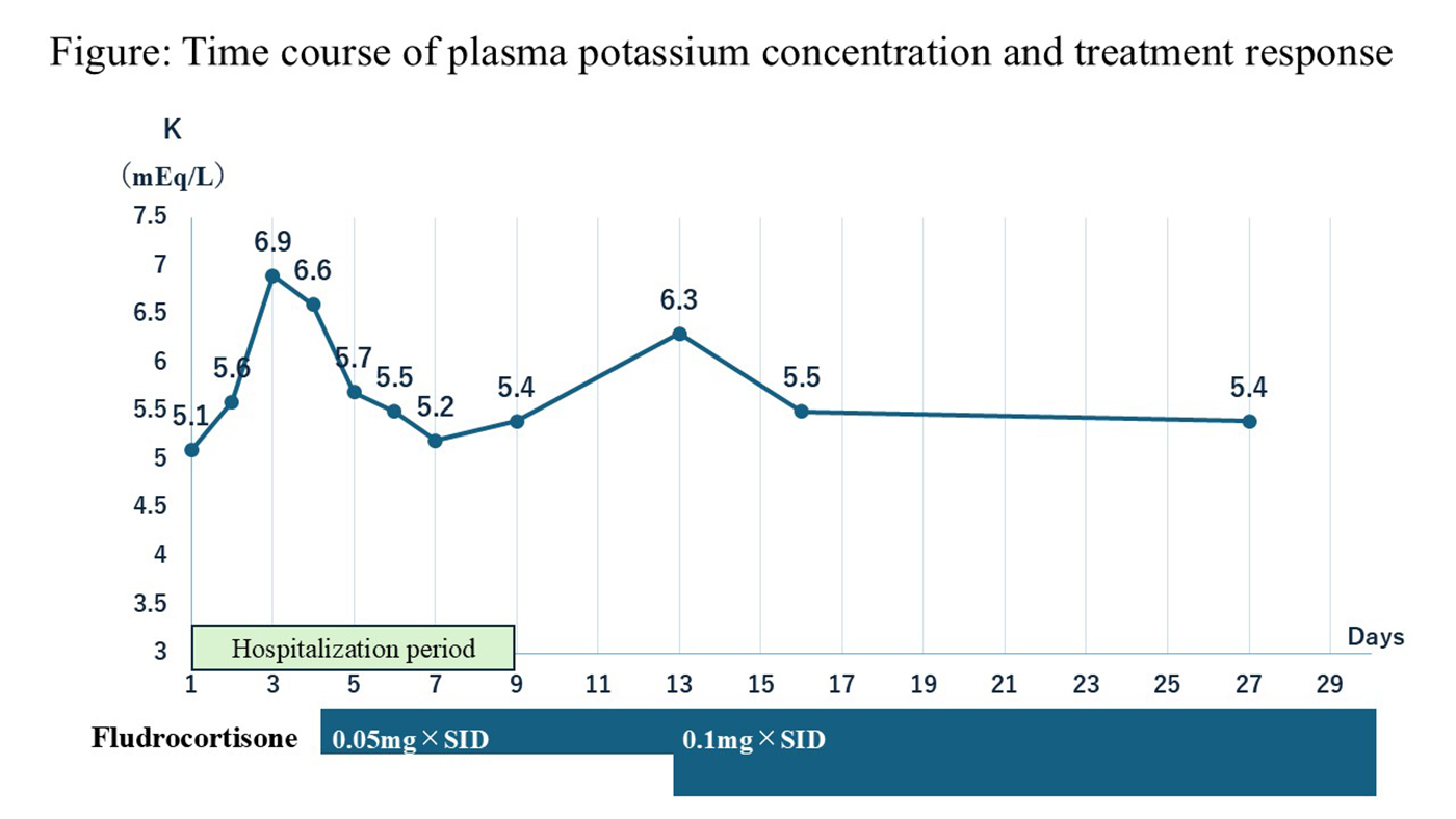
AbstractBackground: Hyperkalemia has multiple etiologies, and although primary adrenocortical insufficiency is a well-known hormonal cause, hyperkalemia may also result from hyporeninaemic hypoaldosteronism, in which impaired renin secretion reduces aldosterone production and mineralocorticoid activity. Case Description: A 15-year-old spayed female Miniature Dachshund presented with persistent hyperkalemia. The dog exhibited systemic hypertension (systolic blood pressure: 192 mmHg) and mild renal insufficiency without evidence of urinary obstruction, hemolysis, cellular lysis, or potassium-sparing drug administration. An adrenocorticotropic hormone (ACTH) stimulation test revealed normal cortisol concentrations (pre: 4.3 µg/dl; post: 20.7 µg/dl), excluding primary hypoadrenocorticism. The plasma renin activity (PRA) and aldosterone concentration were evaluated. PRA was undetectable (<0.2 ng/ml/h), and aldosterone concentrations were low before (<17 pg/ml) and after (124.6 pg/ml) ACTH stimulation. These findings supported hyporeninaemic hypoaldosteronism with concurrent renal insufficiency and hypertension. Fludrocortisone therapy (0.05–0.1 mg/kg PO SID) successfully reduced plasma potassium to 5.2–5.4 mEq/l. Hydralazine (2 mg/kg PO BID) was administered for blood pressure control. The dog remained clinically stable before dying of aspiration pneumonia on day 94. Conclusion: Renal insufficiency, metabolic acidosis, hypertension, suppressed PRA, and low aldosterone levels supported the diagnosis of acquired type IV renal tubular acidosis. Hyperkalemia responded well to mineralocorticoid supplementation. In dogs with renal disease and persistent hyperkalaemia, hyporeninaemic hypoaldosteronism should be considered, and measurement of PRA and aldosterone is recommended when primary adrenal insufficiency is excluded. Keywords: Aldosterone, Hyperkalaemia, Hyporeninaemic hypoaldosteronism, Renin, Type IV renal tubular acidosis. IntroductionHyperkalaemia may arise from impaired renal excretion, tissue breakdown, increased potassium intake, or drug administration. Urinary tract obstruction, acute kidney injury, tumor lysis syndrome, consumption of potassium-rich diets, and administration of potassium-sparing drugs such as trilostane, spironolactone, or angiotensin II receptor blockers are among the common causes. Pseudohyperkalaemia may result from hemolysis or blood collection in ethylenediaminetetraacetic acid-2K-containing tubes (Phillips and Polzin, 1998; Hollander-Rodriguez and Calvert, 2006). The most common cause of hyperkalaemia is aldosterone deficiency, such as in primary hypoadrenocorticism. This condition is characterized by insufficient adrenal cortical function and a lack of cortisol response to adrenocorticotropic hormone (ACTH) stimulation (Brodbelt, 2010; Lathan and Thompson, 2018; Niessen et al., 2025). Because electrolyte abnormalities may result from multiple etiologies, the cortisol response to ACTH is essential for diagnosis confirmation. The renin–angiotensin–aldosterone system (RAAS) regulates aldosterone secretion. Decreased renin secretion results in low aldosterone concentrations despite normal adrenal cortisol production, potentially causing hyperkalaemia (Batlle and Arruda, 2018). We describe a canine case of persistent hyperkalaemia associated with suppressed renin and aldosterone concentrations despite a normal cortisol response. Case DetailsA 15-year-old spayed female Miniature Dachshund was referred for evaluation of vomiting and persistent hyperkalemia. Hyperkalaemia and mild renal impairment were documented 20 days prior, and primary hypoadrenocorticism was suspected; however, the ACTH stimulation test showed a normal cortisol response [pre: 4.3 µg/dl (reference interval (RI): 1–6 µg/dl); post: 20.7 µg/dl, FUJIFILM VET Systems Co., Ltd., Tokyo, Japan]. On presentation, the patient’s systolic blood pressure was high (192 mmHg). Serum biochemistry revealed mild renal insufficiency (International Renal Interest Society stage 1). Blood urea nitrogen was 48.0 mg/dl (RI: 5.4–30.9 mg/dl), creatinine was 1.37 mg/dl (RI: 0.51–1.39 mg/dl), total calcium was 13.2 mg/dl (RI: 8.9–11.3 mg/dl), phosphorus was 6.6 mg/dl (RI: 2.1–5.1 mg/dl), and symmetrical dimethylarginine was elevated (33.7 µg/dl; RI: <14.0 µg/dl). The dog had received intravenous saline for 2 days prior to referral; on admission, potassium was mildly increased (5.1 mEq/l; RI: 4.0–5.0 mEq/l), rising to 6.9 mEq/l after fluid discontinuation. Sodium (146 mEq/l; RI: 134–153 mEq/l) and chloride (111 mEq/l; RI: 105–118 mEq/l) contents were within the RIs. Urinalysis revealed a urine protein–creatinine ratio of 1.26 and a specific gravity of 1.016. Blood gas analysis indicated metabolic acidosis (pH, 7.313; RI: 7.337 ± 0.062; HCO₃⁻ 18.1 mmol/l; RI: 23.4 ± 3.6; base excess −8 mmol/l; RI: −2.5 ± 3.7; anion gap excluding potassium, 17.9 mEq/l; RI: 8–16 mEq/l). Ultrasonography revealed no urinary obstruction; however, the corticomedullary distinction was decreased or absent, and both kidneys contained multiple cysts. Mild enlargement of both adrenal glands (dorsoventral thickness of 8.5 mm) was noted. Electrocardiogram findings and thoracic and abdominal radiographs were unremarkable. Because the previous ACTH stimulation test demonstrated normal cortisol concentrations (pre: 4.3 µg/dl; post: 20.7 µg/dl), we excluded primary hypoadrenocorticism. Plasma renin activity (PRA) and aldosterone concentrations were then measured. PRA and aldosterone concentrations were measured using an animal diagnostic laboratory (Animal Medical Technology, Nagoya, Japan). Aldosterone was measured using a LUMIPULSE L2400 analyzer (Fujirebio Inc., Tokyo, Japan) with the LUMIPULSE Presto Aldosterone reagent (Fujirebio Inc.) by a CLEIA method. PRA was measured using a fully automated microplate enzyme immunoassay (EIA) analyzer AP-X (Canon Medical Diagnostics Corporation, Tokyo, Japan) with the Renin Activity Kit “Yamasa” (Yamasa Corporation, Chiba, Japan) by an EIA method. Analytical procedures until measurement were performed according to the laboratory protocol. After collection, the samples were promptly submitted to the adjacent diagnostic laboratory. After submission, they were separated using a refrigerated centrifuge and stored at −80°C until analysis. The dog was fasted and kept as calm as possible at the time of blood sampling, and blood was collected from the jugular vein with the dog in sternal recumbency. The dog had been fed a low-fat canned gastrointestinal diet (Royal Canin Gastrointestinal Low Fat canned diet; sodium content 0.1%), and no drugs directly suppressing the RAAS had been administered before sampling; however, hydralazine was subsequently administered for hypertension. Samples for cortisol and aldosterone measurement were placed into a serum tube, and those for PRA were placed into an Ethylenediaminetetraacetic acid-2Na tube. PRA was undetectable (<0.2 ng/ml/hr, below detection limit; Animal Medical Technology, Nagoya, Japan). Aldosterone concentrations were markedly reduced both before (<17.1 pg/ml; RI: 25–269; Animal Medical Technology, Nagoya, Japan) and after (124.6 pg/ml; RI: 200–2,000; Animal Medical Technology, Nagoya, Japan) ACTH stimulation. Thus, the dog was diagnosed with hyporeninemic hypoaldosteronism with mild renal failure, hypertension, and metabolic acidosis. On day 4, fludrocortisone (0.05 mg/kg PO SID) was initiated, resulting in a decrease in potassium from 6.3 to 5.2–5.4 mEq/l. When the dose of potassium was increased again to 6.3 mEq/l on day 13, the dose was increased to 0.1 mg/kg PO SID, after which the dose of potassium decreased again to 5.5 mEq/l (Fig. 1). Hydralazine (2 mg/kg PO BID) was administered for the management of hypertension. The dog remained clinically stable until death from aspiration pneumonia on day 94.
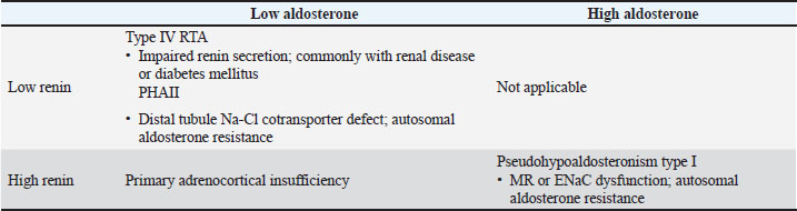
Fig. 1. Time course of plasma potassium concentration and response to treatment. On day 4, fludrocortisone therapy was initiated at 0.05 mg/kg once daily, after which the plasma potassium concentration decreased to 5.2–5.4 mEq/l. Because the patient showed a favorable clinical response, the dog was discharged on Day 9. Subsequently, on Day 13, the plasma potassium concentration increased again to 6.3 mEq/l, prompting an escalation of the fludrocortisone dosage to 0.1 mg/kg once daily. Thereafter, the dog remained clinically stable, and no further increases in the plasma potassium concentration were observed up to Day 81. DiscussionPrimary hypoadrenocorticism is the most common endocrine cause of hyperkalaemia in dogs (Brodbelt, 2010; Lathan and Thompson, 2018). It is diagnosed based on decreased cortisol levels in response to an ACTH stimulation test (Lathan and Thompson, 2018). However, it was excluded in this case based on a normal cortisol response to ACTH stimulation. Markedly reduced PRA and aldosterone levels supported a diagnosis of hyporeninaemic hypoaldosteronism (Lehnhardt and Kemper, 2011). In humans, the differential diagnoses include type IV renal tubular acidosis (RTA) and pseudohypoaldosteronism type II (PHAII) (Table 1). PHAII is a hereditary disorder that affects the thiazide-sensitive sodium–chloride cotransporter, whereas acquired type IV RTA is commonly secondary to renal insufficiency or diabetes mellitus (Sousa et al., 2016; Yaxley and Pirrone, 2016). Table 1. Differential diagnosis of hyperkalaemia using renin activity and aldosterone levels.
This dog had renal insufficiency, hypertension, metabolic acidosis, and suppressed PRA and aldosterone concentrations, which support hyporeninaemic hypoaldosteronism consistent with acquired type IV RTA. In this context, hyporeninaemic hypoaldosteronism describes the underlying pathophysiology, whereas acquired type IV RTA describes the clinical syndrome associated with reduced mineralocorticoid activity. Type IV RTA is classically characterized by hyperkalaemic, hyperchloraemic metabolic acidosis with a normal anion gap. Acidosis is thought to reflect impaired urinary ammonium excretion secondary to reduced mineralocorticoid activity and hyperkalaemia, rather than the accumulation of large amounts of unmeasured anions (Yaxley and Pirrone, 2016; Batlle and Arruda, 2018). Therefore, the persistent hyperkalemia in this case supported type IV RTA rather than other forms of RTA, which are typically associated with hypokalemia. Although the anion gap in this dog was mildly increased, acquired type IV RTA could not be excluded. Normal-anion-gap metabolic acidosis remains the dominant component in chronic kidney disease, whereas a mild increase in the anion gap may be superimposed because of limited retention of unmeasured anions, such as phosphate, sulfate, urate, and other organic anions (Zijlstra and Stegeman, 2023). Accordingly, the acid–base abnormality in the present case was interpreted as predominantly acquired type IV RTA associated with hyporeninaemic hypoaldosteronism, with a possible minor contribution from renal insufficiency to the mild increase in the anion gap. Hypertension may contribute to nephrosclerosis by impairing renin secretion in the juxtaglomerular apparatus. In veterinary medicine, two cats presenting with hyperkalaemia accompanied by hypoaldosteronism have been reported to have type IV RTA (Marino and Foster, 2024). However, PHAII was considered unlikely to be the underlying cause in this case. This disorder is hereditary; however, this patient was geriatric and had no history of hyperkalaemia on previous blood examinations. Moreover, PHAII has never been reported in dogs or cats, and its treatment typically requires thiazide diuretics rather than mineralocorticoid supplementation. In contrast, this dog showed a clear clinical response to fludrocortisone, further supporting the diagnosis of acquired hyporeninaemic hypoaldosteronism consistent with type IV RTA. Based on these factors, the likelihood that PHAII contributed to the hyporeninaemic hypoaldosteronism observed in this case is considered low. Few veterinary cases of hyporeninaemic hypoaldosteronism have been reported, and long-term fludrocortisone therapy has rarely been described (Kreissler and Langston, 2011; Jose and Lucas, 2025). In these reports, hyporeninaemic hypoaldosteronism secondary to renal failure has been suspected, and a similar mechanism was considered in this case. Regarding treatment, previously reported cases received either no specific therapy or only transient administration of fludrocortisone. To the best of our knowledge, this case indicates that mineralocorticoid therapy can provide effective long-term control of hyperkalaemia associated with type IV RTA. However, hydralazine was administered for hypertension after blood sampling. Although hydralazine may influence the RAAS, it is not expected to suppress it; rather, hydralazine monotherapy has been reported to increase angiotensin II and aldosterone concentrations in dogs (Haggstrom et al., 2016). Therefore, it was unlikely to explain the suppressed PRA and aldosterone concentrations observed in this case or to affect the overall pathophysiological interpretation. This report also has the inherent limitation of being a single-case report, and the favorable response to long-term fludrocortisone therapy should not be generalized to all dogs with hyporeninaemic hypoaldosteronism consistent with acquired type IV RTA. In addition, the dog later died of aspiration pneumonia; however, detailed information regarding the terminal episode was unavailable because the patient was treated at another hospital, and the owner only reported the outcome by telephone. Fludrocortisone has some glucocorticoid activity; therefore, treatment contribution cannot be excluded. However, the direct cause of aspiration pneumonia could not be determined, and any association with fludrocortisone treatment remains speculative. Another possible explanation is aspiration associated with chronic kidney disease-related vomiting, but this could not be confirmed. In conclusion, the present findings support hyporeninaemic hypoaldosteronism consistent with acquired type IV RTA. This condition should be considered in dogs presenting with persistent hyperkalaemia, particularly when primary hypoadrenocorticism is excluded, and renal dysfunction is present. PRA and aldosterone measurement are essential for accurate diagnosis. Mineralocorticoid therapy may offer effective long-term management of type IV RTA-associated hyperkalemia. AcknowledgmentsWe would like to thank the staff of the Koganei Animal Emergency Medical Center for their support. Conflict of interestThe authors have no conflicts of interest to declare. FundingNo external funding was received. Authors' contributionsT.I. conceived and designed the study and drafted the manuscript. T.I.-O. summarized the clinical case data. T.K. provided the clinical case data. All authors have revised the manuscript and approved the final version. Data availabilityAll data supporting the findings of this study are available within the manuscript. ReferencesBatlle, D. and Arruda, J. 2018. Hyperkalemic forms of renal tubular acidosis: clinical and pathophysiological aspects. Adv. Chronic Kidney Dis. 25, 321–333. Brodbelt, D. 2010. Feline anesthetic deaths in veterinary practice. Top. Companion. Anim. Med. 25, 189–194. Haggstrom, J., Hansson, K., Karlberg, B.E., Kvart, C., Madej, A. and Olsson, K. 1996. Effects of long-term treatment with enalapril or hydralazine on the renin-angiotensin-aldosterone system and fluid balance in dogs with naturally acquired mitral valve regurgitation. Am. J. Vet. Res. 57(11), 1645–1652. Hollander-Rodriguez, J.C. and Calvert, J.F. 2006. Hyperkalemia. Am. Fam. Physician. 73, 283–290. Jose, M. and Lucas, M. 2025. Hyperkalemia secondary to suspected transient hyporeninemic hypoaldosteronism in a dog. Vet. Rec. Case. Rep. 13, e70121. Kreissler, J.J. and Langston, C.E. 2011. A case of hyporeninemic hypoaldosteronism in the dog. J. Vet. Intern. Med. 25, 944–948. Lathan, P. and Thompson, A. 2018. Management of hypoadrenocorticism in dogs. Vet. Med. Res. Rep. 9, 1–10. Lehnhardt , A. and Kemper, M.J. 2011. Pathogenesis, diagnosis and management of hyperkalemia. Pediatr. Nephrol. 26, 377–384. Marino, C.L. and Foster, J.D. 2024. Pseudohypoaldosteronism and acquired renal aldosterone resistance with hyperkalemic type IV renal tubular acidosis in two cats. J. Vet. Intern. Med. 38, 2344–2347. Niessen, S.J.M., Behrend, E.N., Fracassi, F., Church, D.B., Foster, S.F., Galac, S., Melian, C., Pöppl, A.G., Ramsey, I.K. and Sieber-Ruckstuhl, N.S. 2025. Agreeing language in veterinary endocrinology (ALIVE): cushing’s syndrome and hypoadrenocorticism—a modified Delphi-method-based system to create consensus definitions. Vet. Sci. 12, 761. Phillips, S.L. and Polzin, D.J. 1998. Clinical disorders of potassium homeostasis. Vet. Clin. North Am. Small Anim. Pract. 28, 545–564. Sousa, A.G., Cabral, J.V., El-Feghaly, W.B., De Sousa, L.S. and Nunes, A.B. 2016. Hyporeninemic hypoaldosteronism and diabetes mellitus. World. J. Diabetes. 7, 101–111. Yaxley, J. and Pirrone, C. 2016. Review of the diagnostic evaluation of renal tubular acidosis. Ochsner. J. 16, 525–530. Zijlstra, H.W. and Stegeman, C.A. 2023. The elevation of the anion gap in steady state chronic kidney disease may be less prominent than generally accepted. Clin. Kidney. J. 16, 1684–1690; doi:10.1093/ckj/sfad100 | ||
| How to Cite this Article |
| Pubmed Style Iwanaga T, Iida-ogikubo T, Kazama T. Hyperkalemia caused by hyporeninaemic hypoaldosteronism in a dog. Open Vet. J.. 2026; 16(6): 3590-3594. doi:10.5455/OVJ.2026.v16.i6.30 Web Style Iwanaga T, Iida-ogikubo T, Kazama T. Hyperkalemia caused by hyporeninaemic hypoaldosteronism in a dog. https://www.openveterinaryjournal.com/?mno=305477 [Access: June 26, 2026]. doi:10.5455/OVJ.2026.v16.i6.30 AMA (American Medical Association) Style Iwanaga T, Iida-ogikubo T, Kazama T. Hyperkalemia caused by hyporeninaemic hypoaldosteronism in a dog. Open Vet. J.. 2026; 16(6): 3590-3594. doi:10.5455/OVJ.2026.v16.i6.30 Vancouver/ICMJE Style Iwanaga T, Iida-ogikubo T, Kazama T. Hyperkalemia caused by hyporeninaemic hypoaldosteronism in a dog. Open Vet. J.. (2026), [cited June 26, 2026]; 16(6): 3590-3594. doi:10.5455/OVJ.2026.v16.i6.30 Harvard Style Iwanaga, T., Iida-ogikubo, . T. & Kazama, . T. (2026) Hyperkalemia caused by hyporeninaemic hypoaldosteronism in a dog. Open Vet. J., 16 (6), 3590-3594. doi:10.5455/OVJ.2026.v16.i6.30 Turabian Style Iwanaga, Tomoko, Tomoe Iida-ogikubo, and Takashi Kazama. 2026. Hyperkalemia caused by hyporeninaemic hypoaldosteronism in a dog. Open Veterinary Journal, 16 (6), 3590-3594. doi:10.5455/OVJ.2026.v16.i6.30 Chicago Style Iwanaga, Tomoko, Tomoe Iida-ogikubo, and Takashi Kazama. "Hyperkalemia caused by hyporeninaemic hypoaldosteronism in a dog." Open Veterinary Journal 16 (2026), 3590-3594. doi:10.5455/OVJ.2026.v16.i6.30 MLA (The Modern Language Association) Style Iwanaga, Tomoko, Tomoe Iida-ogikubo, and Takashi Kazama. "Hyperkalemia caused by hyporeninaemic hypoaldosteronism in a dog." Open Veterinary Journal 16.6 (2026), 3590-3594. Print. doi:10.5455/OVJ.2026.v16.i6.30 APA (American Psychological Association) Style Iwanaga, T., Iida-ogikubo, . T. & Kazama, . T. (2026) Hyperkalemia caused by hyporeninaemic hypoaldosteronism in a dog. Open Veterinary Journal, 16 (6), 3590-3594. doi:10.5455/OVJ.2026.v16.i6.30 |


