





| Research Article | ||
Open Vet. J.. 2026; 16(6): 3470-3483 Open Veterinary Journal, (2026), Vol. 16(6): 3470-3483 Research Article Recombinant human erythropoietin corrects anemia in a feline model of chronic kidney diseaseMykola Zhelavskyi1,2*, Mykola Maryniuk3 Serhii Kernychnyi2, Maryna Drobot3, Lesia Klymkovetska3 and Ruslan Kolinchuk21Faculty of Veterinary Medicine, Vinnytsia National Agrarian University, Vinnytsia, Ukraine 2Faculty of Veterinary Medicine, Higher Educational Institution Podillia State University, Kamianets-Podilskyi, Ukraine 3Faculty of Veterinary Medicine, National University of Life and Environmental Sciences of Ukraine, Kyiv, Ukraine *Corresponding Author: Mykola Zhelavskyi. Faculty of Veterinary Medicine, Vinnytsia National Agrarian University, Vinnytsia, Ukraine. Email: nicoladoctor [at] gmail.com Submitted: 09/12/2025 Revised: 05/05/2026 Accepted: 19/05/2026 Published: 05/06/2026 © 2025 Open Veterinary Journal
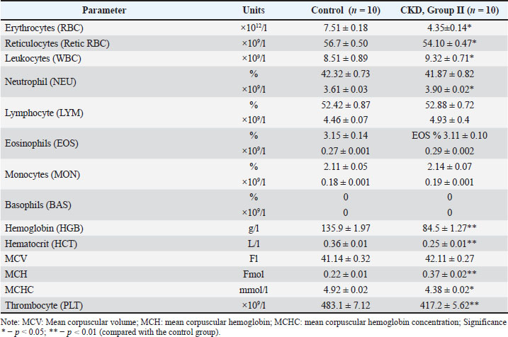
AbstractBackground: Anemia is a common systemic complication of chronic kidney disease (CKD) in cats, arising mainly from insufficient endogenous erythropoietin synthesis and impaired iron utilization. These disturbances reduce oxygen delivery, worsen clinical status, and accelerate CKD progression. Although recombinant human erythropoietin (rHuEPO) is used in selected cases, detailed evaluations of its therapeutic performance in veterinary practice remain limited. Aim: This study investigated the hematologic and biochemical response to an erythropoietin-based treatment protocol in anemic cats diagnosed with chronic kidney disease. Methods: The control group (Control I) consisted of healthy cats (n=10). Following stabilization, the cats were randomly allocated into two equal groups (n=10 per group): Group I: received standard CKD management, including a prescription renal diet, phosphate binders (lanthanum carbonate), and subcutaneous fluid therapy as required; Group II (rHuEPO-treated): received the same standard therapy supplemented with recombinant human erythropoietin (rHuEPO; Eprex®, 1000 IE/0.5 ml, Janssen-Cilag, Switzerland) administered subcutaneously at a dose of 100 IU/kg, three times weekly for 28 days. Clinical evaluations and sample collections were carried out on days 0, 7, 14, 21, and 28. Blood and urine samples were collected at baseline and at subsequent checkpoints. The following parameters were assessed: hematocrit, hemoglobin, erythrocyte count, reticulocytes, serum iron markers (iron, ferritin, Total iron binding capacity), creatinine, blood urea nitrogen, and urine protein-to-creatinine ratio (UPC). Temporal dynamics were analyzed to determine the effectiveness of the treatment. Results: Clinically, cats with CKD presented with dehydration, chronic wasting, and small, irregular kidneys on palpation, with bilateral cortical thinning confirmed by ultrasonography. Hematologically, affected cats exhibited significant normocytic, normochromic, and non-regenerative anemia Red blood cells 4.35 ± 0.14 × 10¹²/l; Hemoglobin 84.5 ± 1.27 g/l; p < 0.01) and reduced thrombocytes. Biochemically, azotemia was evident (urea 11.18 ± 0.22 mmol/l; creatinine 172.4 ± 1.11 µmol/l; p < 0.01). Despite elevated serum iron (16.78 ± 0.07 µmol/l), ferritin (137.30 ± 1.25 µg/l), and endogenous erythropoietin (0.64 ± 0.03 pmol/l) levels were significantly decreased. Urinalysis confirmed proteinuria (UPC 0.68 ± 0.008) and cylindruria (2.74 ± 0.48 casts/hpf). By day 28, rHuEPO therapy (Group II) normalized erythrogram parameters, renal markers, and urinalysis, with efficacy significantly exceeding standard care (Group I). Conclusion: Subcutaneous rHuEPO effectively corrected anemia in feline CKD, resulting in early reticulocyte activation, sustained improvement of red cell indices, normalization of iron parameters, and stabilization of renal biomarkers. Keywords: Anemia, Cats, Chronic kidney disease, Erythropoietin, Hematology. IntroductionAnemia is one of the most frequent systemic complications of chronic kidney disease (CKD) in cats and is a major contributor to the progressive decline in clinical status and quality of life in affected patients (Renard et al., 2021; Tsai et al., 2024). Its reported prevalence rises markedly as renal function deteriorates, reflecting the close physiological relationship between functional nephron mass and erythropoietic capacity (Elliott, 2023; Vaden et al., 2024). Feline renal anemia most often manifests as a normocytic, normochromic, and nonregenerative process and develops through the interaction of several pathophysiological mechanisms. The loss of erythropoietin-producing peritubular interstitial cells, dysregulation of iron metabolism, and sustained inflammatory signaling are considered to be the principal contributors, each of which may variably influence the severity and progression of anemia in individual cats (Uva et al., 2023). The physiological hypoxia-sensing system becomes progressively impaired as kidney damage advances. Under normal conditions, stabilization of hypoxia-inducible factor (HIF) orchestrates a coordinated response to reduced oxygen tension, promoting endogenous erythropoietin synthesis and regulating genes responsible for iron absorption, mobilization, and use. In CKD, however, chronic hypoxia and uremic milieu disrupt this pathway, blunting erythropoietic signaling despite a persistent need for red blood cell production (Hammond et al., 2023; Summers and Quimby, 2024). Immune responses in feline pyometra are accompanied by systemic inflammation, endotoxemia, and cytokine activation, which can negatively affect renal tissue. Such prolonged inflammatory and toxic pressure may be a factor in the development or progression of CRF (Zhelavskyi et al., 2020). Recent metabolomic investigations in feline CKD further suggest that alterations in kynurenine pathway activity, oxidative stress, and systemic inflammation may indirectly impair erythropoiesis or iron availability, thereby compounding the development of anemia (Van Mulders et al., 2025). Exogenous erythropoiesis-stimulating agents (ESAs), including recombinant human erythropoietin and darbepoetin alfa, formed the cornerstone of medical management for many years (Huang et al., 2023; Palmer et al., 2023). Despite their capacity to increase packed cell volume and improve patient vitality, ESAs are associated with well-recognized limitations: induction of anti-EPO antibodies, risk of pure red cell aplasia (PRCA), need for parenteral delivery, and strict hematologic monitoring requirements (Vaden et al., 2023). These drawbacks highlight the need for safer and more physiologic therapeutic options, particularly for cats requiring lifelong treatment. The progressive loss of nephron function leads to the accumulation of uremic toxins, electrolyte imbalances, and a wide range of systemic complications, among which anemia is particularly clinically significant (Chalhoub et al., 2011; Kongtasai et al., 2022). Feline CKD-associated anemia is multifactorial, primarily resulting from decreased renal production of erythropoietin (EPO) but also from iron deficiency, chronic inflammation, and shortened red blood cell survival (Betting et al., 2022; Elliott, 2023). Anemia is an independent predictor of disease progression and mortality in cats with CKD and contributes to lethargy, poor appetite, weight loss, and reduced quality of life (Chakrabarti et al., 2012; Renard et al., 2021). For decades, recombinant human erythropoietin (rHuEPO) has been the standard treatment for severe anemia in CKD. Early studies in dogs and cats have demonstrated that rHuEPO effectively increases hematocrit (HCT) and reduces transfusion requirements (Cowgill et al., 1998). Subsequently, the feline EPO gene was cloned, and recombinant feline EPO (rfEPO) was expressed and purified, showing biological activity in vitro and in vivo (Baldwin et al., 2003; Randolph et al., 2004). However, commercially available rfEPO has not been widely produced, and rHuEPO remains commonly used in veterinary practice despite important safety concerns. The main limitation of rHuEPO in cats is the development of anti-drug antibodies against the human protein, which can lead to PRCA and transfusion dependence (Chalhoub et al., 2011; Elliott, 2023). A recent review by Elliott (2023) emphasized that rHuEPO should therefore be reserved for late-stage CKD (International Renal Interest Society stages 3–4) when the immunological risks outweigh the benefits. Moreover, meta-analyses in human patients with CKD have confirmed that ESAs increase the risk of arterial hypertension by approximately 15%–20% compared with placebo, although they do not significantly raise the incidence of major cardiovascular events or overall mortality (Chung et al., 2023). These findings are consistent with those of veterinary studies. For instance, in a gene therapy trial using AAV-vectored EPO (SB-001) in cats with CKD, hypertension developed in 6 out of 23 animals, and encephalopathy developed in 4 of 12 cats that were already hypertensive at baseline (Vaden et al., 2024). Accumulating evidence suggests that EPO possesses pleiotropic renoprotective properties beyond hematopoiesis. Exogenous EPO exerts anti-apoptotic, anti-inflammatory, and antioxidant effects, which may slow the progression of renal fibrosis (Bartnicki et al., 2013; Nishida et al., 2024). In a mouse model of unilateral ureteral obstruction, EPO treatment reduced interstitial fibrosis and shifted macrophage polarization from a pro-inflammatory M1 to an anti-inflammatory M2 phenotype (Nishida et al., 2024). Clinical studies in humans with CKD have demonstrated that 6 months of rHuEPO therapy significantly decreased urinary levels of L-FABP (a marker of tubular injury) and 8-OHdG (a marker of oxidative stress), indicating reduced kidney damage (Nakamura et al., 2006; Fujiwara et al., 2011). Moreover, EPO modulates complement activation in patients with CKD, potentially contributing to its anti-inflammatory effects (Athanasiadou et al., 2024). In recent years, new therapeutic options for CKD-associated anemia have emerged. These agents have been evaluated in healthy cats and in cats with CKD and have shown efficacy in raising HCT levels with a potentially lower risk of hypertension and antibody formation (Boegel et al., 2024; Charles et al., 2024). Meta-analyses in humans have confirmed that vadadustat is non-inferior to darbepoetin alfa for hemoglobin (HGB) correction, with a similar safety profile (Huang et al., 2023). Nevertheless, the long-term safety and renoprotective effects of HIF-PH inhibitors in cats remain to be fully established. Given the continuing clinical need for effective and safe anemia management in feline CKD and the persistent gaps in knowledge regarding the comparative benefits and risks of rHuEPO versus newer agents, this study focuses on a detailed evaluation of rHuEPO therapy in cats with CKD. Specifically, we analyzed its efficacy in correcting anemia, its safety profile (including hypertension and immunogenicity), and emerging evidence for its renoprotective effects. The goal of this study is to provide a balanced, evidence-based perspective that will help clinicians make informed decisions and guide future research into optimal anemia management in feline patients with CKD. These diagnostic advances, together with renewed interest in endogenous erythropoietic mechanisms, underscore the need to reassess current therapeutic strategies for renal anemia and evaluate novel pharmacologic approaches within the context of contemporary feline nephrology. This study aimed to comprehensively assess the therapeutic efficacy of an erythropoietin-based treatment protocol in feline patients with chronic kidney disease and anemia. Materials and MethodsClinical presentation and selection of casesThe study population consisted of client-owned cats who presented to the Doctor SUPERVET Veterinary Clinic (Kamianets-Podilskyi, Ukraine) between January 2023 and March 2025 with clinical signs indicative of CKD. According to the owners, most animals exhibited a gradual progression of symptoms over several months prior to clinical evaluation. Predominant clinical complaints included polyuria and polydipsia (85%), progressive weight loss (70%), and dull, unkempt hair coat (60%). Approximately half of the cats (55%) demonstrated hyporexia or selective feeding, while 65% of the cats exhibited lethargy and decreased activity levels. Gastrointestinal disturbances, such as intermittent vomiting and mild diarrhea, occurred in approximately 30% of the patients, typically in those with more advanced uremic changes. The initial diagnostic evaluation included complete blood count (CBC), serum biochemical profiling, and urinalysis to establish baseline clinical and metabolic status (Nassar et al., 2025). All cats were clinically stabilized using intravenous or subcutaneous fluid therapy to correct dehydration and restore hemodynamic balance before enrollment. This ensured the accurate assessment of renal function and erythropoietic status at baseline. Importantly, none of the animals had received erythropoiesis-stimulating agents (ESAs), iron supplementation, or related medications within the preceding 6 months, eliminating potential confounding effects on hematologic parameters. The combination of clinical, biochemical, and imaging findings, including anemia, azotemia, dehydration, and reduced endogenous erythropoietin synthesis, provided the rationale for selecting this cohort as a clinically representative model for evaluating rHuEPO therapy under controlled veterinary conditions (Segev et al., 2024). Throughout the treatment and observation period, owners maintained structured daily records documenting water intake, appetite, urination frequency, and behavioral changes, which complemented laboratory monitoring to provide a comprehensive evaluation of clinical response. All enrolled cats tolerated blood sampling and erythropoietin administration well, with no significant adverse effects or treatment-related complications. Experimental animals and their designTwenty adult domestic cats (Felis catus) aged 6–10 years and weighing 3.4–5.2 kg were enrolled in the study. All animals were privately owned and diagnosed with stage III CKD according to the International Renal Interest Society (IRIS, 2023) classification criteria. Before study initiation, each cat underwent rehydration and stabilization to correct uremic manifestations and electrolyte imbalances. The control group (Control I) consisted of healthy cats (n=10). Following stabilization, the cats were randomly allocated into two equal groups (n=10 per group): Group I: received standard CKD management, including a prescription renal diet, phosphate binders (lanthanum carbonate), and subcutaneous fluid therapy as required; Group II (rHuEPO-treated): received the same standard therapy supplemented with recombinant human erythropoietin (rHuEPO; Eprex®, 1000 IE/0.5 ml, Janssen-Cilag, Switzerland) administered subcutaneously at a dose of 100 IU/kg, three times weekly for 28 days. Clinical evaluations and sample collections were performed on days 0, 7, 14, 21, and 28 (De Lombaert et al., 2024; Vaden et al., 2024). The control group consisted of healthy cats (n=10). Infectious diseases, neoplastic disorders, autoimmune pathologies, non-renal anemia, and other hematological disorders were considered in determining and differentiating the diagnosis. Animals were not selected for the experimental group. Sample collection and handlingAll biological samples were obtained between 08:00 and 10:00 am following an 8–10 hour overnight fast. Venous blood was collected aseptically from the cephalic vein using 22G needles into K₂EDTA tubes (for hematologic assessment) and plain tubes (for biochemical and erythropoietin analysis). Urine samples were collected using ultrasound-guided cystocentesis under aseptic conditions. The samples were processed within 1 hour of collection. Serum was separated by centrifugation at 1,500 × g for 10 minutes at +4°C (Centric 100, Tehtnica, Slovenia) and subsequently stored at −80°C (UFV 850, Arctiko, Denmark) to preserve analyte stability (Boegel et al., 2024). Hematological evaluationComplete blood counts were obtained using a VetScan HM5 analyzer (Abaxis, USA). The evaluated indices included Red blood cells (RBC) count, Hemoglobin (HGB), hematocrit (HCT), mean corpuscular volume (MCV), mean corpuscular hemoglobin concentration (MCHC), reticulocyte count, and reticulocyte of red blood cells (Retic RBC) (Betting et al., 2022). Additional leukocyte and platelet indices, such as white blood cell (WBC) count, neutrophil-to-lymphocyte ratio, platelet count, and platelet-to-lymphocyte ratio, were also determined. Peripheral blood smears were prepared immediately after sampling, air-dried, and stained using Wright–Giemsa stain (Sigma-Aldrich, USA) on an Aerospray Hematology Slide Stainer (Wescor, USA). The morphological evaluation of erythrocytes and leukocytes was conducted with a Zeiss Axio Laboratory. An A1 microscope (Carl Zeiss, Germany) under oil immersion at 100× magnification to identify potential cytological abnormalities associated with uremia or anemia (Charles et al., 2024). Biochemical analysesSerum biochemical parameters were assessed using a VetScan VS2 analyzer (Zoetis, USA) equipped with dry chemistry rotors (VetScan Comprehensive Diagnostic Profile). Serum iron: Concentration was determined using the ferrozine method on the Cobas c 501 clinical chemistry analyzer (Roche Diagnostics, Switzerland). Ferritin: Levels were measured via an immunoturbidimetric assay performed on an Architect i2000SR immunoassay analyzer (Abbott Laboratories, USA). Total iron binding capacity (TIBC): This parameter was calculated based on standard diagnostic protocols using measured serum iron and unsaturated iron-binding capacity values obtained from the Cobas c 501 analyzer (Roche Diagnostics, Switzerland; Girelli et al., 2025). Erythropoietin assaySerum erythropoietin (EPO) concentrations were measured using a feline-validated enzyme-linked immunosorbent assay (ELISA) kit (Cat. No. SEA005Ca (Cluster-Clone Corp., USA). Standards and diluted serum samples were incubated in precoated microplate wells for 2 hours at 37°C, followed by sequential incubation with a biotinylated detection antibody and streptavidin–HRP conjugate (Vaden et al., 2023). The chromogenic reaction was developed using a TMB substrate, halted with sulfuric acid, and the optical density was measured at 450/620 nm using an ELx808 microplate reader (BioTek, Agilent Technologies, USA). The assay exhibited a sensitivity of 2.3 mIU/ml, with intra- and inter-assay coefficients of variation below 6% and 8%, respectively. To ensure precision and reproducibility, all samples were analyzed in duplicate (R&D Systems, 2023; Van Mulders et al., 2025). UrinalysisUrine evaluation was conducted using Combur-Test® reagent strips (Roche Diagnostics, Switzerland) and analyzed using a Crisis 1100 Urine Analyzer (Roche Diagnostics, Switzerland). Urine specific gravity was determined using a MISCO PA202X refractometer (USA). For sediment analysis, urine samples were centrifuged at 400 × g for 5 minutes, and the pellet was resuspended in 0.5 ml of supernatant for microscopic examination under 100 × and 400 × magnification using a Zeiss Axio Laboratory. A1 microscope (Germany) Proteinuria was quantified by calculating the urine protein-to-creatinine ratio (UPC), where protein concentration was measured via the pyrogallol red method and Jaffe reaction (Chew et al., 2023). Statistical analysisAll statistical computations were performed using Statistica® 12.6 (TIBCO Software Inc., USA) and GraphPad Prism 10.0 (GraphPad Software, USA). Results were expressed as mean ± standart error of means (SEM), and statistical significance was defined as p < 0.05. Effect sizes were calculated to assess the magnitude of observed differences, ensuring a robust interpretation of treatment outcomes. Quality control and ethical considerationsAll experimental and clinical procedures were conducted by licensed veterinary clinicians who strictly adhered to aseptic techniques to minimize procedural complications and animal discomfort. Analytical reliability was ensured through daily quality control runs using standardized calibration materials and by applying the Westgard multirule criteria for the internal validation of hematological and biochemical assays. Adverse reactions were monitored, and routine physical examinations were performed. The study protocol complied with the ARRIVE 2.0 guidelines to promote methodological transparency, reproducibility, and ethical use of animals in research. Pain management protocols were implemented as necessary, and all animals were monitored post-intervention until full recovery to ensure that welfare standards were met. Ethical approvalAll experimental procedures were conducted in accordance with the principles of the European Convention for the Protection of Vertebrate Animals Used for Experimental and Other Scientific Purposes (ETS No. 123, Council of Europe, 1986). The Institutional Animal Care and Use Committee of the Faculty of Veterinary Medicine, Higher Educational Institution Podillia State University reviewed and approved the study protocol (Protocol No. 4/2021). Research and clinical interventions were conducted at the Doctor SUPERVET Veterinary Clinic (Kamianets-Podilskyi, Ukraine). ResultsClinical manifestationsOn physical examination, most cats exhibited mild to moderate dehydration, characterized by reduced skin elasticity and tacky mucous membranes. The Body Condition Score (BCS) ranged between 3 and 5 of 9, indicating varying degrees of chronic wasting. Oral lesions, including uremic halitosis, mucosal ulceration, and stomatitis, were recorded in approximately one-quarter of the animals. Cardiac auscultation revealed soft systolic murmurs in 3 cats, later attributed to anemia-related hemodynamic alterations. Renal palpation commonly revealed small, firm, irregularly contoured kidneys with decreased mobility, consistent with chronic INA (International Renal Interest Society, IRIS, 2023). Ultrasonographic assessment (SonoSite 180 plus, USA) revealed bilateral cortical thinning, increased echogenicity, and loss of corticomedullary distinction in all cats, which are characteristic of chronic nephropathy. No obstructive or neoplastic lesions were identified. Hematological parametersThe hematological analysis revealed a pronounced normocytic, normochromic, and non-regenerative anemia in cats diagnosed with CKD compared with the healthy control group. Erythrocyte counts (Table 1) were significantly decreased in the CKD group (mean 4.35 ± 0.14 ×10¹²/l) compared to controls (7.51 ± 0.18 ×10¹²/l; p < 0.05). This was accompanied by a marked reduction in Hb concentration (84.5 ± 1.27 g/l in patients with CKD vs. 135.9 ± 1.97 g/l in controls; p < 0.01) and a lower HCT value (0.25 ± 0.01 L/L in patients with CKD vs. 0.36 ± 0.01 L/L in controls; p < 0.01). Regarding regenerative indicators, the absolute reticulocyte count in cats with CKD (54.10 ± 0.47 ×10⁹/l) did not differ significantly from that in the control group (56.70 ± 0.50 ×10⁹/l; p > 0.05). Table 1. Hematological parameters of cats with chronic kidney disease (CKD) and anemia [mean ± SEM].
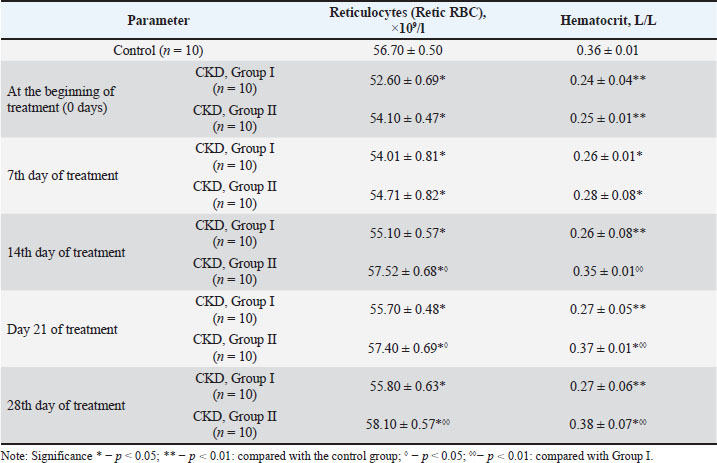
Leukocyte analysis revealed no significant differences in the overall leukogram between the groups. However, a slight but statistically significant increase in the absolute neutrophil count was observed in the CKD group (mean 3.90 ± 0.02×10⁹/l) compared with that in the control group (p < 0.01). Conversely, the platelet counts were lower in cats with renal pathology (417.2 ± 5.62 × 10⁹/l) relative to the control group (483.1 ± 7.12 × 10⁹/l; p < 0.01). Following the initiation of therapy (group II, receiving rHuEPO), a progressive restoration of red blood cell parameters was observed, with values approaching the reference range by the end of the observation period (day 28). The erythrocyte count increased steadily from baseline. Values rose from 4.35 ± 0.05 × 10¹²/l pre-treatment to 4.64 ± 0.04 × 10¹²/l by Day 7. A marked improvement was noted by day 14 (5.49 ± 0.05 × 10¹²/l), with further increases on day 21 (6.30 ± 0.03 × 10¹²/l). By day 28, the RBC count had reached 6.73 ± 0.03 × 10¹²/l, approaching the levels of the healthy control group. In Group І (standard care), the reticulocyte count showed a progressive increase over the treatment period. The baseline count was 52.60 ± 0.69 × 10⁹/l, which gradually increased to 54.01 ± 0.81 × 10⁹/l by day 7, 55.10 ± 0.57 × 10⁹/l by day 14, 55.70 ± 0.48 × 10⁹/l by day 21, and reached 55.80 ± 0.63 × 10⁹/l by day 28. However, this increase was less pronounced than that observed in Group 2, which received adjunctive recombinant human erythropoietin (rhEPO) therapy (Table 3). Table 3. Dynamics of reticulocyte count and hematocrit during rHuEPO therapy in cats with CKD-associated anemia (mean ± SEM).
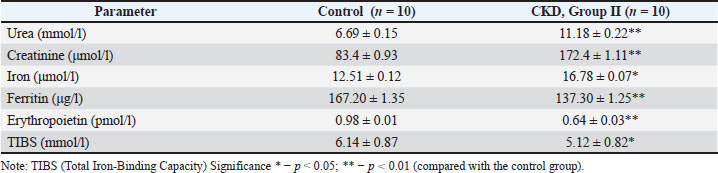
The reticulocyte (Group ІІ) count exhibited a moderately upward trend throughout the treatment period. Values increased progressively from a baseline of 54.10 ± 0.47 × 10⁹/l to 54.71 ± 0.82 × 10⁹/l (Day 7) and 57.52 ± 0.68 × 10⁹/l (Day 14). The count stabilized thereafter, reaching 57.40 ± 0.69 × 10⁹/l on Day 21 and peaking at 58.10 ± 0.57 × 10⁹/l on Day 28 (Table 3). Hemoglobin concentrations showed a robust recovery. Levels increased incrementally from a baseline of 84.50 ± 1.27 g/l to 86.40 ± 1.37 g/l (day 7) and 90.60 ± 1.92 g/l (day 14). A substantial elevation was recorded on day 21 (118.60 ± 1.37 g/l), peaking at 138.70 ± 1.20 g/l by day 28, consistent with the control group’s physiological norms. The HCT in Group І exhibited a gradual upward trend. Similar to the reticulocyte response, this increase was less pronounced than that observed in Group ІІ (Table 3). The HCT (Group ІI) values followed a similar normalization trajectory. The baseline value (0.25 ± 0.01 L/L) increased incrementally to 0.28 ± 0.08 L/L (day 7), 0.35 ± 0.01 L/L (Day 14), 0.37 ± 0.01 L/L (day 21), and finally to 0.38 ± 0.07 L/L by Day 28, aligning with the control values (0.36 ± 0.01 L/L). Biochemical parametersThe baseline biochemical analysis revealed significant alterations in renal function markers and iron metabolism parameters in cats with CKD compared with those in the healthy control group (Table 2). Table 2. Biochemical parameters in cats with CKD and anemia (mean ± SEM).
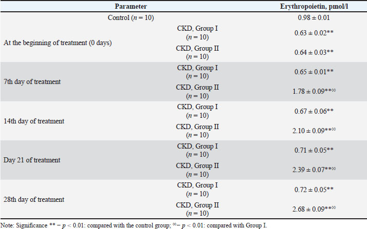
Serum urea concentration was significantly elevated in the CKD group (11.18 ± 0.22 mmol/l) compared with that in the control group (6.69 ± 0.15 mmol/l; p < 0.01). Serum creatinine levels were markedly higher in the affected animals (172.4 ± 1.11 µmol/l) than in the healthy controls (83.4 ± 0.93 µmol/l; p < 0.01). The serum iron concentration was unexpectedly higher in cats with CKD (16.78 ± 0.07 µmol/l) than in the control group (12.51 ± 0.12 µmol/l; p < 0.05). Conversely, ferritin levels were significantly reduced in patients with CKD (137.30 ± 1.25 µg/l) versus healthy animals (167.20 ± 1.35 µg/l; p < 0.01). The baseline serum EPO concentration in cats with CKD was significantly reduced (0.64 ± 0.03 pmol/l) compared with the control values (0.98 ± 0.01 pmol/l; p < 0.01). Progressive normalization of renal and iron metabolism parameters was observed throughout the 28-day treatment period following the initiation of erythropoietin therapy (Group II). A consistent and marked reduction in serum urea concentration was observed during the therapeutic period. Values decreased progressively from the baseline elevation of 11.18 ± 0.22 mmol/l to 10.26 ± 0.06 mmol/l by day 7, 9.52 ± 0.05 mmol/l by day 14, and 8.12 ± 0.03 mmol/l by day 21. Urea levels had effectively normalized by day 28 of treatment, reaching 6.27 ± 0.03 mmol/l, which is comparable to control values (p > 0.05). The dynamics of serum iron concentration during therapy (group II, receiving rHuEPO) followed a biphasic pattern. Following the elevated baseline level (16.78 ± 0.07 µmol/l), a transient decrease to 15.36 ± 0.06 µmol/l was observed by day 7. A modest, statistically insignificant increase to 16.28 ± 0.05 µmol/l occurred on day 14, followed by a consistent decline to 14.38 ± 0.06 µmol/l on day 21. By the end of the observation period (day 28), serum iron concentration had decreased to 12.51 ± 0.05 µmol/l, aligning precisely with the values of the control group. In contrast to serum iron, ferritin levels showed a progressive and sustained increase throughout the treatment period. From the reduced baseline of 137.30 ± 1.25 µg/l, the concentrations increased to 152.80 ± 0.58 µg/l (day 7), 167.50 ± 0.50 µg/l (day 14), and 175.90 ± 0.51 µg/l (day 21). By day 28, ferritin levels had reached 187.20 ± 0.42 µg/l. Serum EPO levels in Group І demonstrated a modest but steady increase throughout the study. The concentration rose from a baseline of 0.63 ± 0.02 pmol/l on day 0 to 0.65 ± 0.01 pmol/l on day 7, 0.67 ± 0.06 pmol/l on day 14, 0.71 ± 0.05 pmol/l on day 21, and finally to 0.72 ± 0.05 pmol/l on day 28. This kinetic profile was significantly different from that observed in Group II rats. Administration of exogenous erythropoietin resulted in a rapid and sustained elevation of serum EPO concentrations (Table 4). Levels increased from the deficient baseline of 0.64 ± 0.03 pmol/l to 1.78 ± 0.09 pmol/l by day 7, representing an approximately 2.8-fold increase (p < 0.01). Further progressive elevations were documented on days 14 (2.10 ± 0.09 pmol/l), 21 (2.39 ± 0.07 pmol/l), and 28 (2.68 ± 0.09 pmol/l). Table 4. Dynamics of serum erythropoietin concentration in cats with chronic kidney disease-associated anemia during rHuEPO therapy mean ± SEM).
Urinalysis parametersUrinalysis revealed significant proteinuria and increased sediment activity in cats with CKD compared with the healthy control group (Table 5). Table 5. Urinalysis parameters in cats with CKD and anemia (mean ± SEM).
Urinary Protein-to-Creatinine Ratio: The baseline UPC ratio was markedly elevated in patients with CKD (0.68 ± 0.008) relative to that in control animals (0.18 ± 0.009; p < 0.01). Urinary Casts: Microscopic examination of urine sediment revealed a substantial increase in cast excretion in cats with CKD (2.74 ± 0.48 casts/hpf) compared with that in healthy controls (0.40 ± 0.03 casts/hpf; p < 0.01). Therapeutic intervention resulted in progressive improvement in both proteinuria and urinary sediment abnormalities throughout the 28-day observation period (Group II, receiving rHuEPO). From the elevated baseline of 0.681 ± 0.003, values decreased progressively to 0.625 ± 0.004 by day 7; 0.527 ± 0.003 by day 14, and 0.273 ± 0.003 by day 21 (p < 0.01 for all time points vs. baseline). By day 28, the UPC ratio had normalized to 0.177 ± 0.003, which was comparable to the control values (p > 0.05). Concurrently, urinary cast counts exhibited a progressive decline throughout the treatment period. From the elevated baseline of 2.70 ± 0.15 casts/hpf, the counts decreased sequentially to 2.40 ± 0.16 casts/HPF (day 7), 2.20 ± 0.13 casts/hpf (day 14), and 1.70 ± 0.21 casts/HPF (day 21). Complete resolution of cylindruria was observed by day 28, with cast counts (0.40 ± 0.02 casts/hpf indistinguishable from those of the healthy control group (p > 0.05). The normalization of urological parameters was more dynamic in Group II, and their values reached the reference ranges more rapidly than those in Group I. DiscussionThe management of chronic kidney disease-associated anemia in affected cats represents a complex clinical challenge requiring integrated therapeutic strategies targeting multiple pathophysiological pathways (Nealon et al., 2024). Kidney disease in dogs induces systemic inflammation and hormonal imbalance, which directly disrupts the hypothalamic–pituitary axis. These metabolic shifts lead to reduced fertility, anovulation, and spermatogenesis pathologies, even in the early, asymptomatic stages of renal dysfunction (Zhelavskyi et al., 2025). Our findings substantiate that systematic intervention can simultaneously influence several key mechanisms, including erythropoiesis, iron metabolism, inflammatory response, and renal tissue functional status (Li et al., 2024). The observed progressive normalization of hematological, biochemical, and urinary parameters aligns with contemporary evidence supporting the efficacy of multimodal treatment approaches (Vaden et al., 2024; Elliott et al., 2025). The sequential improvement in regenerative blood parameters demonstrated a characteristic "cascade" pattern typical of erythropoietic recovery: initial reticulocyte elevation preceded subsequent increases in HGB and HCT. This temporal profile indicates early activation of the bone marrow, followed by maturation of erythroid cells and their subsequent release into the circulation (Elliott, 2023). The elevation in reticulocyte counts during the initial treatment weeks serves as a valuable indicator of adequate erythropoietic stimulation and enables early identification of patients with suboptimal response (De Lombaert et al., 2024). The development of rfEPO was a logical step to overcome the immunogenicity associated with the use of human analogs. Baldwin et al. (2003) demonstrated that the resulting protein indeed exhibited the expected biological activity in vitro. However, despite the initial success in restoring erythropoiesis in most cats, the pivotal clinical study by Randolph et al. (2004) revealed a critical limitation: a significant proportion of animals that initially responded to therapy subsequently developed anemia due to PRCA, confirming the impossibility of completely avoiding this serious complication even with a species-specific preparation (Randolph et al., 2004). Currently, rfEPO can only be used for research. The progressive increase in erythropoietin concentration throughout the treatment period confirms the effectiveness of replacement therapy or endogenous hormone synthesis activation, which correlates with the restoration of hematological parameters (IRIS, 2023). Similar trends have been reported in other studies, indicating reduced uremic suppression of bone marrow under conditions of improved tissue oxygenation (Summers and Quimby, 2024). The temporal association between erythropoietin elevation and hematological improvement provides compelling evidence for the central role of erythropoietin in resolving renal anemia. The biochemical alterations observed during treatment reflected iron redistribution and normalization of its use for erythropoiesis. Elevated serum iron concentrations concurrent with reduced ferritin levels are associated with dysfunctional iron usage in CKD (Charles et al., 2024; Chen et al., 2024). The gradual decrease in iron levels alongside increasing ferritin suggests optimization of storage mobilization and efficient use in erythropoiesis (Boegel et al., 2024). The emergence of Hypoxia-Inducible Factor Prolyl Hydroxylase (HIF-PH) inhibitors in veterinary medicine offers new therapeutic possibilities for anemia correction, as these agents simultaneously stimulate endogenous EPO production and improve iron bioavailability (Charles et al., 2024). Their ability to reduce immunogenicity and provide more physiological restoration of erythropoiesis already defines their promising application in long-term treatment regimens (Boegel et al., 2024; De Lombaert et al., 2024). The dual mechanism of action targeting erythropoietin synthesis and iron metabolism represents a significant advancement over traditional ESA therapies. Recent advances in metabolomic profiling have revealed profound disturbances in the metabolism of tryptophan in feline CKD patients. Our findings align with emerging evidence demonstrating significant alterations in kynurenine pathway metabolites, particularly 3-hydroxykynurenine, which shows promise as an early diagnostic biomarker (Vanden Broecke et al., 2025). The application of machine learning algorithms incorporating these metabolic signatures has demonstrated remarkable accuracy in early CKD detection, potentially enabling intervention before irreversible renal damage occurs. The notable decrease in proteinuria ratio observed in our study is clinically significant, as persistent proteinuria is a well-established negative prognostic marker in feline chronic kidney disease. According to the IRIS guidelines, a proteinuria ratio consistently above 0.4 is classified as proteinuria and is linked to a fourfold higher risk of mortality in cats with CKD (Syme et al., 2009). The therapeutic response we observed aligns with clinical trials of Renin-Angiotensin-Aldosterone System inhibitors, with Han et al. (2018) reporting a significant reduction in proteinuria with telmisartan. Similarly, benazepril therapy was shown to significantly decrease proteinuria across all patient subgroups, with the strongest effects seen in cats with baseline proteinuria values greater than 1.0 (King et al., 2006). The concurrent improvement in azotemia and proteinuria supports the idea that treatments targeting both anemia and proteinuria may work together to slow CKD progression because anemia and proteinuria are independent markers of more advanced renal disease (Chakrabarti et al., 2012; Chakrabarti et al., 2013). Regarding the erythropoietin dynamics documented in our study, it is important to clarify that the measured serum EPO concentrations reflect total immunoreactive erythropoietin detected with a commercially available enzyme-linked immunosorbent assay kit (Quantikine IVD Human EPO Immunoassay, R&D Systems). This assay uses antibodies raised against human EPO that cross-react significantly with rHuEPO but cannot distinguish between endogenous feline EPO and externally administered human EPO (R&D Systems, 2023). Therefore, the progressive rise in serum EPO levels during treatment mainly indicates the buildup of exogenous rHuEPO detectable at administration time, rather than an increase in endogenous EPO production. This aligns with the known pharmacology of rHuEPO, which peaks in serum within 24 hours of injection and remains detectable for 3–4 days afterward (Cowgill et al., 1988). Our results show effective systemic absorption and sustained presence of exogenous EPO throughout the 28-day treatment, matching the observed hematological recovery. Future studies using species-specific enzyme-linked immunosorbent assays that differentiate endogenous and exogenous EPO will help gain further understanding of the remaining synthetic capacity of feline kidneys during CKD progression (Chalhoub et al., 2011). Despite the high efficacy of rhEPO in correcting anemia, its safety profile—particularly in the context of CKD—remains a key concern (Athanasiadou et al., 2024). A recent network meta-analysis (Chung et al., 2023) confirmed that the use of erythropoietin and its analogues is associated with an increased risk of arterial hypertension (odds ratio increase of approximately 15%–20% compared with placebo), but it did not detect a significant increase in the frequency of major cardiovascular events or overall mortality. These findings are mirrored in veterinary practice. In a study (De Lombaert et al., 2024) using AAV-vectored gene therapy (SB-001) in cats with CKD, hypertension developed in 6 out of 23 animals, and among the 12 cats that were already hypertensive at baseline, 4 developed encephalopathy. Furthermore, a review (Elliott, 2023) highlights the risk of antibody formation against the foreign human protein, which can lead to PRCA and transfusion dependence in cats. Consequently, the use of rhEPO in veterinary medicine should be reserved for late-stage CKD. Thus, patient stratification based on baseline blood pressure and careful HCT monitoring is critical to minimize risks. Beyond its hematological action, accumulating evidence points to erythropoietin’s potential renoprotective properties. A systematic review (Fernandez Jimenez and Fernandez Romero, 2023) summarized data from 21 experimental studies, showing that exogenous rhEPO exerts its protective effects through anti-apoptotic, proliferative, antioxidant, and anti-inflammatory mechanisms, which may slow the progression of CKD. In a mouse model of unilateral ureteral obstruction, rhEPO treatment significantly reduced interstitial fibrosis and myofibroblast accumulation by shifting macrophage polarization from a pro-inflammatory M1 phenotype to an anti-inflammatory M2 phenotype, without affecting HCT levels (Nishida et al., 2024). Bartnicki and co-authors (Bartnicki et al., 2013) confirmed that the effect of rhEPO on renal interstitial fibrosis is mediated by reduced ischemia, decreased oxidative stress, and an anti-apoptotic effect on the tubular epithelium. In a clinical study of patients with CKD, 6 months of rhEPO therapy resulted in a significant decrease in the levels of L-FABP (a marker of tubular injury) and 8-OHdG (a marker of oxidative stress), indicating reduced kidney damage and oxidative stress along with improvement in anemia. These findings open perspectives for further research into the use of rhEPO not only as an anemia corrector but also as an agent that may slow renal fibrosis and reduce systemic oxidative burden. The role of the kynurenine pathway in CKD pathophysiology extends beyond mere biomarker utility. The substantial accumulation of kynurenine and its metabolites in renal insufficiency has been documented experimentally, with altered peripheral distribution patterns suggesting systemic metabolic disruption (Tsai et al., 2024; Van Mulders et al., 2025). These findings are further corroborated by clinical investigations in feline patients, where rapidly progressive CKD is associated with distinct metabolic alterations involving tryptophan-kynurenine pathway intermediates (Van Mulders et al., 2025). The implications of these metabolic disturbances are multifaceted. The activation of the kynurenine pathway contributes to the pro-inflammatory milieu characteristic of advanced CKD, potentially worsening both renal parenchymal damage and anemia through multiple mechanisms. These include enhanced oxidative stress generation, immune system modulation, and direct nephrotoxic effects. Furthermore, the gut-kidney axis appears to be significantly involved, as the intestinal microbiota substantially influence tryptophan metabolism, suggesting potential therapeutic avenues targeting microbial composition and function (Tsai et al., 2024). Anemia is closely associated with chronic inflammation in cats with CKD, supported by evidence of elevated hemogram-derived inflammatory indices and specific mediators (Uva et al., 2023). The notable improvement in erythropoiesis observed in our study, despite potentially persistent inflammation, underscores the need for simultaneous intervention targeting multiple pathogenic mechanisms. This aligns with the concept of multimodal therapy incorporating dysbiosis correction, inflammatory response reduction, and gut–kidney axis function support (Summers and Quimby, 2024). Growing evidence from human nephrology and, more recently, veterinary medicine has shifted attention toward agents that modulate the HIF pathway upstream of erythropoiesis. HIF prolyl hydroxylase inhibitors (HIF-PHIs enhance the natural hypoxic response by preventing degradation of HIF-α subunits, thereby stimulating endogenous erythropoietin production and downregulating hepcidin, the central hormone that limits iron availability (Charles et al., 2024; Vaden et al., 2024). Molidustat, the first HIF-PHI specifically authorized for veterinary use, represents a significant development in this context. Its mechanism of action closely resembles physiologic erythropoietic regulation and offers the additional advantage of oral administration, which may help overcome adherence challenges commonly encountered in the long-term management of feline CKD. The most recent guidelines of the International Renal Interest Society (IRIS, 2023) acknowledge this drug class’s therapeutic potential, especially for cats in CKD stages 2–4 with documented anemia and inadequate endogenous erythropoietin concentrations. The increasing availability of advanced diagnostic tools, including biomarkers of tubular injury, metabolomic profiling, and machine learning, has enhanced our understanding of the broader systemic consequences of renal disease in cats (Kongtasai et al., 2022; Kornya et al., 2025; Vanden Broecke et al., 2025). The management of comorbid conditions presents additional challenges in patients with CKD. Geddes and Aguiar (2022) highlighted that the simultaneous presence of hyperthyroidism and CKD creates a complex clinical scenario requiring careful therapeutic balancing. Thyroid dysfunction can significantly influence erythropoietic response, iron metabolism, and renal hemodynamics, necessitating integrated treatment approaches that simultaneously address both conditions. This complexity is further compounded by the emerging understanding of adrenal gland involvement in CKD progression, with recent evidence suggesting dysregulation of the hypothalamic–pituitary–adrenal axis in feline renal disease (Marques et al., 2025). The reduction in urea levels and normalization of proteinuria detected in our study indicate improved metabolic status and reduced uremic burden. The gradual decrease in proteinuria and urinary cast counts suggests restoration of nephron structure and function (Uva et al., 2023; Charles et al., 2024). Improved filtration capacity may be associated with enhanced oxygen delivery to renal tissue and reduced hypoxic and inflammatory damage following anemia correction (De Lombaert et al., 2024). Recent advances in renal biomarker research have identified several promising candidates for the early detection and monitoring of CKD progression. Urinary kidney injury molecule-1 has demonstrated particular utility in feline patients, with Kornya et al. (2025) reporting significant elevations and correlations with disease severity in cats with established CKD. This biomarker appears to more sensitively reflect tubular damage than conventional parameters, potentially enabling earlier intervention and more precise monitoring of treatment response. Management of hypertension and proteinuria remains a crucial aspect of comprehensive CKD care. De Freitas et al. (2025) emphasized the importance of regular BP monitoring and proteinuria assessment in older feline populations, demonstrating significant associations between these parameters and renal function decline. Their findings support the integration of cardiovascular risk assessment into standard CKD management protocols, particularly given the high prevalence of hypertension in this patient population. Minor deviations in leukogram and platelet counts in cats with CKD align with the absence of systemic inflammatory leukocytosis in this form of anemia (Uva et al., 2023). Uremic dysfunction may explain mild thrombocytopenia at treatment initiation, while partial recovery during therapy demonstrates hemostatic stabilization (De Lombaert et al., 2024). This confirms that erythropoietin and related agents do not substantially stimulate thrombopoiesis; however, improved systemic status may indirectly contribute to platelet function normalization. The stability of leukocyte parameters throughout the treatment period suggests that the therapeutic interventions did not induce significant inflammatory or stress responses, supporting the safety profile of the administered treatments. The results demonstrated a broad therapeutic window for erythropoiesis stimulators and HIF agonists, confirming their key role in correcting CKD-associated anemia (Vaden et al., 2024). ConclusionThe findings of this study demonstrate that subcutaneous administration of recombinant human erythropoietin is an effective approach to correcting anemia in cats with chronic kidney disease. The treatment produced a clear and consistent hematological response: reticulocyte numbers were the first to rise, indicating early activation of erythropoiesis, followed by progressive increases in Hb concentration and HCT values. Parallel improvements in biochemical indicators further support the restoration of endogenous red blood cell production and more efficient iron utilization within the organism. Collectively, these results confirm the therapeutic value of rHuEPO in stabilizing the hematologic status of feline patients with CKD under controlled veterinary conditions. AcknowledgmentsThe authors would like to express their sincere gratitude to the faculty and staff of the Higher educational institution Podillia State University, Kamianets-Podilskyi, Ukraine, the Vinnytsia National Agrarian University, Vinnytsia, Ukraine, and National University of Life and Environmental Sciences of Ukraine, Kyiv, Ukraine for their invaluable support and assistance throughout this research. Their contributions were essential to the successful completion of this study. Conflict of interestThe authors declare no conflicts of interest. FundingThis research was not supported by any funding organization. Authors' contributionsMZ: conceived and designed the study, coordinated data collection, performed primary data acquisition, and performed the initial interpretation of hematological, biochemical, and urinalysis results. MZ also drafted the first full version of the manuscript and substantially contributed to the refinement of the Results and Discussion sections. MZ and SK: Supervised the overall research workflow, ensured methodological accuracy, and oversaw laboratory diagnostic procedures. MM and LK verified the hematological and biochemical datasets, contributed to data interpretation, and actively participated in the critical revision and academic editing of the manuscript. MD and RK: performed the clinical examination of feline patients, ensured proper case selection and clinical monitoring throughout the study, and contributed to clinical dynamics interpretation. They assisted in preparing the manuscript text, improving the clarity of research descriptions, and refining the results and discussion sections. All authors critically reviewed the manuscript, provided substantial intellectual input during multiple revision rounds, and approved the final version for submission. Data availabilityAll data supporting this study’s findings are available within the manuscript. ReferencesAthanasiadou, V., Ampelakiotou, K., Grigoriou, E., Psarra, K., Tsirogianni, A., Valsami, S., Pittaras, T., Grapsa, E. and Detsika, M.G. 2024. Erythropoietin effect on complement activation in chronic kidney disease. Biomedicines 12(8), 1746; doi:10.3390/biomedicines12081746 Baldwin, S.L., Powell, T.D., Wonderling, R.S., Keiser, K.C.L., Morales, T., Hunter, S., Mcdermott, M., Radecki, S.V. and Milhausen, M.J. 2003. Transient and stable transfection of Chinese hamster ovary cells with the recombinant feline erythropoietin gene and expression, purification, and biological activity of feline erythropoietin protein. Am. J. Vet. Res. 64(12), 1465–1471; doi:10.2460/ajvr.2003.64.1465 Bartnicki, P., Kowalczyk, M. and Rysz, J. 2013. The influence of the pleiotropic action of erythropoietin and its derivatives on nephroprotection. Med. Sci. Monitor. Int. Med. J. Exp. Clin. Res. 19, 599–605; doi:10.12659/MSM.889023 Betting, A., Schweighauser, A. and Francey, T. 2022. Diagnostic value of reticulocyte indices for the assessment of the iron status of cats with chronic kidney disease. J. Vet. Internal. Med. 36(2), 619–628; doi:10.1111/jvim.16367 Boegel, A., Flamme, I., Krebber, R., Settje, T., Schmidt, F., Kruedewagen, E., Mangold-Gehring, S., Lainesse, C., Moritz, A. and Beddies, G. 2024. Pharmacodynamic effects of molidustat on erythropoiesis in healthy cats. J. Vet. Intern. Med. 38(3), e16827; doi:10.1111/jvim.16827 Chakrabarti, S., Syme, H.M. and Elliott, J. 2012. Clinicopathological variables predicting progression of azotemia in cats with chronic kidney disease. J. Vet. Intern. Med. 26(2), 275–281; doi:10.1111/j.1939-1676.2011.00874.x Chakrabarti, S., Syme, H.M., Brown, C.A. and Elliott, J. 2013. Histomorphometry of feline chronic kidney disease and correlation with markers of renal dysfunction. Vet. Pathol. 50(1), 147–155; doi: 10.1177/0300985812453176 Chalhoub, S., Langston, C. and Eatroff, A. 2011. Anemia of renal disease: what it is, what to do and what's new. J. Feline. Med. Surg. 13(9), 629–640; doi:10.1016/j.jfms.2011.07.016 Charles, S., Süssenberger, R., Settje, T., Langston, C. and Lainesse, C. 2024. Use of molidustat, a hypoxia-inducible factor prolyl hydroxylase inhibitor, in chronic kidney disease-associated anemia in cats. J. Vet. Intern. Med. 38(1), 197–204; doi:10.1111/jvim.16807 Chen, C.H., Hsu, W.L., Tsai, P.J., Lai, C.F., Wu, M.T. and Lee, Y.J. 2024. Evaluation of hypoxia-inducible factor-1α and urine non-transferrin-bound iron in cats with chronic kidney disease. Front. Vet. Sci. 11, 1482998; doi:10.3389/fvets.2024.1482998 Chew, D.J., Schenck, P.A., Chew, D.J. and Schenck, P.A. 2023. Examination of urinary sediment. In Urinalysis in the Dog and Cat. Chew, D.J. and Schenck, P.A Hoboken, NJ: Wiley-Blackwell, pp: 161–298. Council of Europe. 1986. European Convention for the Protection of Vertebrate Animals used for Experimental and Other Scientific Purposes. European Treaty Series No. 123, Strasbourg. Available via https://www.coe.int/en/web/conventions/fulllist? module=treaty-detail&treatynum=123 Chung, E.Y., Palmer, S.C., Saglimbene, V.M., Craig, J.C., Tonelli, M. and Strippoli, G.F. 2023.Erythropoiesisstimulating agents for anaemia in adults with chronic kidney disease: a network meta-analysis. Cochrane Database Syst. Rev. 2(2), CD010590; doi: 10.1002/14651858.CD010590.pub3 Cowgill, L.D., James, K.M., Levy, J.K., Browne, J.K., Miller, A., Lobingier, R.T. and Egrie, J.C. 1998. Use of recombinant human erythropoietin for management of anemia in dogs and cats with renal failure. J. Am. Vet. Med. Assoc. 212(4), 521–528. De Freitas, M.N., De Freitas, M.S., Da Silva, T.F., Lopes, J.M., Gonçalves, J.A.C.N., Mendonça, A.J., Bortoloni, J., Néspoli, P.E.B., De Almeida, A.D.B.P.F. and Sousa, V.R.F. 2025. Blood pressure and proteinuria in older cats and cats with chronic kidney disease. Vet. World 18(2), 527–533; doi:10.14202/vetworld.2025.527-533 De Lombaert, M.C.M., Schmiedt, C.W., Lourenço, B.N., Tarigo, J.L., Hodges, K.M. and Brainard, B.M. 2024. Evaluation of darbepoetin therapy on platelet count and function in healthy cats and cats with surgically induced chronic kidney disease. Am. J. Vet. Res. 85(8), ajvr.24.03.0077; doi:10.2460/ajvr.24.03.0077 Elliott, J., Reyes-Hughes, H., Hibbert, A., Blackwell, E. and Finch, N.C. 2025. Owners' experiences of caring for cats with chronic kidney disease in the UK. J. Feline. Med. Surg. 27(4), e251314769; doi:10.1177/1098612X251314769 Elliott, J. 2023. Therapeutics of managing reduced red cell mass associated with chronic kidney disease—Is there a case for earlier intervention?. J. Vet. Pharmacol. Ther. 46(3), 145–157; doi:10.1111/jvp.13127 Fernández Jiménez, M.E. and Fernández Romero, T. 2023. Renoprotective effect of erythropoietin in animal models of kidney injury. A systematic review. Rev. Haban. Cienc. Méd. 22(1), e4699. Available via http://scielo.sld.cu/scielo.php?script=sci_arttext&pid=S1729-519X2023000100004 Fujiwara, N., Nakamura, T., Sato, E., Kawagoe, Y., Hikichi, Y., Ueda, Y. and Node, K. 2011. Renovascular protective effects of erythropoietin in patients with chronic kidney disease. Intern. Med. 50(18), 1929–1934; doi:10.2169/internalmedicine.50.5145 Geddes, R. and Aguiar, J. 2022. Feline comorbidities: balancing hyperthyroidism and concurrent CKD. J. Feline. Med. Surg. 24(7), 641–650; doi:10.1177/1098612X221090390 Girelli, D., Marchi, G., Busti, F., Chesini, F. and Castagna, A. 2025. Diagnostics: markers of Body Iron Status. Adv. Exp. Med. Biol. 1480, 387–398; doi:10.1007/978-3-031-92033-2_25 Hammond, H. and Pierce, K.V. 2023. Treatment of high-output cardiac failure secondary to anemia in three cats. J. Feline. Med. Surg. Open. Rep. 9(1), 20551169231154178; doi:10.1177/20551169231154178 Han, D., Lee, D.G. and Jung, D.I. 2018. Evaluation of effect over time after oral administration of telmisartan for chronic kidney disease in cats. J. Biomed. Transl. Res. 19(4), 86–91; doi:10.12729/jbtr.2018.19.4.086 Huang, Q., Liao, Z., Liu, X., Xia, Y. and Wang, J. 2023. Efficacy and safety of vadadustat compared to darbepoetin alfa on anemia in patients with chronic kidney disease: a meta-analysis. Int. Urol. Nephrol. 55(2), 325–334; doi:10.1007/s11255-022-03316-z IRIS (International Renal Interest Society). 2023. Guidelines for the diagnosis and treatment of CKD in cats and dogs. Available via https://www.iris-kidney.com/iris-guidelines-1https://www.iris-kidney.com/iris-guidelines-1 King, J.N., Gunn-Moore, D.A., Tasker, S., Gleadhill, A. and Strehlau, G. 2006. Tolerability and efficacy of benazepril in cats with chronic kidney disease. J. Vet. Intern. Med. 20(5), 1054–1064. Kongtasai, T., Paepe, D., Meyer, E., Mortier, F., Marynissen, S., Stammeleer, L., Defauw, P. and Daminet, S. 2022. Renal biomarkers in cats: a review of the current status in chronic kidney disease. J. Vet. Internal Med. 36(2), 379–396; doi:10.1111/jvim.16377 Kornya, M., Defarges, A. and Bienzle, D. 2025. Evaluation of urinary kidney injury molecule-1 in cats with CKD. J. Feline. Med. Surg. 27(4), doi: 10.1177/1098612X251314778 Li, Q., Holzwarth, J.A., Smith, B., Karaz, S., Membrez, M., Sorrentino, V., Summers, S., Spears, J. and Migliavacca, E. 2024. Impaired renal transporter gene expression and uremic toxin excretion as aging hallmarks in cats with naturally occurring chronic kidney disease. Aging 16(22), 13588–13607; doi:10.18632/aging.206176 Marques, P.L., Galac, S., Mateus, L. and Leal, R.O. 2025. Bridging the gap−Rethinking the adrenal gland in feline CKD. Domest. Anim. Endocrinol. 93, 106955; doi:10.1016/j.domaniend.2025.106955 Mulders, L., Vanden Van Broecke, E., Mortier, F., De Paepe, E., Vanhaecke, L. and Daminet, S. 2025. Metabolic Alterations Associated With Rapidly Progressive Chronic Kidney Disease in Cats. J. Vet. Intern. Med. 39(4), e70170; doi:10.1111/jvim.70170 Nakamura, T., Sugaya, T., Kawagoe, Y., Suzuki, T., Ueda, Y. and Koide, H. 2006. Effect of erythropoietin on urinary liver-type fatty-acid-binding protein in patients with chronic renal failure and anemia. Am. J. Nephrol.26(3), 276–280; doi: 10.1159/000093934 Nassar, G.E., Mahmoud, A.R.M., Mohamed, S.E.M., El-Sheikh, A.K. and Bayoumi, Y.H. 2025. Chronic kidney disease in cats. Egypt. J. Vet. Sci. 56(13), 109–119; doi:10.21608/ejvs.2025.366943.2688 Nealon, N.J., Summers, S., Quimby, J. and Winston, J.A. 2024. Untargeted metabolomic profiling of serum from client-owned cats with early and late-stage chronic kidney disease. Sci. Rep. 14, 4755; doi:10.1038/s41598-024-55249-5 Nishida, A., Nishida, M. and Iehara, T. 2024. Delayed treatment with erythropoietin attenuates renal fibrosis in mouse model of unilateral ureteral obstruction. Int. J. Urol. 31(6), 682–689; doi:10.1111/iju.15427 R&D Systems (Bio-Techne). 2023. Erythropoietin and analogs for veterinary use. US Patent Application No. US20230416323, filed June 6, 2017, and published May 30, 2023. Available via https://patents.justia.com/patent/20230416323 Randolph, J.E., Scarlett, J.M., Stokol, T., Saunders, K.M. and MacLeod, J.N. 2004. Expression, bioactivity, and clinical assessment of recombinant feline erythropoietin. Am. J. Vet. Res. 65(10), 1355–1366; doi:10.2460/ajvr.2004.65.1355 Renard, J., Faucher, M.R., Combes, A., Concordet, D. and Reynolds, B.S. 2021. Machine-learning algorithm as a prognostic tool in non-obstructive acute-on-chronic kidney disease in the cat. J. Feline. Med. Surg. 23(12), 1140–1148; doi:10.1177/1098612X211001273 Segev, G., Cortellini, S., Foster, J.D., Francey, T., Langston, C., Londoño, L., Schweighauser, A. and Jepson, R.E. 2024. International Renal Interest Society best practice consensus guidelines for the diagnosis and management of acute kidney injury in cats and dogs. Vet. J. 305, 106068; doi:10.1016/j.tvjl.2024.106068 Summers, S. and Quimby, J. 2024. Insights into the gut-kidney axis and implications for chronic kidney disease management in cats and dogs. Vet. J. 306, 106181; doi:10.1016/j.tvjl.2024.106181 Syme, H.M. 2009. Proteinuria in cats. Prognostic marker or mediator?. J. Feline Med. Surg. 11(3), 211–218; doi:10.1016/j.jfms.2009.01.003 Tsai, C.W., Huang, H.W., Lee, Y.J. and Chen, M.J. 2024. Investigating the Efficacy of Kidney-Protective Lactobacillus Mixture-Containing Pet Treats in Feline Chronic Kidney Disease and Its Possible Mechanism. Animals. Open Access J. From MDPI 14(4), 630; doi:10.3390/ani14040630 Uva, A., Cavalera, M.A., Gusatoaia, O., Donghia, R., Gernone, F., Silvestrino, M. and Zatelli, A. 2023. Inflammatory Status and Chronic Kidney Disease in Cats: old and New Inflammatory Markers-A Pilot Prospective Study. Animals Open. Access. J. From. MDPI. 13(23), 3674; doi:10.3390/ani13233674 Vaden, S.L., Kendall, A.R., Foster, J.D., New, H.L., Eagleson, J.S., May, J.L., Traas, A.M., Wilson, M.J., Mcintyre, B.H., Hinderer, C.J., Olenick, L.K. and Wilson, J.M. 2023. Adeno-associated virus-vectored erythropoietin gene therapy for anemia in cats with chronic kidney disease. J. Vet. Intern. Med. 37(6), 2200–2210; doi:10.1111/jvim.16900 Vaden, S.L., Quimby, J. and Langston, C.E. 2024. New Therapeutic Approaches to Management of Anemia and Iron Metabolism in Chronic Kidney Disease. Adv. Small Anim. Care 5(1), 179–188; doi:10.1016/j.yasa.2024.06.013 Van Mulders, L., Vanden Broecke, E., Mortier, F., De Paepe, E., Vanhaecke, L. and Daminet, S. 2025. Metabolic alterations in rapidly progressive CKD in cats. J. Vet. Intern. Med. 39(4), e70170; doi:10.1111/jvim.70170 Vanden Broecke, E., Van Mulders, L., De Paepe, E., Paepe, D., Daminet, S. and Vanhaecke, L. 2025. Early detection of feline CKD via 3-hydroxykynurenine and machine learning. Sci. Rep. 15(1), 6875; doi:10.1038/s41598-025-90019-x Zhelavskyi, M., Maryniuk, M., Drobot, M., Kostenko, V., Boiko, N. and Paliukh, T. 2025. Luteal insufficiency in canines: assessment of progesterone dynamics and efficacy of combined hormonal treatment. Open Vet. J. 15(6), 2671–2681; doi:10.5455/OVJ.2025.v15.i6.36 Zhelavskyi, M., Shunin, I. and Midyk, S. 2020. Extracellular antibacterial defense mechanisms of neutrophil granulocytes and their role in pathogenesis of pyometra (cases) in cats. Polish J. Natural Sci. 35(3), 363–378. Available via http://www.uwm.edu.pl/polish-journal/sites/default/files/issues/articles/09-zhelavsky.pdf | ||

| How to Cite this Article |
| Pubmed Style Zhelavskyi M, Maryniuk M, Kernychnyi S, Drobot M, Klymkovetska L, Kolinchuk R. Recombinant human erythropoietin corrects anemia in a feline model of chronic kidney disease. Open Vet. J.. 2026; 16(6): 3470-3483. doi:10.5455/OVJ.2026.v16.i6.18 Web Style Zhelavskyi M, Maryniuk M, Kernychnyi S, Drobot M, Klymkovetska L, Kolinchuk R. Recombinant human erythropoietin corrects anemia in a feline model of chronic kidney disease. https://www.openveterinaryjournal.com/?mno=302574 [Access: June 26, 2026]. doi:10.5455/OVJ.2026.v16.i6.18 AMA (American Medical Association) Style Zhelavskyi M, Maryniuk M, Kernychnyi S, Drobot M, Klymkovetska L, Kolinchuk R. Recombinant human erythropoietin corrects anemia in a feline model of chronic kidney disease. Open Vet. J.. 2026; 16(6): 3470-3483. doi:10.5455/OVJ.2026.v16.i6.18 Vancouver/ICMJE Style Zhelavskyi M, Maryniuk M, Kernychnyi S, Drobot M, Klymkovetska L, Kolinchuk R. Recombinant human erythropoietin corrects anemia in a feline model of chronic kidney disease. Open Vet. J.. (2026), [cited June 26, 2026]; 16(6): 3470-3483. doi:10.5455/OVJ.2026.v16.i6.18 Harvard Style Zhelavskyi, M., Maryniuk, . M., Kernychnyi, . S., Drobot, . M., Klymkovetska, . L. & Kolinchuk, . R. (2026) Recombinant human erythropoietin corrects anemia in a feline model of chronic kidney disease. Open Vet. J., 16 (6), 3470-3483. doi:10.5455/OVJ.2026.v16.i6.18 Turabian Style Zhelavskyi, Mykola, Mykola Maryniuk, Serhii Kernychnyi, Maryna Drobot, Lesia Klymkovetska, and Ruslan Kolinchuk. 2026. Recombinant human erythropoietin corrects anemia in a feline model of chronic kidney disease. Open Veterinary Journal, 16 (6), 3470-3483. doi:10.5455/OVJ.2026.v16.i6.18 Chicago Style Zhelavskyi, Mykola, Mykola Maryniuk, Serhii Kernychnyi, Maryna Drobot, Lesia Klymkovetska, and Ruslan Kolinchuk. "Recombinant human erythropoietin corrects anemia in a feline model of chronic kidney disease." Open Veterinary Journal 16 (2026), 3470-3483. doi:10.5455/OVJ.2026.v16.i6.18 MLA (The Modern Language Association) Style Zhelavskyi, Mykola, Mykola Maryniuk, Serhii Kernychnyi, Maryna Drobot, Lesia Klymkovetska, and Ruslan Kolinchuk. "Recombinant human erythropoietin corrects anemia in a feline model of chronic kidney disease." Open Veterinary Journal 16.6 (2026), 3470-3483. Print. doi:10.5455/OVJ.2026.v16.i6.18 APA (American Psychological Association) Style Zhelavskyi, M., Maryniuk, . M., Kernychnyi, . S., Drobot, . M., Klymkovetska, . L. & Kolinchuk, . R. (2026) Recombinant human erythropoietin corrects anemia in a feline model of chronic kidney disease. Open Veterinary Journal, 16 (6), 3470-3483. doi:10.5455/OVJ.2026.v16.i6.18 |


