





| Research Article | ||
Open Vet. J.. 2026; 16(6): 3456-3463 Open Veterinary Journal, (2026), Vol. 16(6): 3456-3463 Research Article Comparison of aqueous tear film measurements obtained using different endodontic paper point brands in laboratory rats (Rattus norvegicus)Isabela Canto1, André Saldanha2, Claudia Sayuri Saçaki3 and Fabiano Montiani-Ferreira1*1Veterinary Medicine Department, Federal University of Parana, Rua dos Funcionários, Curitiba, Brazil 2Positivo University, Veterinary Medicine Department, R. Prof. Pedro Viriato Parigot de Souza, Curitiba, Brazil 3The Pelé Pequeno Príncipe Research Institute, Department of Advanced Therapy and Cellular Biotechnology in Regenerative Medicine, Curitiba, Brazil *Corresponding Author: Fabiano Montiani-Ferreira. Veterinary Medicine Department, Federal University of Parana, Rua dos Funcionários, Curitiba, Brazil. Email: montiani [at] ufpr.br Submitted: 20/10/2025 Revised: 30/03/2026 Accepted: 10/04/2026 Published: 05/06/2026 © 2025 Open Veterinary Journal
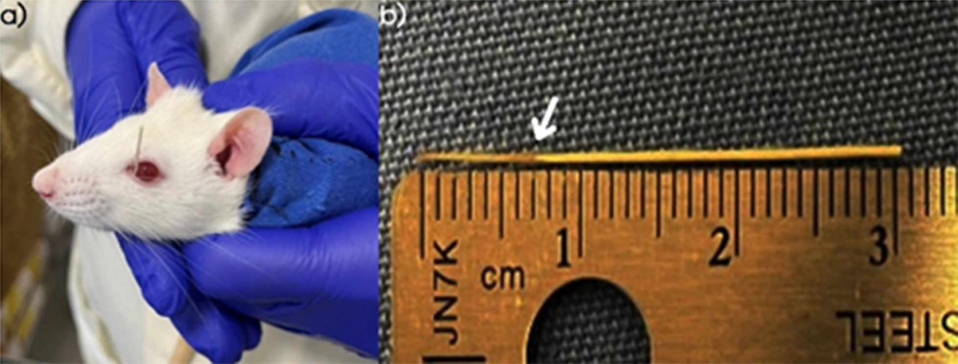
AbstractBackground: The evaluation of aqueous tear film production in laboratory rodents is limited by the small palpebral fissure and globe size. Aim: This study aimed to compare aqueous tear film measurements obtained using different commercially available endodontic paper points (EPPs) in laboratory rats. Methods: Aqueous tear production was measured in 22 adult Rattus norvegicus using six size-30 EPP brands applied for 15 and 60 s. Brand comparisons were performed at each time point using the Friedman test with multiplicity-adjusted post hoc comparisons. Paired time comparisons (15 vs. 60 s) were analyzed using the Wilcoxon signed-rank test. The effect size was reported as r (Z/√N). The central 90% reference intervals (5th–95th percentile) were calculated for each brand and time point. Results: Brand-related differences were identified at 15 s (Friedman χ²=38.56; p=2.91 × 10⁻⁷) and 60 s (Friedman χ²=31.79; p=6.55 × 10⁻⁶). Measurements were higher at 60 s than at 15 s for all brands (all p ≤ 0.0057; all r ≥ 0.589). The values ranged from 4.39 ± 0.83 to 6.50 ± 1.46 mm at 15 s and from 6.73 ± 1.91 to 10.45 ± 3.00 mm at 60 s. Conclusion: EPPs are practical, simple, and minimally invasive tools for evaluating aqueous tear film in laboratory rats; however, to ensure reproducibility and accurate interpretation, brand-specific and time-specific reference intervals are recommended. Keywords: Aqueous tear production test; Endodontic paper points; Laboratory rats; Tear film; Veterinary ophthalmology. IntroductionOphthalmic research using animal models continues to evolve, driven by the ongoing development of novel products and techniques tailored and refined for a wide range of species, including companion animals, livestock, and laboratory animals (Lange et al., 2013; Montiani-Ferreira et al., 2022). The production of the aqueous tear film is an essential parameter to assess during ophthalmic examination across species (Beaumont, 2002; Manning, 2014). The Schirmer Tear Test (STT) type I is the most widely used and standardized method in both human and veterinary ophthalmology. This test involves placing a strip of absorbent filter paper (Whatman No. 40) into the conjunctival sac of each eye for 60 s (Lange et al., 2013; Lima et al., 2014; Manning, 2014). The moistened length of the strip is measured in millimeters after this period, providing an estimate of aqueous tear production (Williams, 2005; White et al., 2011; Richards and Trbolová, 2016; Bolzanni et al., 2020). However, due to their small globe size, certain species present challenges, which make conventional STT application difficult (Lange et al., 2013). The standard STT strip measures 5 mm in width—often exceeding the total palpebral fissure width in small animals. In response, adaptations such as the modified STT (using manually halved strips) and the phenol red thread test have been proposed to facilitate aqueous tear measurement in smaller species (Vashisht and Singh, 2011; Lange et al., 2013; Lima et al., 2014; Rajaei et al., 2014; Somma et al., 2019). Despite these adaptations, significant limitations remain when evaluating aqueous tear production in small-eyed animals. Several studies have investigated the use of endodontic paper points (EPPs) as a viable alternative to obtain precise measurements of aqueous tear production in such species (Lange et al., 2012; Oriá et al., 2013; Lima et al, 2014; Rajaei et al., 2014; Oriá et al., 2015; Somma et al., 2015; Monção-Silva et al., 2016; Sayed et al., 2016; Richards et al., 2016; Kilic et al., 2016; Oliveira et al., 2017; Fornazari et al, 2018; Limón et al., 2019; Pelych et al., 2019; Bustamante et al., 2020; Colby et al., 2020; Martin De Bustamante et al., 2020), hereafter referred to as the paper point tear test (PPTT). The laboratory rat (Rattus norvegicus) is widely used as a research model and an exotic pet worldwide. Although rats are well-established models in ophthalmologic research, including experimental dry eye studies, the literature describing the use of EPPs in this species remains limited. Existing studies are sparse and lack consistent reference values (Beaumont, 2002; Rocha et al., 2008; Huang et al., 2021; Zhu et al., 2022). To date, no study has systematically compared multiple commercially available EPP brands in laboratory rats while establishing brand-specific reference intervals. This study aimed to compare six different brands of EPPs for aqueous tear film measurement in Rattus norvegicus at two time intervals (15 and 60 s), evaluate inter-brand variability, and establish brand- and time-specific reference intervals. We hypothesized that (1) different brands would demonstrate significantly different absorptive capacities and (2) measurements obtained at 60 s would be significantly higher than those at 15 s. Materials and MethodsForty-four globes from 22 adult laboratory rats (Rattus norvegicus), 13 males and 9 females, weighing between 200 and 230 g, were evaluated. Animals were obtained from the Pelé Pequeno Príncipe Research Institute. Each animal contributed one averaged measurement per brand and time point, obtained from the mean of both eyes; therefore, the unit of analysis was the animal (n=22), resulting in 22 measurements per brand at each time interval. The rats were housed in pairs in standard laboratory cages containing wood shavings bedding. The animals had free access to species-specific feed and water. Environmental conditions were maintained at a 12:12 h light-dark cycle (lights on at 8:00 AM and off at 8:00 PM), with a controlled temperature of 22°C ± 2°C. The air exchange was managed via exhaust ventilation, ensuring 20 air changes per hour with 100% renewal. Six commercial brands of endodontic paper points (EPPs), all size 30 (0.30 mm in diameter), were selected for comparison: Dentsply Sirona® (Dentsply Sirona Inc., York, PA, USA), DiaDent® (DiaDent Group International, Burnaby, BC, Canada), GAP® (GAP Dental Products S.A., São Paulo, Brazil), MK Life® (MK Life Medical and Dental Products, Porto Alegre, Brazil), Tanari® (Tanariman Industrial Ltda., Manacapuru, Brazil), and RoeckoColor® (Langenau, Germany). Rats and globes were numbered, and experimental groups were assigned to each eye and animal using a random number generator (https://www.random.org/) to ensure unbiased allocation and eliminate potential selection bias. For aqueous tear film measurements, all animals were manually restrained by the same experienced handler using a cloth and a standardized two-hand technique: one hand secured the body while the other stabilized the head and neck. Following proper restraint, an EPP was gently placed into the lower conjunctival fornix to absorb the aqueous tear film (Fig. 1a). The timer was then started, and the EPP was removed after either 15 or 60 s. Wetted length was measured in millimeters using a ruler (Fig. 1b). To minimize potential circadian variation in aqueous tear production, all measurements were performed between 9:00 AM and 12:00 PM. Wetted length readings were obtained from a single experienced examiner who was blinded to brand identity during measurement recording.
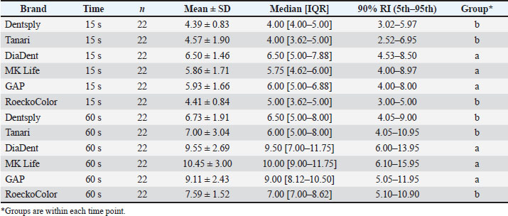
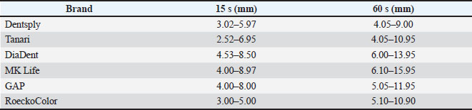
Fig. 1. Manual restraint and application of endodontic paper point (EPP) for tear film measurement in a rat (Rattus norvegicus) (a), followed by millimetric reading of the wetted portion (b). The study was conducted in two phases. Aqueous tear production was measured using all six EPP brands in the first phase, with each test performed for 60 seconds. Each day, two EPP brands were randomly applied per animal—one to each eye—selected randomly. Because measurements were obtained from both eyes, values were averaged per animal prior to analysis to avoid pseudoreplication. The procedure was repeated with two new brands after a 24-hour interval, and another 24 hours later, the final two brands were tested. Two weeks after the initial phase, the same procedure was repeated with a shorter test duration of 15 s. EPP brands were applied randomly to each eye at 24-hour intervals until all six brands had been tested across all animals. This approach minimized bias and allowed for robust comparison of absorptive performance across brands and time points. The aqueous tear production values for each animal and brand were recorded in Microsoft Excel (Microsoft Corporation, Redmond, WA, USA). Because the study employed a repeated-measures (within-subject) design, each animal contributed measurements across all brands and both time points. The final sample size of 22 rats provided complete paired data for all comparisons and was considered appropriate for detecting within-subject differences and estimating brand- and time-specific reference intervals. Ocular surface health was assessed by gross ophthalmic examination 24 hours after each measurement session, including evaluation of conjunctival hyperemia, ocular discharge, corneal opacity, and blepharospasm. No clinically detectable abnormalities were observed. Statistical analysis Normality was assessed using the Shapiro–Wilk test. Inter-brand comparisons were performed at each time point (six repeated measurements within animals) using the Friedman test with multiplicity-adjusted post hoc comparisons. Paired time comparisons (15 vs. 60 s) for each brand were performed using the Wilcoxon signed-rank test. The effect size was calculated as r=Z/√N and interpreted as small (0.10–0.29), medium (0.30–0.49), or large (≥0.50). Significance was set at p < 0.05. The central 90% reference intervals (5th–95th percentile) were calculated for each brand and time point. (Geffré et al., 2011) Ethical approvalAll procedures were conducted in accordance with the ethical guidelines approved by the Animal Care and Use Committee of Faculdade Pequeno Príncipe (Protocol No. 077-2024). ResultsAll 22 laboratory rats were successfully evaluated in both experimental phases, ensuring complete paired data for all brands and time intervals. The results are summarized in Table 1. For enhanced usability and rapid clinical reference, the central 90% reference intervals are summarized in Supplementary Table S1. Table 1. Aqueous tear film measurements (mm) using six EPP brands in laboratory rats (n=22). Data shown as Mean ± SD, median [IQR], and central 90% reference interval (RI; 5th–95th percentile). Different superscript letters within each time point indicate significant inter-brand differences (post hoc, adjusted p < 0.05).
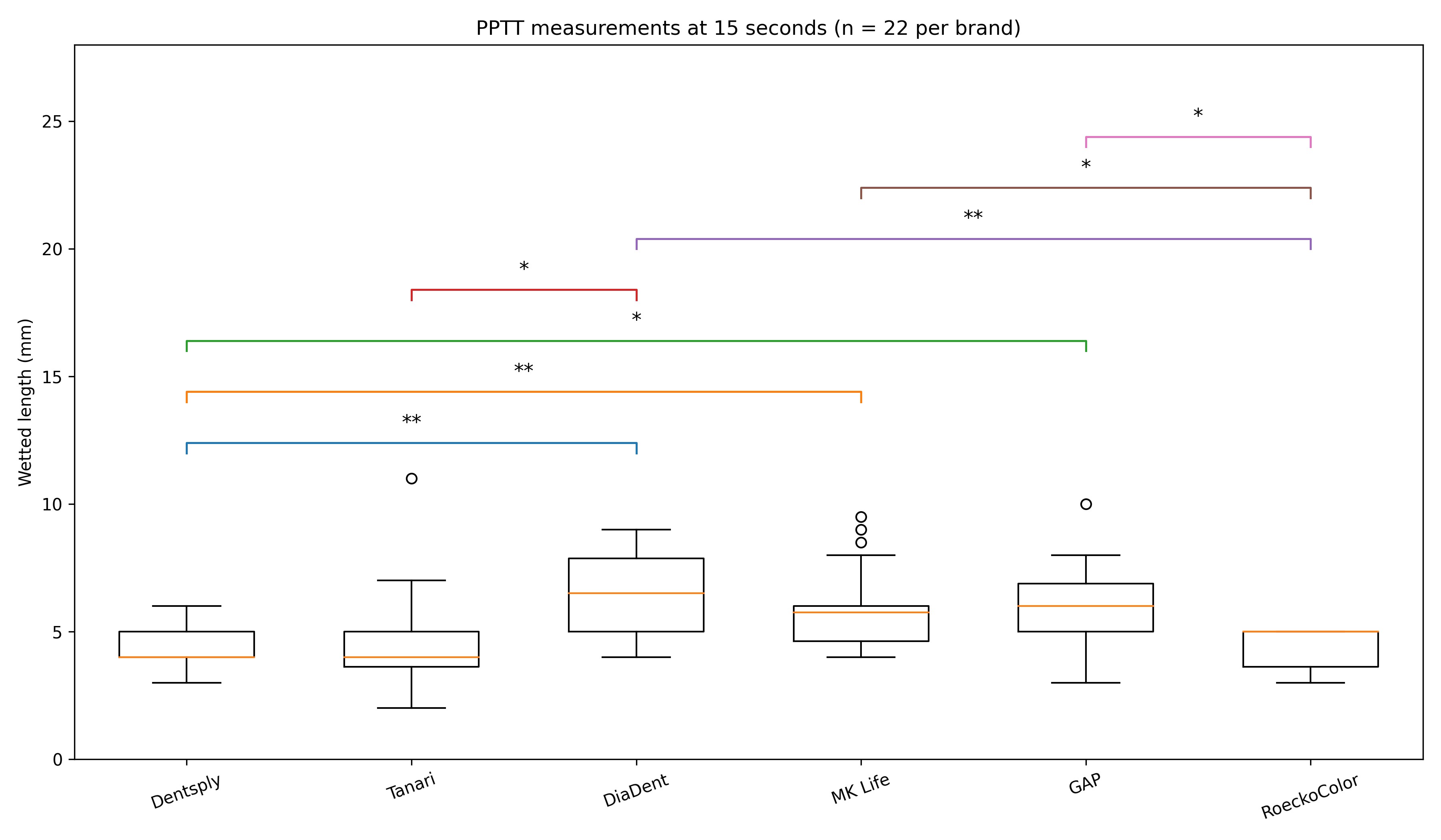
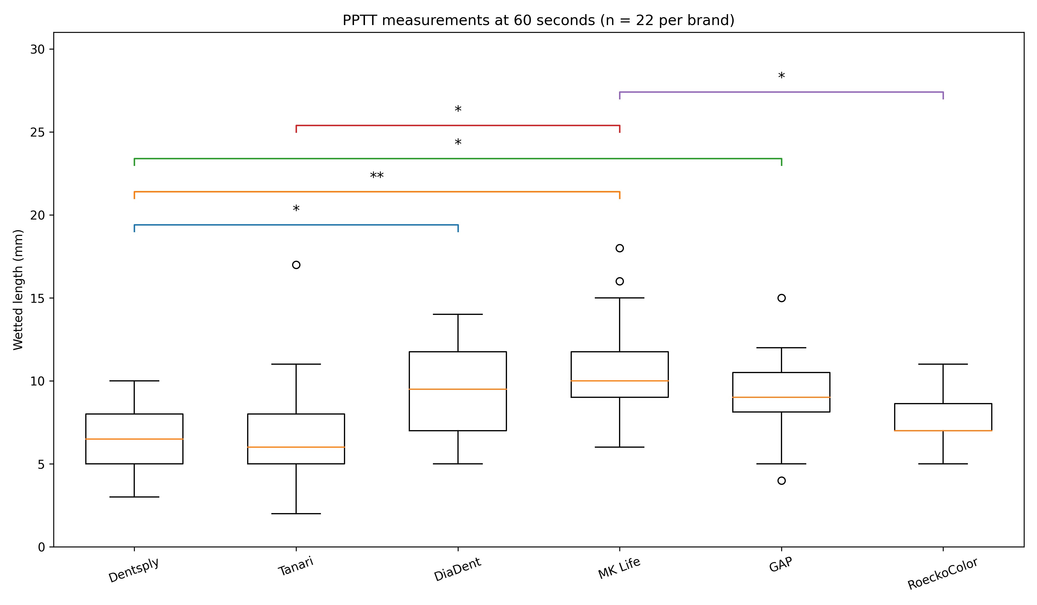
Aqueous tear volume after 15 sAt 15 s, measurements differed significantly among brands (Friedman χ²=38.56; p=2.91 × 10⁻⁷). Mean ± SD values ranged from 4.39 ± 0.83 mm (Dentsply) to 6.50 ± 1.46 mm (DiaDent). Table 1 presents the median [IQR] values and 90% reference intervals. Aqueous tear volume in 60 sAt 60 seconds, inter-brand differences remained significant (Friedman χ²=31.79; p=6.55 × 10⁻⁶). Mean ± SD values ranged from 6.73 ± 1.91 mm (Dentsply) to 10.45 ± 3.00 mm (MK Life). Comparison between 15 vs 60 sFor every brand, the 60-second measurements were significantly higher than the 15-second measurements (Wilcoxon signed-rank tests; all p ≤ 0.0057), with large effect sizes (all r ≥ 0.589), indicating a significant difference between the time points. When averaged across brands within animals, aqueous tear production was significantly higher at 60 s (p=4.00 × 10⁻⁵; r=0.876). Inter-brand comparisonsDiaDent, MK Life, and GAP formed the higher absorbency group, whereas Dentsply, Tanari, and RoeckoColor formed the lower absorbency group at both time points (Table 1; Figs. 2–3).
Fig. 2. Box plot of aqueous tear film measurements (PPTT) for different brands of endodontic paper points applied for 15 seconds. Brands sharing the same letters did not differ statistically (p < 0.05).
Fig. 3. Box plot of aqueous tear film measurements (PPTT) for different brands of endodontic paper points applied for 60 seconds. Brands sharing the same letters did not differ statistically (p < 0.05). Sex-related analysesNo significant differences were observed between male and female rats for any brand at either time point (p > 0.05). DiscussionEndodontic paper points (EPPs) were used to assess the aqueous phase of tear film in laboratory rats, which proved to be an effective and feasible technique for this species. Although this study did not include direct comparisons with the Schirmer tear test (STT), modified STT, or phenol red thread test, the authors’ experience suggests that, despite notable differences observed among the evaluated brands, EPPs are less stressful and more easily applied in rats. Significant differences in aqueous tear volume were observed between the 15- and 60-s time points, with large effect sizes (all r ≥ 0.589). Specifically, the 60-second readings were 50%–100% higher than those at 15 seconds, consistent with the continuous secretion of tears by the lacrimal glands throughout the evaluation period. Generally, aqueous tear production tests, regardless of the material used (STT or EPP), are commonly conducted with 60-s durations across a wide range of species (Lange et al., 2013; Lima et al., 2014; Kilic and Kulualp, 2016; Fornazari et al., 2018). However, 15-second methodologies have also been reported, particularly in phenol red thread testing and in dry eye models in rats (Kilic and Kulualp, 2016). Shorter evaluation times may be advantageous for uncooperative species or individuals that are intolerant of materials placed in the conjunctival fornix. Nevertheless, evaluators must refer to reference values aligned with the chosen methodology, given the significant discrepancies observed between time points. Although sex-based anatomical differences, such as larger acinar areas in male lacrimal glands, have been reported in rats (Cornell-Bel et al., 1985), these differences do not appear to influence aqueous tear production. This is supported by the absence of statistically significant differences between males and females across all brands and evaluation times in this study. Significant differences were identified regarding EPP brand performance. Dentsply Sirona and Tanari exhibited lower absorptive capacity, whereas DiaDent, GAP, and MK Life exhibited higher absorbency. RoeckoColor showed lower-to-intermediate values and did not differ statistically from the lower absorbency group. These inter-brand differences were more pronounced at the 60-second time point. As all EPPs tested were of the same diameter (size 30), the variations in absorption are likely attributable to each brand’s physical and material-specific characteristics. Previous dental studies have reported similar variability in fluid absorption among EPP brands (Pumarola-Suñé et al., 1998; Renjini et al., 2020). The current study provides the first reference to this behavior in an ophthalmologic context, supporting further exploration of EPP use in both rats and other species. In addition to absorptive capacity, differences in handling were observed among brands. Due to their rigidity, Tanari EPPs were less tolerated and often caused increased agitation. Such behavior, combined with tear absorption, may heighten ocular sensitivity and injury risk (Meng et al., 2015). However, no signs of ocular inflammation were observed after 24 h. In contrast, DiaDent EPPs offered better firmness and ease of placement, especially during 60-s tests, reducing the need for repeated attempts. Other brands are occasionally dislodged prematurely. From a methodological perspective, investigators using groups with EPP in rats should select a single brand and maintain consistency throughout the study. Switching between brands without adjusting reference intervals may lead to an inaccurate interpretation of aqueous tear production values. Establishing brand- and time-specific reference intervals may facilitate future experimental dry eye studies and translational ophthalmic research in laboratory rats (Leonard et al., 2019; Huang et al., 2021; Zhu et al., 2022). This study has several limitations. First, this study did not include a direct comparison of the results of the Schirmer tear test, modified STT, or phenol red thread testing in the same animals. Second, intraobserver repeatability and test–retest reliability were not assessed. Only clinically healthy rats of a single strain and weight range were evaluated; therefore, reference intervals may not be generalizable to other strains or pathological conditions. Diagnostic cutoff values for dry eye or inflammatory ocular disease were not established and warrant further investigation. ConclusionDifferent EPP brands demonstrated significantly different absorptive capacities in laboratory rats, and the 60-second protocols yielded consistently higher values than the 15-second protocols with large effects. Therefore, to ensure accurate interpretation and reproducibility, brand- and time-specific reference intervals should be used consistently. This methodology supports more refined, welfare-conscious ophthalmic diagnostics for small laboratory animals. AcknowledgmentsThe authors are grateful to Instituto Pelé Pequeno Príncipe for their availability in receiving the researchers and supporting the project. Conflict of interestThe authors declare no conflicts of interest. FundingThis research did not receive any grant from funding agencies in the public, commercial, or not-for-profit sectors. FMF wishes to thank the Brazilian National Council for Scientific and Technological Development (CNPq) for personal financial support in the form of a research grant, process 306872/2022-6. Authors' contributionsIC: study design, data collection and analysis, statistical analysis, and preparation of the manuscript. AS: study design and analysis. CSS: data collection. FMF: study design, statistical analysis, and manuscript review. Data availabilityInterested parties can contact to request access to the data by contacting the corresponding author. ReferencesBeaumont, S.L. 2002. Ocular disorders of pet mice and rats. Vet. Clin. North Am. Exot. Anim. Pract. 5(2), 311–324; doi:10.1016/S1094-9194(01)00009-3 Bolzanni, H., Oriá, A.P., Raposo, A.C.S. and Sebbag, L. 2020. Aqueous tear assessment in dogs: impact of cephalic conformation, inter-test correlations, and test-retest repeatability. Vet. Ophthalmol. 23(1), 1–10; doi: 10.1111/vop.12751 Colby, L.A., Nowland, M.H. and Kennedy, L.H. 2020. Rats. In Colby, L.A., Nowland, M.H., and Kennedy, L.H. (eds), Clinical Laboratory Animal Medicine. (Ames, IA: Wiley-Blackwell). Cornell-Bell, A.H., Sullivan, D.A. and Allansmith, M.R. 1985. Gender-related differences in the morphology of the lacrimal gland. Invest. Ophthalmol. Vis. Sci. 26(1), 1170–1175. Fornazari, G., Ferreira, T.A.C., Santin, E., Panisson, J.C., Maiorka, A. and Montiani-Ferreira, F. 2018. Schirmer’s I, modified Schirmer’s I, phenol red thread, and paper point tests: a comparative study for tear production measurement techniques in broiler chicks (Gallus gallus domesticus). Poult. Sci. 97(9), 3258–3263; doi:10.3382/ps/pey159 Geffré, A., Concordet, D., Braun, J.P. and Trumel, C. 2011. Reference Value Advisor: a new freeware set of macroinstructions to calculate reference intervals with Microsoft Excel. Vet. Clin. Pathol. 40(1), 107–112; doi:10.1111/j.1939-165X.2011.00287.x Huang, W., Tourmouzis, K., Perry, H., Honkanen, R.A. and Rigas, B. 2021. Animal models of dry eye disease: useful, varied and evolving. Exp. Ther. Med. 22(6), 1394; doi:10.3892/etm.2021.10830 Kilic, S. and Kulualp, K. 2016. Tear production rate in a mouse model of dry eye according to the phenol red thread and endodontic absorbent paper point tear tests. Comp. Med. 66(5), 367–372. Lange, R.R., Lima, L. and Montiani‐Ferreira, F. 2012. Measurement of tear production in black-tufted marmosets (Callithrix penicillata) using three different methods: modified Schirmer’s I, phenol red thread and standardized endodontic absorbent paper points. Vet. Ophthalmol. 15(6), 376–382; doi:10.1111/j.1463-5224.2012.00998.x Lange, R.R., Lima, L., Przydzimirski, A.C. and Montiani‐Ferreira, F. 2013. Reference values for the production of the aqueous fraction of the tear film measured by the standardized endodontic absorbent paper point test in different exotic and laboratory animal species. Vet. Ophthalmol. 17(1), 1–5; doi:10.1111/vop.12038 Leonard, B.C., Stewart, K.A., Shaw, G.C., Hoehn, A.L., Stanley, A.A., Murphy, C.J. and Thomasy, S.M. 2019. Comprehensive clinical, diagnostic, and advanced imaging characterization of the ocular surface in spontaneous aqueous-deficient dry eye disease in dogs. Cornea 38(12), 1568–1575; doi:10.1097/ICO.0000000000002081 Lima, L., Lange, R.R., Turner-Giannico, A. and Montiani-Ferreira, F. 2014. Evaluation of standardized endodontic paper point tear test in New Zealand white rabbits and comparison between corneal sensitivity and tear tests. Vet. Ophthalmol. 17(1), 1–6; doi:10.1111/vop.12178 Limón, D., Vásquez, C. and Czaplewski, R. 2019. Measurement of tear production using Schirmer tear test and standardized endodontic absorbent paper points in ferrets (Mustela putorius furo). J. Exot. Pet Med. 29(1), 10–14. Manning, S. 2014. BSAVA Manual of Canine and Feline Ophthalmology. In Gould, D. and McLellan, G.J. (eds), Chapter 9. Gloucester: BSAVA. pp: 133–66; doi:10.22233/9781910443170.9 Martin De Bustamante, M.G., Johnson, A.N., Shippy, S.G., Allgood, H. and Plummer, C.E. 2020. Ophthalmic examination, biometry and histologic findings in captive inland bearded dragons (Pogona vitticeps). Vet. Ophthalmol. 23(1), 1–11; doi:10.1111/vop.12779 Meng, I.D., Barton, S.T., Mecum, N.E. and Kurose, M. 2015. Corneal sensitivity following lacrimal gland excision in the rat. Invest. Ophthalmol. Vis. Sci. 56(5), 3347–3354; doi:10.1167/iovs.15-16717 Monção-Silva, R.M., Ofri, R., Raposo, A.C.S., Libório, F.A., Estrela-Lima, A. and Oriá, A.P. 2016. Ophthalmic parameters of blue-and-yellow macaws (Ara ararauna) and Lear’s macaws (Anodorhynchus leari). Avian Biol. Res. 9(4), 240–249; doi:10.3184/175815516X14725499175746 Montiani-Ferreira, F., Moore, B.A. and Ben-Shlomo, G. 2022. Wild and Exotic Animal Ophthalmology.In Montiani-Ferreira, F., Moore, B.A., and Ben-Shlomo, G. (eds), Volume 2: mammal. 2 (Cham, Switzerland: Springer). Oliveira, M.M.S., Araújo, N.L.L.C., Raposo, A.C.S., Torezani, J.C., Menezes, I.D.S. and Oriá, A.P. 2017. Reference values for selected ophthalmic diagnostic tests in the Caatinga parakeet (Eupsittula cactorum) and yellow-chevroned parakeet (Brotogeris chiriri). Avian Biol. Res. 10(4), 211–217. Oriá, A.P., Monção Silva, R.M., Pinna, M.H., Oliveira, A.V.D., Ferreira, P.R.B., Martins Filho, E.F., Meneses, I.D.S., Requião, K.G. and Ofri, R. 2015. Ophthalmic diagnostic tests in captive red-footed tortoises (Chelonoidis carbonaria) in Salvador, northeast Brazil. Vet. Ophthalmol. 18(Suppl. 1), 46–52. Pelych, L.N., Shellabarger, W., Vassallo, M., Noland, E., Sledge, D. and Aquino, S.E. 2019. Ophthalmic findings in a captive population of Panamanian golden frogs (Atelopus zeteki). Vet. Ophthalmol. 22(1), 1–10. Pumarola-Suñé, J., Solá-Vicens, L., Senís-Vilalta, J., Canalda-Sahlis, C. and Brau-Aguadé, E. 1998. Absorbency properties of different brands of standardized endodontic paper points. J. Endod. 24(12), 796–803. Rajaei, S.M., Mood, M.A., Ghaffari, M.S. and Williams, D.L. 2014. Measurement of tear production using phenol red thread and standardized endodontic absorbent paper points in European pond turtles (Emys orbicularis). J. Zoo. Wildl. Med. 45(4), 825–829. Renjini, T.R., James, B., Devadathan, A., Nair, M., Sreekumar, A.A. and George, E. 2020. Comparative evaluation of absorbing capacity of standardized endodontic paper points: an in vitro study. Int. J. Sci. Res. 9(11), 62–64. Richards, M. and Trbolová, A. 2016. Reference values for the ophthalmic Schirmer tear test and the intraocular pressure in healthy chinchillas. Folia. Vet. 60(3), 29–33. Rocha, E.M., Alves, M., Rios, J.D. and Dartt, D.A. 2008. The aging lacrimal gland: changes in structure and function. Ocul. Surf. 6(4), 162–174. Somma, A.T., Coimbra, C.M., Lange, R.R., Moore, B.A. and Montiani‐Ferreira, F. 2019. Reference values for selected ophthalmic diagnostic tests in two species of microchiroptera bats (Artibeus lituratus and Anoura caudifer). Vet. Ophthalmol. 22(1), 1–6. Somma, A.T., Lima, L., Lange, R.R., Turner‐Giannico, A. and Montiani‐Ferreira, F. 2015. The eye of the red-eared slider turtle: morphologic observations and reference values for selected ophthalmic diagnostic tests. Vet. Ophthalmology 18(Suppl. 1), 61–70. Vashisht, S. and Singh, S. 2011. Evaluation of phenol red thread test versus Schirmer test in dry eyes: a comparative study. Int. J. Appl. Basic Med. Res. 1(1), 40–42. White, J.S., Grundon, R.A., Hardman, C., O’Reilly, A. and Stanley, R.G. 2011. Tear production measurement techniques in dogs using standardized methods. Vet. Ophthalmol. 14(1), 1–5. Williams, D.L. 2005. Analysis of tear uptake by the Schirmer tear test strip in the canine eye. Vet. Ophthalmol. 8(5), 325–330. Zhu, J., Inomata, T., Shih, K.C., Okumura, Y., Fujio, K., Huang, T., Nagino, K., Akasaki, Y., Fujimoto, K., Yanagawa, A., Miura, M., Midorikawa-Inomata, A., Hirosawa, K., Kuwahara. M., Shokirova, H., Eguchi, A., Morooka, Y., Chen, F. and Murakami A. 2022. Application of animal models in interpreting dry eye disease. Front Med (Lausanne). 9, 830592; doi: 10.3389/fmed.2022.830592 Supplementary MaterialSupplementary Table S1. Central 90% reference intervals (5th–95th percentile) for aqueous tear film measurements in laboratory rats (n=22) using six endodontic paper point brands.
| ||
| How to Cite this Article |
| Pubmed Style Canto I, Saldanha A, Sacaki CS, Montiani-ferreira F. Comparison of aqueous tear film measurements obtained using different endodontic paper point brands in laboratory rats (Rattus norvegicus). Open Vet. J.. 2026; 16(6): 3456-3463. doi:10.5455/OVJ.2026.v16.i6.16 Web Style Canto I, Saldanha A, Sacaki CS, Montiani-ferreira F. Comparison of aqueous tear film measurements obtained using different endodontic paper point brands in laboratory rats (Rattus norvegicus). https://www.openveterinaryjournal.com/?mno=291595 [Access: June 26, 2026]. doi:10.5455/OVJ.2026.v16.i6.16 AMA (American Medical Association) Style Canto I, Saldanha A, Sacaki CS, Montiani-ferreira F. Comparison of aqueous tear film measurements obtained using different endodontic paper point brands in laboratory rats (Rattus norvegicus). Open Vet. J.. 2026; 16(6): 3456-3463. doi:10.5455/OVJ.2026.v16.i6.16 Vancouver/ICMJE Style Canto I, Saldanha A, Sacaki CS, Montiani-ferreira F. Comparison of aqueous tear film measurements obtained using different endodontic paper point brands in laboratory rats (Rattus norvegicus). Open Vet. J.. (2026), [cited June 26, 2026]; 16(6): 3456-3463. doi:10.5455/OVJ.2026.v16.i6.16 Harvard Style Canto, I., Saldanha, . A., Sacaki, . C. S. & Montiani-ferreira, . F. (2026) Comparison of aqueous tear film measurements obtained using different endodontic paper point brands in laboratory rats (Rattus norvegicus). Open Vet. J., 16 (6), 3456-3463. doi:10.5455/OVJ.2026.v16.i6.16 Turabian Style Canto, Isabela, André Saldanha, Claudia Sayuri Sacaki, and Fabiano Montiani-ferreira. 2026. Comparison of aqueous tear film measurements obtained using different endodontic paper point brands in laboratory rats (Rattus norvegicus). Open Veterinary Journal, 16 (6), 3456-3463. doi:10.5455/OVJ.2026.v16.i6.16 Chicago Style Canto, Isabela, André Saldanha, Claudia Sayuri Sacaki, and Fabiano Montiani-ferreira. "Comparison of aqueous tear film measurements obtained using different endodontic paper point brands in laboratory rats (Rattus norvegicus)." Open Veterinary Journal 16 (2026), 3456-3463. doi:10.5455/OVJ.2026.v16.i6.16 MLA (The Modern Language Association) Style Canto, Isabela, André Saldanha, Claudia Sayuri Sacaki, and Fabiano Montiani-ferreira. "Comparison of aqueous tear film measurements obtained using different endodontic paper point brands in laboratory rats (Rattus norvegicus)." Open Veterinary Journal 16.6 (2026), 3456-3463. Print. doi:10.5455/OVJ.2026.v16.i6.16 APA (American Psychological Association) Style Canto, I., Saldanha, . A., Sacaki, . C. S. & Montiani-ferreira, . F. (2026) Comparison of aqueous tear film measurements obtained using different endodontic paper point brands in laboratory rats (Rattus norvegicus). Open Veterinary Journal, 16 (6), 3456-3463. doi:10.5455/OVJ.2026.v16.i6.16 |


