


| Research Article | ||
Open Vet. J.. 2025; 15(12): 6572-6580 Open Veterinary Journal, (2025), Vol. 15(12): 6572-6580 Research Article Impact of phytochemical constituents of milk thistle extract on body score and hematological and biochemical parameters in dromedary camels (Camelus dromedarius)Asem Al Shehri1, Basel Hamoud2, Ahmed M. A. Meligy1, Mohammad Asvad3 and Turke Shawaf1*1Department of Clinical Sciences, College of Veterinary Medicine, King Faisal University, Al-Ahsa, Saudi Arabia 2Department of Surgery and Gynecology, College of Veterinary Medicine, Hama University, Hama, Syria 3Department of Animal Nutrition, Al Emar International—Arasco co, Riyadh, Saudi Arabia *Corresponding Author: Turke Shawaf. Department of Clinical Sciences, College of Veterinary Medicine, King Faisal University, Al-Ahsa, Saudi Arabia. Email: tshawaf [at] kfu.edu.sa Submitted: 09/09/2025 Revised: 05/11/2025 Accepted: 15/11/2025 Published: 31/12/2025 © 2025 Open Veterinary Journal
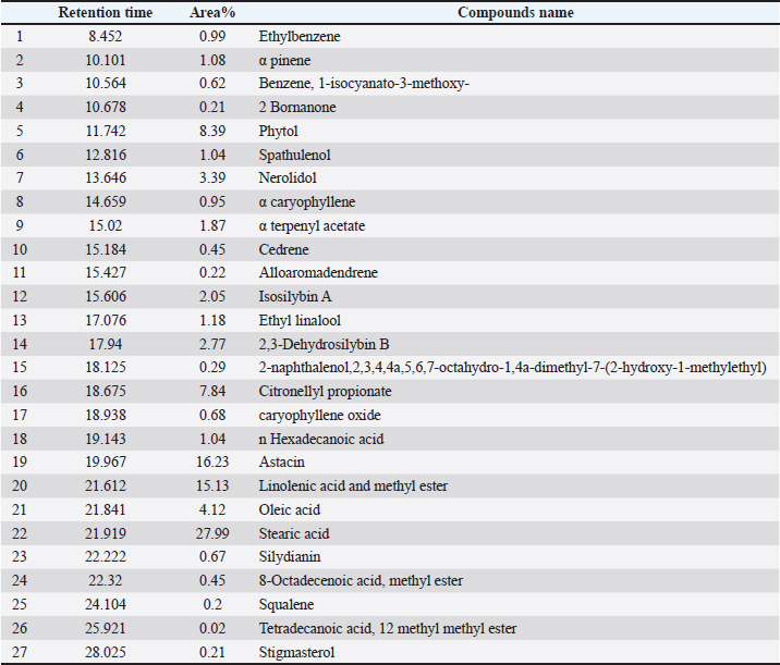
AbstractBackground: Herbal extracts are increasingly being utilized in veterinary medicine, with milk thistle extract being recognized for its significant role in animal health, and milk thistle extract is widely acknowledged for its crucial contribution in supporting liver function. Camels are among the animals whose liver enzymes are often altered, which can have a detrimental effect on their health. Aim: This study investigated the therapeutic effects of milk thistle extract on body scores and hematological and biochemical parameters in dromedary camels with elevated liver enzyme levels. Methods: This study was conducted on 18 dromedary camels (eight males and ten females). The camels were divided into two groups: the first group consisted of five healthy camels with normal liver enzyme levels, and the second group consisted of 13 camels with elevated liver enzyme levels. The prepared milk thistle extract from seeds was subjected to phytochemical and Gas Chromatography-Mass Spectrometry (GC-MS) MS analyses. The camel in the second group orally received milk thistle extract, which was calculated based on Silymarin concentration, for 6 weeks. The clinical score and hematological and biochemical analyses were performed in control and diseased camels before and after treatment. Results: The health and appetite scores showed significant improvements after treatment. The percentage of lymphocytes increased in diseased camels following treatment, whereas the percentage of eosinophils decreased in healthy camels. After 42 days of milk thistle treatment, liver enzymes, including alkaline phosphatase, gamma glutamyl transferase, total bilirubin, and bilirubin urea nitrogen, improved in diseased camels. However, other biochemical parameters, including total protein and albumin, decreased in the affected animals compared with the healthy animals, whereas these parameters improved closer to normal values after treatment. Conclusion: Our data provide important evidence for the therapeutic efficacy of milk thistle extract in dromedary camels with liver disorders. Keywords: Blood, Camel, Liver, Milk thistle, Silymarin. IntroductionThe Arabian camel (Camelus dromedarius) is an important and adaptable animal that provides valuable meat and milk in arid and semi-arid regions (Manee et al., 2024; Padalino and Menchetti, 2024). Camel use has evolved over the past decades, with their use as a sporting activity in racing and beauty contests (Leue, 1971; Tharwat and Al-Hawas, 2021). Camel management has been modified as a result of the evolution of camel use, removing them from their natural environment, where they were fed thorny pasture plants, which camels are unique among animals in their ability to consume (Tharwat and Al-Hawas, 2021; Kena, 2022; Nagy et al., 2022; Laameche et al., 2024). Many of these pasture plants have nutritional and medicinal benefits for camels (Habte et al., 2021; Jayamanna Mohottige et al., 2025). Modern camel rearing in confined spaces and feeding them concentrated feed has led to the emergence of numerous health problems in camels, some of which may be due to changes in nutritional quality and deprivation of the pasture plants they have consumed for thousands to hundreds of thousands of years, which have provided them with healthy adaptations (Rahimi et al., 2022; Naji et al., 2024; Tharwat et al., 2024). In recent years, there has been increased interest in plant-based supplements to improve livestock health, driven by the demand for sustainable and cost-effective alternatives to synthetic drugs (Yurdakok-Dikmen et al., 2018; Schlittenlacher et al., 2022). Among these botanical options, Silybum marianum milk thistle has emerged as a promising contender due to the bioactive flavonolignan compound, silymarin, known for its hepatoprotective, antioxidant, and anti-inflammatory properties (Zholobenko et al., 2016; Dockalova et al., 2021; Guerrini and Tedesco, 2023). Although the therapeutic potential of silymarin has been extensively documented in humans and monogastric animals (El-Far et al., 2018; Dockalova et al., 2021; Khazaei et al., 2022) use in camels remains relatively unexplored, necessitating comprehensive investigations into its effects on hematological and biochemical parameters. In camels, changes in blood parameters are often associated with the development of organ disorders, certain nutritional deficiencies, infectious diseases, or toxins (Lamo et al., 2020; Lamraoui et al., 2023). Liver dysfunction, which is common in camels grazing toxic plants, can result in elevated aspartate aminotransferase (AST) and aspartate aminotransferase (ALT) levels, indicating cellular damage (Lee et al., 2016; Maharem et al., 2020; Ghavipanje et al., 2022). The ability of silymarin to stabilize cell membranes, scavenge free radicals, and stimulate liver regeneration suggests its potential as a mitigator of these imbalances (Amjad et al., 2024; Dhande et al., 2024). Milk thistle supplements improve liver function in dairy cows by reducing oxidative stress and normalizing enzyme activities (Tedesco et al., 2004; Garavaglia et al., 2015). Although these findings suggest the potential applicability of milk thistle supplements, data specific to camels remain limited. This study aimed to evaluate the effects of milk thistle extract’s phytochemical constituents on hematological and biochemical parameters in Arabian camels with elevated liver enzymes. Materials and MethodsSample extractionDried milk thistle seeds (100 g) were pulverized and homogenized, and subsequently extracted with ethyl acetate using a shaker apparatus for 48 hours. The extracts were filtered through the Whatman filter paper No. 1 using a Buchner funnel. Solvent removal was performed via rotary evaporation under reduced pressure at 35ºC (El Sherif et al., 2020). Phytochemical analysisThe phytochemical constituents of fresh dry seeds of Milk Thistle (Silybum marianum L.) plants were analyzed at the Central Laboratory, Clinical Sciences Department, Faculty of Veterinary Medicine, King Faisal University. The extract was evaluated for the presence of terpenoids, alkaloids, flavonoids, Tannins, Phenols, and Saponins was evaluated using standard colorimetric methods described by (Mahmoud and Selim, 2025), (Kennedy and Thorley, 2000), and (Sultan et al., 2025). GC MS analysisThe crude extracts were analyzed using a Shimadzu GCMS QP2010 Plus system Japan. High-purity (99.9999%) helium served as the carrier gas, with a flow rate of 1 ml/minute. The analysis employed a capillary column (DB-5MS, 30-m length, 0.25-mm film thickness, and 0.25-μm film diameter). A 1-μl aliquot of the sample was injected in split mode (10:1) at 260°C. The GC temperature program commenced at 60°C, held for 5 minutes, then increased at a rate of 10°C minutes to 240°C, where it was maintained for 15 minutes. The interface temperature was set to 250°C, and the ion source temperature was maintained at 220°C. Data acquisition was performed in scan mode over a mass range of 50–550 amu. The relative abundance of each constituent was determined by integrating the peak areas and expressed as a percentage of the total ion current (Khattab et al., 2023). Animals and clinical examinationThis study was conducted on 18 dromedary camels (eight males and ten females) between May 2023 and December 2024. Camels had a median age ± SEM of 9 ± 6.5 years and a median weight ± SEM of 335 ± 125 kg. The clinical score of the general condition of camels with elevated liver enzymes before and after treatment was assessed based on a description of their health, changes in appetite, and changes in weight, based on previous studies (Menchetti et al., 2021; Padalino and Menchetti, 2024). The camels were divided into two groups: the first group consisted of five healthy camels with normal liver enzyme levels, and the second group consisted of 13 camels with elevated liver enzyme levels. The camels in the second group received orally milk thistle extract, which was calculated based on laboratory analysis, with Silymarin at a dose of 5 mg kg per day for 6 weeks (Karimi et al., 2011). Blood sample collectionBlood samples were collected from the jugular vein into two tubes Guangzhou Improve Medical, China), one containing EDTA for hematological analysis and the other devoid of anticoagulants Becton Dickinson, Franklin Lakes, USA for biochemical analysis. All samples were transferred into the laboratory as quickly as possible in ice. The samples were collected before and 42 days after the start of treatment. Blood samples for hematological examination were analyzed within 45 minutes of collection using CELL DYN 3700 analyzer for total and differential WBC count (WBC), total RBC count (RBC), hemoglobin (Hb), hematocrit, mean corpuscular volume (MCV), mean corpuscular hemoglobin (MCH), mean corpuscular hemoglobin concentration (MCHC), and mean platelet volume. The blood samples for biochemical analysis were placed in a centrifuge immediately after the sampling and centrifuged at 3,200 × g at room temperature for 10 minutes. Serum was collected and kept frozen at −20ºC until used for biochemical analysis. Serum biochemistry was carried out by using Vet scan vs 2 analyzer (ABAXIS, USA for total protein (TP), albumin (ALB), globulin, alkaline phosphatase (ALP), AST, gamma glutamyl transferase (GGT), ALT, total bilirubin (TBIL), urea nitrogen (BUN), creatine kinase, creatinine (CRE), and glucose (GLU). Statistical analysisFor the statistical analysis, Graph Pad Prism 7 software was used to calculate the minimum and maximum values to determine the mean range, mean, and standard deviation. Additionally, the Shapiro-Wilk normality test was used to evaluate the normal distribution of the values; the p-value significance thresholds were determined (p < 0.05). Ethical approvalThe Ethics Committee at King Faisal University in Saudi Arabia approved the study for research purposes Approval number: KFU254116. ResultsQualitative phytochemical analysis of milk thistle seeds showed high levels of anthraquinones, tannins, and flavonoids, while steroids, terpenoids, alkaloids, and saponins were moderately detectable as 27 compounds (Table 1). The main components of the milk thistle extract were as follows: stearic acid 27.99 casticin, 16.23 linoleic acid, methyl ester, 15.3 phytol, 8.39 citronellyle propionate, 7.84 oliec acid, 4.12 nerolidol 3. 39, 2.2,3-dehydrosilbin B 2.77, and isosilybin A 2.05 (Table 1). In addition, the extract contained different terpenoids such as α pinene 1. 08 and sesquiterpenoid, such as nerolidol 3.39 spathulenol, 1.04 α caryophyllene 0.95, and squalene 0% 2. Also contains Stigmasterol (0.21%) as a sterol. Table 1. GC–MS analysis of the ethyl acetate extract of milk thistle.
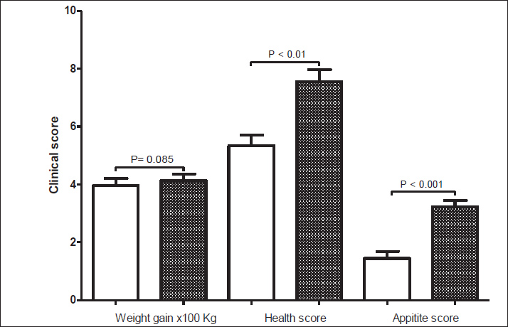
Figure 1 shows the clinical scores related to body parameters, including weight gain, health score, and appetite score, before and after treatment with milk thistle extract in dromedary camels with high liver enzyme levels. After treatment with milk thistle extract, there was an obvious improvement in these parameters. The weight gain score showed an increase (p=0.085). However, the health score significantly improved (p < 0.01), and the appetite score also showed a significant improvement (p < 0.001) after treatment.
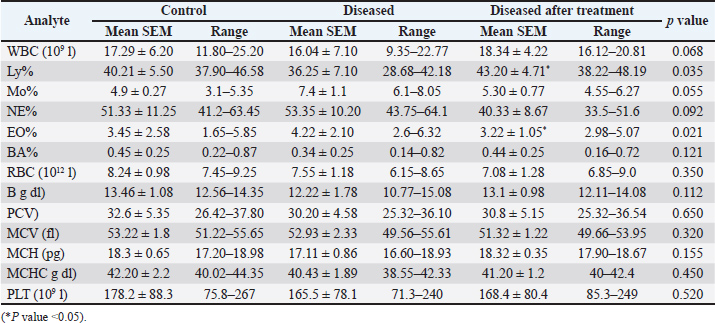
Fig. 1. Clinical scores before and 6 weeks after treatment with milk thistle extract in dromedary camel suffering from elevated levels of liver enzymes in the blood. Table 2 details the changes in hematological values in control, diseased, and treated camels. Notably, the diseased camels showed some alterations in WBC, Ly%, Mo%, and NE%, and RBC, Hb, PCV, MCV, MCH, MCHC, and PLT. After 42 days of treatment with milk thistle extract, many of these values showed a trend toward regulation. For instance, Ly% increased significantly from 36.25 ± 7.10 in diseased camels to 43.20 ± 4.71 after treatment, approaching the control group's 40.21 ± 5.50. Similarly, EO% decreased from 4.22 ± 2.10 to 3.22 ± 1.05, nearing its value in healthy camels of 3.45 ± 2.58. NE% also showed a notable decrease, but not of statistical significance, from 53.35 ± 10.20 to 40.33 ± 8.67, which is lower than the control group's 51.33 ± 11.25. Table 2. Mean, SEM, and range of hematological values in control and diseased dromedary camels before and after 42 days of treatment with milk thistle extract. p values calculated between parameters in diseased camels before and after treatment.
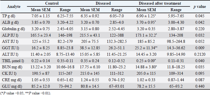
Table 3 presents the biochemical parameters in control, diseased, and post-treatment camels. Specifically, ALP increased from 165.5 ± 23.4 IU/l in control to 215.5 ± 43.1 IU/l in diseased camels, GGT from 16.2 ± 8.25 IU/l to 38.5 ± 12.85 IU/l, TBIL from 0.22 ± 0.14 µmol/l to 0.35 ± 0.24 µmol/l, and BUN from 13.22 ± 3.20 mg/dl to 17.75 ± 6.10 mg/dl. After 42 days of milk thistle treatment, a significant improvement was observed in these liver enzymes. ALP decreased to 173.1 ± 32.2 IU/l, GGT to 25.2 ± 11.34 IU/l, TBIL to 0.25 ± 0.09 µmol/l, and BUN to 14.88 ± 3.80 mg/dl. Table 3. Mean, standard error of the mean, and range of biochemical parameters in control and diseased camels before and after 42 days of treatment with milk thistle extract. p values calculated between parameters in diseased camels before and after treatment.
DiscussionA healthy liver is central for metabolism, nutrient absorption, and detoxification, all of which directly affect an animal’s weight, appetite, and health status (Tharwat, 2020). Milk thistle is a very important plant because it contains highly effective active ingredients (Aida et al., 2022; Amjad et al., 2024). Our analysis of the components of milk thistle seeds aligned with previously reported findings, indicating that milk thistle contains a variety of secondary metabolites, including flavonoids, terpenoids, tannins, saponins, alkaloids, and steroids (Yurdakok-Dikmen et al., 2018; Rasha Eldalawy et al., 2022). The most important components for liver function in the extract were 2,3-dihydrosilbin B, silydianin, and Isosilybin A (Karimi et al., 2011; Fried, 2012; El-Far et al., 2018). These compounds, all classified as flavonolignans, are responsible for the antioxidant, anti-inflammatory, and liver-protective properties of silymarin (Poltavets et al., 2021). However, the terpenoids, including nerolidol, spathulenol, α caryophyllene, and squalene in the extract which are considered natural antioxidants that support cellular health (Rasha Eldalawy et al., 2022). Moreover, casticin was present at a high level in our extract and could have potential benefits in the attenuation and treatment of liver fibrosis (Zhou et al., 2017). Previous studies on other animal species, such as the one by (Tedesco and Guerrini, 2023), have also reported that milk thistle can lead to enhanced animal performance and health status (Garavaglia et al., 2015; Janocha et al., 2021; Gousias et al., 2025). Similar results for improvements in weight gain, health score, and appetite in our study were reported by (Hashemi Jabali et al., 2018; Amjad et al., 2024; Gousias et al., 2025), which support the benefits of milk thistle extracts in animals suffering from liver problems. Similar results of lymphocytosis in animals after silymarin treatment have been reported previously (Lukanov et al., 2018; Mohammed et al., 2022). These changes suggest that milk thistle has a positive impact on the immune response and overall blood health (Wilasrusmee et al., 2002). Previous research on the effects of milk thistle on animal hematological parameters is varied. A review by (Tedesco and Guerrini, 2023) indicated that milk thistle can influence blood parameters, whereas other studies have shown changes in blood parameters (Mohammadi et al., 2019; Ahammad et al., 2024; Imbabi et al., 2024; Cong et al., 2025). Although direct comparisons to camel studies are limited, our results align with the general understanding that silymarin, the active compound in milk thistle, possesses properties that can modulate physiological responses, including those related to blood immunity parameters. The improvement in lymphocyte and monocyte percentages, coupled with a decrease in neutrophil percentage in the present study, could indicate a decrease in inflammatory processes associated with liver dysfunction (Del Campo et al., 2018). These values are much closer to those of the control group, indicating that the milk thistle extract exerts a hepatoprotective effect. However, Tedesco et al. (2004) demonstrated that silymarin improved liver function in periparturient dairy cows by reducing oxidative stress and normalizing enzyme activities. Moreover, a review by (Vargas-Mendoza, 2014) the ability of silymarin to protect against many toxic models of experimental liver diseases in laboratory animals. Other biochemical parameters, such as TP and ALB, decreased in affected animals compared to healthy animals, while these parameters improved closer to normal values after treatments, which was in agreement with Sun et al. (2019), who reported a decrease in albumin in patients with liver dysfunction. This is a positive indicator because albumin is synthesized in the liver and its levels often reflect the capacity of the liver to synthesize it. The improvement in total protein and albumin levels in diseased camels after silymarin treatment, which is consistent with weight gain and improved appetite, has been reported in previous studies (Eminzade et al., 2008; Dockalova et al., 2021; Ghazaghi et al., 2024). The observed increase in TP and ALB suggests an improvement in the liver’s synthetic function, further supporting the hepatoprotective effects of milk thistle extract, similar to its effects in other species (Tedesco et al., 2004; Dockalova et al., 2021; Amjad et al., 2024; Dhande et al., 2024; Lambo et al., 2024). While the study provides compelling evidence for the therapeutic benefits of milk thistle extract in dromedary camels, some limitations should be acknowledged. The sample size was relatively small, particularly for the healthy control group. Future studies with larger cohorts and longer treatment durations would provide more robust data. Additionally, exploring different dosages and formulations of milk thistle extract could optimize its therapeutic efficacy. Further research could also delve into the specific mechanisms by which each identified phytochemical constituent contributes to the overall therapeutic effect. ConclusionIn conclusion, the current study provides promising data on milk thistle in dromedary camels. Our data provide important evidence for the therapeutic efficacy of milk thistle extract in dromedary camels with liver disorders. The observed improvements in clinical scores, hematological parameters, and biochemical markers highlight the hepatoprotective, anti-inflammatory, and immune modulating properties of the hepatocytes. These findings support the use of milk thistle extract as a natural and effective intervention to improve liver health and overall well-being in camels. Further research is warranted to fully elucidate the underlying mechanisms and optimize its application in veterinary practice. AcknowledgmentsThis work was supported by the Deanship of Scientific Research, Vice Presidency for Graduate Studies and Scientific Research, King Faisal University, Saudi Arabia, Grant No. Grant Nr: KFU254116. Many thanks to the TAMIVET factory for providing milk thistle extracts to conduct the study. Conflict of interestThe authors declare that they have no competing interests. Authors' contributionsAA, TS, and BH performed the examination; MA, AM, and ST performed the laboratory work. TS, AM, and MA drafted the manuscript. The final manuscript was approved by all authors. Data availabilityAll data are within the manuscript. ReferencesAhammad, G.S., Jang, S.Y. and Kim, I.H. 2024. Effects of micelle silymarin in corn-soybean meal-based diet on laying hens' performance, egg quality, and blood profile, with comparative assessment of blood absorption rates between powdered and micelle silymarin. Poult. Sci. 103, 104029. Amjad, M.Z., Hassan, M.U., Rehman, M., Shamim, A., Zeeshan, H.M., Iftikhar, Z., Ahmed, A. and Jamil, M.I. 2024. Impact of Silymarin supplementation on liver function and enzyme profiles in diverse chronic liver disease etiologies. Cureus 16, e76313. Cong, G., Xia, S., Liu, C., Li, J., Hung, I., Zhang, L., Guo, S. and Zhao, B. 2025. Effects of silymarin supplementation in late pregnancy and lactation on reproductive performance, colostrum quality, blood biochemistry and inflammation levels of sows. Trop. Anim. Health Prod. 57, 66. Dhande, D., Dhok, A., Anjankar, A. and Nagpure, S. 2024. Silymarin as an antioxidant therapy in chronic liver diseases: a comprehensive review. Cureus 16, e67083. Dockalova, H., Zeman, L. and Horky, P. 2021. Influence of milk thistle (Silybum marianum) seed cakes on biochemical values of equine plasma subjected to physical exertion. Animals (Basel) 210, 11. Doostkam, A., Fathalipour, M., Anbardar, M.H., Purkhosrow, A. and Mirkhani, H. 2022. Therapeutic Effects of Milk Thistle (Silybum marianum L.) and Artichoke (Cynara scolymus L.) on Nonalcoholic Fatty Liver Disease in Type 2 Diabetic Rats. Can. J. Gastroenterol. Hepatol. 2022, 1–8. El Sherif, F., Albotnoor, N., Yap, Y.K., Meligy, A. and Khattab, S. 2020. Enhanced bioactive compounds composition in Lavandula officinalis in-vitro plantlets using NaCl and Moringa oleifera, Aloe vera and Spirulina platensis extracts. Ind. Crops. Prod. 157, 112890. El-Far, M., Salah, N., Essam, A., Abd El-azim, A.O. and El-Sherbiny, I.M. 2018. Silymarin nanoformulation as potential anticancer agent in experimental Ehrlich ascites carcinoma-bearing animals. Nanomed. (Lond). 13, 1865–1858. Eminzade, S., Uraz, F. and Izzettin, F.V. 2008. Silymarin protects liver against toxic effects of anti-tuberculosis drugs in experimental animals. Nutr. Metab. (Lond). 5, 18. Fried, M.W. 2012. Effect of silymarin (milk thistle) on liver disease in patients with chronic hepatitis C unsuccessfully treated with interferon therapy: a randomized controlled trial. JAMA 308, 274–282. Garavaglia, L., Galletti, S. and Tedesco, D. 2015. Silymarin and lycopene administration in periparturient dairy cows: effects on milk production and oxidative status. N. Z. Vet. J. 63, 313–318. Ghavipanje, N., Vargas-Bello-Pérez, E., Afshin, M., Hosseini, S.A., Aghashahi, A. and Vatankhah, A.M. 2022. The Inclusion of Alhagi maurorum in Growing Camel Diet: effect on Performance, Liver-Related Blood Metabolites, and Antioxidant Status. Front. Vet. Sci. 9, 863121. Ghazaghi, M., Isazaei, A., Bagherzadeh-Kasmani, F. and Mehri, M. 2024. Regression-derived optimal milk thistle in growing quail's diet. Poult. Sci. 103, 103465. Gousias, F., Stylianaki, I., Giannenas, I., Kallitsis, T., Papaioannou, N., Chaitidis, E., Squires, C., Arsenos, G., Tsiouris, V. and Papadopoulos, G.A. 2025. Effects of Milk Thistle Extract Supplementation on Performance, Egg Quality, and Liver Pathology of Laying Hens' Fed Diets Lacking Supplemental Choline Chloride. Vet. Sci. 12, 1-21. Guerrini, A. and Tedesco, D.E.A. 2023. Restoring Activity of Milk Thistle (Silybum marianum L.) on Serum Biochemical Parameters, Oxidative Status, Immunity, and Performance in Poultry and Other Animal Species, Poisoned by Mycotoxins: a Review. Animals (Basel) 3,13. Habte, M., Eshetu, M., Andualem, D., Maryo, M. and Legesse, A. 2021. The inventory of camel feed resource and the evaluation of its chemical composition in south-east rangelands of Ethiopia. Vet. Med. Sci. 7, 1172–1184. Hashemi Jabali, N.S., Mahdavi, A.H., Ansari Mahyari, S., Sedghi, M. and Akbari Moghaddam Kakhki, R. 2018. Effects of milk thistle meal on performance, ileal bacterial enumeration, jejunal morphology and blood lipid peroxidation in laying hens fed diets with different levels of metabolizable energy. J. Anim. Physiol. Anim. Nutr. (Berl). 102, 410–420. Imbabi, T.A., El-Sayed, A.I., El-Habbak, M.H., Nasr, M.A. and Halawa, E.H. 2024. Ameliorative effects of silymarin on aflatoxin B1 toxicity in weaned rabbits: impact on growth, blood profile, and oxidative stress. Sci. Rep. 14, 21666. Ivelina Pavlova, H.L. and Yoana Petrova, Nadya Bozakova, V.I.T.S. 2018. Effect of silymarin supplementation on some productive and hematological parameters in meat type male Japanese quails. Emirates. J. Food. Agriculture. 30, 984–989. Janocha, A., Milczarek, A. and Pietrusiak, D. 2021. Impact of Milk Thistle (Silybum marianum L. Gaertn.) Seeds in Broiler Chicken Diets on Rearing Results, Carcass Composition, and Meat Quality. Animals. (Basel) 6, 11. Jayamanna Mohottige, M.W., Juhász, A., Nye-Wood, M.G., Farquharson, K.A., Bose, U. and Colgrave, M.L. 2025. Beyond nutrition: exploring immune proteins, bioactive peptides, and allergens in cow and Arabian camel milk. Food Chem. 467, 142471. Karimi, G., Vahabzadeh, M., Lari, P., Rashedinia, M. and Moshiri, M., 2011. "Silymarin", a promising pharmacological agent for treatment of diseases. Iran J Basic Med Sci. 14, 308-17. Kena, D. 2022. Review on camel production and marketing status in Ethiopia. Pastoralism 12, 38. Kennedy, J.F. and and Thorley, M., 2000. Carbohydrate polymers. Pharmacognosy, Photochemistry, Medicinal Plants. Paris: Lavoisier Publishing. 2, 428–429. Khattab, S., Alkuwayti, M.A., Yap, Y.K., Meligy, A.M.A., Bani Ismail, M. and El Sherif, F. 2023. Foliar Spraying of ZnO Nanoparticals on Curcuma longa Had Increased Growth, Yield, Expression of Curcuminoid Synthesis Genes, and Curcuminoid Accumulation. Horticulturae 9, 355. Khazaei, R., Seidavi, A. and Bouyeh, M. 2022. A review on the mechanisms of the effect of silymarin in milk thistle (Silybum marianum) on some laboratory animals. Vet. Med. Sci. 8, 289–301. Laameche, F., Chehma, A., Mastori, H., Guerrida, F., Tobchi, M. and Faye, B. 2024. Nutritional factors affecting camel (Camelus dromedarius) milk composition. Trop. Anim. Health. Prod. 56, 108. Lambo, M.T., Liu, R., Zhang, X., Zhang, Y., Li, Y. and Sun, M. 2024. Nutritional Evaluation of Milk Thistle Meal as a Protein Feedstuff for Diets of Dairy Cattle. Animals. (Basel). 13, 14. Lamo, D., Gahlawat, G., Kumar, S., Bharti, V.K., Ranjan, P., Kumar, D. and Chaurasia, O.P. 2020. Morphometric, haematological and physio-biochemical characterization of Bactrian (Camelus bactrianus) camel at high altitude. BMC Vet. Res. 16, 291. Lamraoui, M., De Almeida, A.M., Khelef, Y., Boukhalfa, F., Lamraoui, I. and Sahraoui, N. 2023. Establishment of biochemical and hematological profiles of dromedary camel (Camelus dromedarius) under extensive and intensive production systems. Trop. Anim. Health. Prod. 56, 25. Lee, G.H., Tan, B.H., Chi-yuan Teo, E., Lim, S.G., Dan, Y.Y., Wee, A., Kim Aw, P.P., Zhu, Y., Hibberd, M.L., Tan, C.K., Purdy, M.A. and Teo, C.G. 2016. Chronic Infection With Camelid Hepatitis E Virus in a Liver Transplant Recipient Who Regularly Consumes Camel Meat and Milk. Gastroenterology 150, 355–367. Leue, G. 1971. First camel race in Europe 1969 at the Cologne race course from the veterinary-physiological, genetic and biomedical viewpoint. Dtsch. Tierarztl. Wochenschr. 78, 500–502. Maharem, T.M., Emam, M.A. and Said, Y.A. 2020. Purification and characterization of l-glutaminase enzyme from camel liver: enzymatic anticancer property. Int. J. Biol. Macromol. 150, 1213–1222. Mahmoud, N.N. and Selim, M.T. 2025. Phytochemical analysis and antimicrobial activity of Silybum marianum L. via multi-solvent extraction. AMB Express 15, 122. Manee, M.M., Al-Shomrani, B.M. and Alqahtani, F.H. 2024. Mitochondrial DNA of the Arabian Camel Camelus dromedarius. Animals (Basel). 17, 14. Menchetti, L., Faye, B. and Padalino, B. 2021. New animal-based measures to assess welfare in dromedary camels. Trop. Anim. Health. Prod. 53, 533. Mohammadi, H., Hadi, A., Arab, A., Moradi, S. and Rouhani, M.H. 2019. Effects of silymarin supplementation on blood lipids: a systematic review and meta-analysis of clinical trials. Phytother. Res. 33, 871–880. Mohammed, I.A., Shaban, K.A. and Albadrany, Y.M. 2022. Hepato-renal and hematological effects of flunixin and silymarin coadministration in rats. Iraqi. J. Vet. Sci. 36, 367–373. Nagy, P.P., Skidmore, J.A. and Juhasz, J. 2022. Intensification of camel farming and milk production with special emphasis on animal health, welfare, and the biotechnology of reproduction. Anim. Front. 12, 35–45. Naji, R., Kandeel, M. and Mahmoud, M. 2024. A bibliometric analysis of six decades of camel research in North Africa: trends, collaboration, and emerging themes. Open Vet. J. 14, 3505–3524. Pavlova, I., Petrova, Y. and Bozakova, N. 2019. Effect of silymarin supplementation on some productive and hematological parameters in meat type male Japanese quails. Emir. J. Food Agric. 30, 984–989. Padalino, B. and Menchetti, L. 2024. The first protocol for assessing the welfare of dromedary camels (Camelus dromedarius) kept under nomadic pastoralism. Front. Vet. Sci. 11, 1416714. Poltavets, Y.I., Kuznetsov, S.L., Tubasheva, I.A., Murav’Eva, A.I. and Gukasova, N.V. 2021. Nano- and Microsized Forms of Silymarin and Silybin. Nanotechnol. Russia 16, 115–137. Rahimi, J., Fillol, E., Mutua, J.Y., Cinardi, G., Robinson, T.P., Notenbaert, A.M.O., Ericksen, P.J., Graham, M.W. and Butterbach-Bahl, K. 2022. A shift from cattle to camel and goat farming can sustain milk production with lower inputs and emissions in north sub-Saharan Africa's drylands. Nat. Food. 3, 523–531. Rasha Eldalawy., Wasan Abdul Kareem. and Widad M K Al-Ani. 2022. GC-MS analysis of Iraqi Silybum marianum Flowers, Leaves and Seeds Extracts. Al Mustansiriyah J. Pharm. Sci. 20, 93–112. Schlittenlacher, T., Knubben-Schweizer, G., Dal Cero, M., Vogl, C.R., Maeschli, A., Hamburger, M. and Walkenhorst, M. 2022. What can we learn from past and recent Bavarian knowledge for the future development of European veterinary herbal medicine? An ethnoveterinary study. J. Ethnopharmacol. 288, 114933. Sultan, F., Meligy, A.M.A. and Sherief, M.A. 2025. Phytochemical profiling, bioactive compound isolation, and animal health implications of Calotropis procera in Al-Ahsa, Saudi Arabia. Open. Vet. J. 15, 2722–2728. Sun, L., Yin, H., Liu, M., Xu, G., Zhou, X., Ge, P., Yang, H. and Mao, Y. 2019. Impaired albumin function: a novel potential indicator for liver function damage?. Ann. Med. 51, 333–344. Tedesco, D., Tava, A., Galletti, S., Tameni, M., Varisco, G., Costa, A. and Steidler, S. 2004. Effects of silymarin, a natural hepatoprotector, in periparturient dairy cows. J. Dairy. Sci. 87, 2239–2247. Tedesco, D.E.A. and Guerrini, A. 2023. Use of Milk Thistle in Farm and Companion Animals: a Review. Planta. Med. 89, 584–607. Tharwat, M. and Al-Hawas, A. 2021. Ultrasound detection of cosmic filler injection of lips in camel beauty pageants: first report in veterinary medicine. Trop. Anim. Health. Prod. 53, 53. Tharwat, M., Ghareeb, W. and Almundarij, T. 2024. Depraved appetite in dromedary camels: clinical, ultrasonographic, and postmortem findings. Open Vet. J. 14, 652–663. Tharwat. and M. 2020. Ultrasonography of the liver in healthy and diseased camels (Camelus dromedaries). J. Vet. Med. Sci. 82, 399–407. Vargas-Mendoza, N. 2014. Hepatoprotective effect of silymarin. World. J. Hepatol. 6, 144–149. Wilasrusmee, C., Kittur, S., Shah, G., Siddiqui, J., Bruch, D., Wilasrusmee, S. and Kittur, D.S. 2002. Immunostimulatory effect of Silybum Marianum (milk thistle) extract. Med. Sci. Monit. 8, BR439–BR443. Yurdakok-Dikmen, B., Turgut, Y. and Filazi, A. 2018. Herbal Bioenhancers in Veterinary Phytomedicine. Front. Vet. Sci. 5, 249. Zholobenko, A., Mouithys-Mickalad, A., Modriansky, M., Serteyn, D. and Franck, T. 2016. Polyphenols from Silybum marianum inhibit in vitro the oxidant response of equine neutrophils and myeloperoxidase activity. J. Vet. Pharmacol. Ther. 39, 592–601. Zhou, L., Dong, X., Wang, L., Shan, L., Li, T., Xu, W., Ding, Y., Lai, M., Lin, X., Dai, M., Bai, X., Jia, C. and Zheng, H. 2017. Casticin attenuates liver fibrosis and hepatic stellate cell activation by blocking TGF-beta/Smad signaling pathway. Oncotarget 8, 56267–56280. | ||
| How to Cite this Article |
| Pubmed Style Al-shehri A, Hamoud B, Meligy AMA, Asvad M, Shawaf T. Impact of phytochemical constituents of milk thistle extract on body score and hematological and biochemical parameters in dromedary camels (Camelus dromedarius). Open Vet. J.. 2025; 15(12): 6572-6580. doi:10.5455/OVJ.2025.v15.i12.41 Web Style Al-shehri A, Hamoud B, Meligy AMA, Asvad M, Shawaf T. Impact of phytochemical constituents of milk thistle extract on body score and hematological and biochemical parameters in dromedary camels (Camelus dromedarius). https://www.openveterinaryjournal.com/?mno=289306 [Access: January 25, 2026]. doi:10.5455/OVJ.2025.v15.i12.41 AMA (American Medical Association) Style Al-shehri A, Hamoud B, Meligy AMA, Asvad M, Shawaf T. Impact of phytochemical constituents of milk thistle extract on body score and hematological and biochemical parameters in dromedary camels (Camelus dromedarius). Open Vet. J.. 2025; 15(12): 6572-6580. doi:10.5455/OVJ.2025.v15.i12.41 Vancouver/ICMJE Style Al-shehri A, Hamoud B, Meligy AMA, Asvad M, Shawaf T. Impact of phytochemical constituents of milk thistle extract on body score and hematological and biochemical parameters in dromedary camels (Camelus dromedarius). Open Vet. J.. (2025), [cited January 25, 2026]; 15(12): 6572-6580. doi:10.5455/OVJ.2025.v15.i12.41 Harvard Style Al-shehri, A., Hamoud, . B., Meligy, . A. M. A., Asvad, . M. & Shawaf, . T. (2025) Impact of phytochemical constituents of milk thistle extract on body score and hematological and biochemical parameters in dromedary camels (Camelus dromedarius). Open Vet. J., 15 (12), 6572-6580. doi:10.5455/OVJ.2025.v15.i12.41 Turabian Style Al-shehri, Asem, Basel Hamoud, Ahmed M. A. Meligy, Mohammad Asvad, and Turke Shawaf. 2025. Impact of phytochemical constituents of milk thistle extract on body score and hematological and biochemical parameters in dromedary camels (Camelus dromedarius). Open Veterinary Journal, 15 (12), 6572-6580. doi:10.5455/OVJ.2025.v15.i12.41 Chicago Style Al-shehri, Asem, Basel Hamoud, Ahmed M. A. Meligy, Mohammad Asvad, and Turke Shawaf. "Impact of phytochemical constituents of milk thistle extract on body score and hematological and biochemical parameters in dromedary camels (Camelus dromedarius)." Open Veterinary Journal 15 (2025), 6572-6580. doi:10.5455/OVJ.2025.v15.i12.41 MLA (The Modern Language Association) Style Al-shehri, Asem, Basel Hamoud, Ahmed M. A. Meligy, Mohammad Asvad, and Turke Shawaf. "Impact of phytochemical constituents of milk thistle extract on body score and hematological and biochemical parameters in dromedary camels (Camelus dromedarius)." Open Veterinary Journal 15.12 (2025), 6572-6580. Print. doi:10.5455/OVJ.2025.v15.i12.41 APA (American Psychological Association) Style Al-shehri, A., Hamoud, . B., Meligy, . A. M. A., Asvad, . M. & Shawaf, . T. (2025) Impact of phytochemical constituents of milk thistle extract on body score and hematological and biochemical parameters in dromedary camels (Camelus dromedarius). Open Veterinary Journal, 15 (12), 6572-6580. doi:10.5455/OVJ.2025.v15.i12.41 |

