





| Research Article | ||
Open Vet. J.. 2026; 16(6): 3384-3400 Open Veterinary Journal, (2026), Vol. 16(6): 3384-3400 Research Article Molecular characterization and phylogenetic analysis of lumpy skin disease virus detected in Tanzania between 2023 and 2024Paskalina Badi Niima1,2*, Alexanda Danford Mzula3, Philemon Nyangi Wambura3 and Gabriel Mkilema Shirima11The Nelson Mandela African Institution of Science and Technology, School of Life Sciences and Bioengineering, Arusha, Tanzania 2Mbeya University of Science and Technology, College of Science and Technical Education, Mbeya, Tanzania 3Sokoine University of Agriculture, College of Veterinary Medicine and Biomedical Sciences, Morogoro, Tanzania *Corresponding Author: Paskalina Badi Niima. The Nelson Mandela African Institution of Science and Technology, School of Life Sciences and Bioengineering, Arusha, Tanzania. Email: niimap [at] nm-aist.ac.tz Submitted: 21/09/2025 Revised: 14/04/2026 Accepted: 27/04/2026 Published: 05/06/2026 © 2025 Open Veterinary Journal
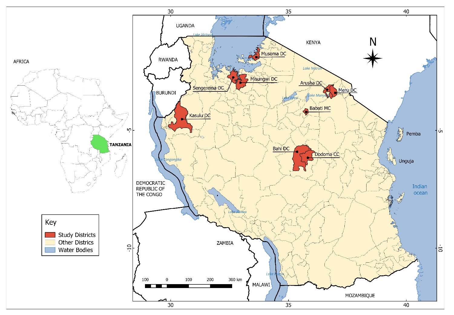
AbstractBackground: Lumpy skin disease virus (LSDV), a double-stranded DNA virus of the genus Capripoxvirus, causes Lumpy skin disease in cattle, leading to significant economic and production losses. In Tanzania, information on the molecular epidemiology of LSDV is limited, as its circulation has only been investigated in the Tanga, Pwani, and Rukwa regions. Aim: This study aimed to characterize circulating LSDV strains in Dodoma, Arusha, Manyara, Kigoma, Mwanza, and Mara regions, which are densely populated with cattle in Tanzania, to understand genetic diversity in these areas. Methods: Blood and skin biopsy samples (n=33 each) were collected from nine districts in Tanzania. Molecular detection was performed by targeting the P32 gene, and genetic variability was assessed by amplifying and sequencing the G protein-coupled chemokine receptor (GPCR) gene. Nucleotide sequences were translated into amino acid sequences using the ExPASy Translate tool, and then both nucleotide and amino acid sequences were aligned, followed by phylogenetic analysis. Results: Of the 33 blood and 33 skin biopsy samples tested, 20 skin biopsy and 3 blood samples were polymerase chain reaction-positive for LSDV based on the GPCR gene. Multiple sequence alignment revealed nucleotide substitutions (A→C) at positions 10 and 34 and an amino acid substitution (T→P) at position 12 in some Tanzania field isolates, while others exhibited unique amino acid signatures at positions A11, T12, T34, S99, and P199. Phylogenetic analysis demonstrated that the LSDV isolates obtained from Tanzania clustered closely with one another, as well as with reference strains from Africa, Asia, Europe, and Eurasia. Additionally, the Tanzania field isolates formed a different cluster from most reference vaccine strains, suggesting notable genetic variation between circulating field viruses and vaccine-derived strains. However, an exception was observed with the Kenya vaccine strains (KP663708 and KJ818282), which clustered closely with the Tanzania field isolates, indicating a close genetic relationship with these particular vaccine strains. Conclusion: Tanzanian LSDV isolates are genetically similar to the reference strains but distinct from most vaccine strains, confirming the circulation of wild-type viruses and highlighting the need for targeted control measures, including monitoring emerging variants and regulating animal movement. Keywords: G protein-coupled chemokine receptor gene, Lumpy skin disease virus, Molecular detection, Phylogenetic analysis, Tanzania. IntroductionLumpy skin disease virus (LSDV) is a causative agent of LSD, which primarily infects cattle, leading to huge economic losses in livestock industries (Sharawi and Abd El-Rahim, 2011; Molla et al., 2017). The LSDV, a double-stranded DNA virus, belongs to the Capripoxvirus genus (family Poxviridae, subfamily Chordopoxvirinae) together with sheep pox virus (SPPV) and goat pox virus (GTPV) (Tulman et al., 2001). The virus is mainly transmitted through arthropod vectors, such as Aedes aegypti mosquitoes, biting flies, and tick species (Lubinga et al., 2014; Farah Gumbe, 2018). Non-vector transmission occurs through artificial insemination and shared food and water between infected and susceptible cattle (Annandale et al., 2014; Aleksandr et al., 2020). Lumpy skin disease (LSD) is clinically characterized by skin nodules, fever, lesions in the mouth, nasal secretion, loss of appetite, swelling of the limbs, lachrymation, and reluctance to move (Davies, 1991; Ayelet et al., 2014). Additionally, LSD negatively affects livestock production through skin damage, reduced milk production, weight loss, infertility in bulls, abortion, trade restrictions, and loss of draught power, resulting in substantial economic losses (Lubinga et al., 2014; Bich et al., 2024). Furthermore, LSD exhibits a high morbidity rate ranging from 3% to 85%, with a mortality rate of approximately 10%, which occurs as a consequence of a secondary infection (Davies, 1991; Ayelet et al., 2014; Mulatu and Feyisa, 2018). The morbidity and mortality rates of LSD varied widely between regions due to host susceptibility, viral virulence, and vector abundance (Mulatu and Feyisa, 2018). Several strategies, including early detection of outbreaks, restrictions on animal movement, vector control, quarantine measures, and mass vaccination campaigns, have been used to prevent the spread of LSD (Gupta et al., 2020). However, vaccination is the most effective method for managing the disease in endemic areas (Mulatu and Feyisa, 2018). Live attenuated vaccines (LAVs) are used globally to prevent LSD in cattle; however, some studies have documented challenges such as reversion to virulence, accessibility, and cost-effectiveness (Abutarbush et al., 2016; Sprygin et al., 2018; Haegeman et al., 2021; Tuppurainen et al., 2021). In Tanzania, LSD vaccination mainly uses LAVs, which are locally produced and imported from neighboring countries, including Kenya. In Tanzania, vaccination is highly coordinated by individual farmers, although its coverage is not well established (Makoga et al., 2024). However, effective disease control relies on the presence of molecular epidemiological information, which guides control strategies (Snow, 2011). The LSDV genome, which is relatively stable, can be detected in various specimens, including skin nodules, blood, ulcerations, semen, and milk secretions (Annandale et al., 2014; Bedeković et al., 2018). Various diagnostic techniques used in LSDV detection include the virus neutralization test, enzyme-linked immunosorbent assay technique, transmission electron microscopy, virus isolation, and polymerase chain reaction (PCR) (Mulatu and Feyisa, 2018; Gupta et al., 2020). Different LSDV gene targets, including the G protein-coupled chemokine receptor (GPCR) gene, P32 gene, RPO30 gene, Fusion gene, and EEV glycoprotein gene, have been characterized and sequenced to identify LSDV strains. The GPCR gene is a key target for distinguishing Capripoxviruses and has been widely used for LSDV characterization and phylogenetic analysis (Le Goff et al., 2009; El‐Tholoth and El‐Kenawy, 2016; Abdallah et al., 2018; Ochwo et al., 2020; Seerintra et al., 2022; Abdelsalam et al., 2023; Bich et al., 2024; Tran et al., 2024). Previous studies utilizing GPCR gene analysis revealed different LSDV variants circulating in Vietnam, while strains in Uganda showed a close relationship to reference sequences from Africa and Europe, highlighting its use for molecular epidemiology and subgrouping (Ochwo et al., 2020; Tran et al., 2024). The LSDV strains were 100% identical when analyzed using the P32 and RPO30 genes, despite the variation observed in the GPCR gene (Tran et al., 2024). Therefore, the information obtained from molecular epidemiology is useful in decision-making for improving LSD management and control (Ochwo et al., 2020). Previous molecular studies conducted in Tanzania documented the circulation of LSDV strains in Tanga (Northern zone), Pwani (Eastern zone), and Rukwa (Southern Highland zone) (Makoga et al., 2024). However, there is no information on the LSDV circulating strain in regions highly populated with cattle in Tanzania, including Dodoma (Central zone), Mwanza and Mara (Lake zone), Arusha and Manyara (Northern zone), and Kigoma (Western zone), which are explored in the present study. The current study focused on the highly conserved P32 gene for detecting the presence of LSDV and the variable GPCR gene for identifying genetic variability, as adopted from previous studies (Seerintra et al., 2022; Bich et al., 2024). This study aimed to generate the first molecular data on LSDV strains circulating in cattle in highly populated regions in Tanzania by characterizing the GPCR gene and analyzing its phylogenetic relationships. The findings of this study highlight the genetic diversity and phylogenetic relationships of LSDV strains circulating in Tanzania, contributing to a better understanding of disease dynamics and informing the development of effective surveillance and control strategies. Materials and MethodsDescription of the study areaAn outbreak-based study was conducted in nine districts of Tanzania, spanning four zones (Lake, Central, Northern, and Western), where the LSD outbreaks were reported between 2023 and 2024 (Fig. 1). In the Northern Zone, an outbreak study was conducted in the Meru District Council (3.336904° S, 36.763634° E) and Arusha District Council (3.457402° S, 36.805629° E), both located in the Arusha region, and in the Babati District Council (4.260240° S, 35.789764° E) in the Manyara region. These districts are characterized by a tropical savanna climate with notable geographical features, including Mount Meru in Meru District and Mount Hanang in Babati District, as well as proximity to Tarangire National Park and Lake Babati. The area experiences bimodal rainfall, with long rains from March to May and short rains from October to December, with annual rainfall ranging from 500 to 1,200 mm and mean annual temperatures ranging between 15°C and 34°C (Ghiglieri et al., 2012; Maleko et al., 2012). Together with topographical features and proximity to water bodies, these climatic conditions may play a role in shaping the distribution and abundance of vectors across the districts. For example, in Babati District, the malaria vector Anopheles arabiensis feeds on both humans and livestock and exhibits seasonal peaks in abundance following the rainy seasons (Mwanziva et al., 2011). The study was conducted across the Central Zone in the Bahi District (6.099329° S, 35.490185° E) and Dodoma City Council (6.1807206° S, 35.7495265° E), both located in the Dodoma Region. These districts experience a semi-arid climate with a unimodal rainfall pattern, receiving approximately 500–700 mm of rainfall annually, mainly between November and April, while mean annual temperatures range from 18°C to 30°C. The districts are characterized by swampy areas, seasonal wetlands, permanent water bodies, savanna vegetation, seasonal rivers, wells, and dams, which provide favorable conditions for the breeding of LSDV-associated insect vectors.
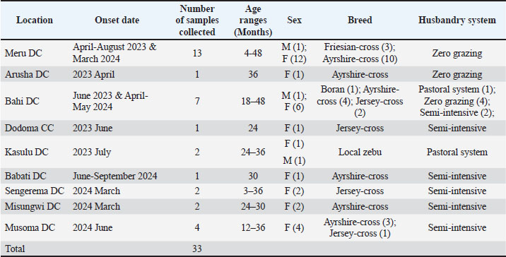
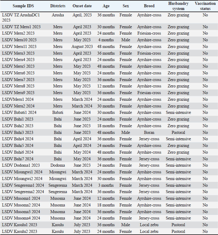
Fig. 1. Map showing the study locations in Tanzania from which the samples were obtained. The study was conducted in three districts in the Lake Zone: Sengerema (2.528289° S, 32.791196° E) and Misungwi (2.977130° S, 33.178453° E), both located in the Mwanza Region, and Musoma (1.898378° S, 33.736069° E) in the Mara Region. These districts experience a tropical savanna climate with bimodal rainfall ranging from 900 to 1,200 mm per year, with rains occurring from October to May, and temperatures ranging between 21°C and 32°C, influenced by their proximity to Lake Victoria. Their climatic conditions favor prolonged breeding of insect vectors, thereby contributing to the transmission of LSDV. The outbreak-based study was conducted in the Kasulu district (4.518115° S, 30.051556° E) of the Kigoma region within the western zone. The district is characterized by a tropical savanna climate with two rainy seasons, with the major rainy season occurring between March and May, and temperatures ranging from 20°C to 30°C. The climatic condition is influenced by Lake Tanganyika moisture, which contributes to significant rainfall. Sampling and collection of samplesBlood and skin biopsy samples (n=33 each) were purposively collected from clinically affected cattle, the natural host of LSDV, shortly after disease onset in the herds following LSD outbreaks. District Veterinary Officers informed the research team of outbreak locations, and Livestock Field Officers (LFOs) facilitated access to the affected herds. During sample collection, cattle owners were interviewed to obtain information about each affected animal, including vaccination history, age, sex, breed, and husbandry system. All cattle sampled during the outbreaks had no history of prior LSDV vaccination, and their demographic information was recorded and summarized in Table 1, with detailed data for each animal provided in Supplementary Table S1. Blood samples were collected in labeled sterile Ethylenediaminetetraacetic acid (EDTA) tubes packed in a cool box with ice packs, transported to the laboratory, and stored at −20°C. The skin biopsy samples were surgically collected under aseptic conditions using a sterile scalpel blade and local anesthesia (2ml of 2% lignocaine). The collected skin biopsy samples were placed in a labeled sterile 15-ml Falcon tube containing phosphate-buffered saline (PBS) with antibiotics and immediately stored in a cool box with ice packs. The samples were then transported to the Molecular Biology Laboratory at the Nelson Mandela African Institution of Science and Technology, stored at −80°C for further molecular analysis. Table 1. Summary of animal biodata sampled during 2023–2024 across nine districts of Tanzania.
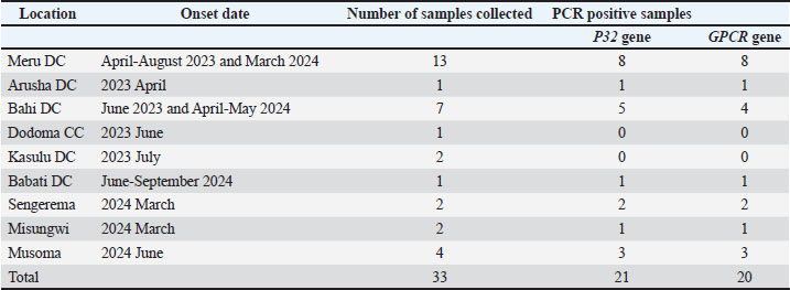
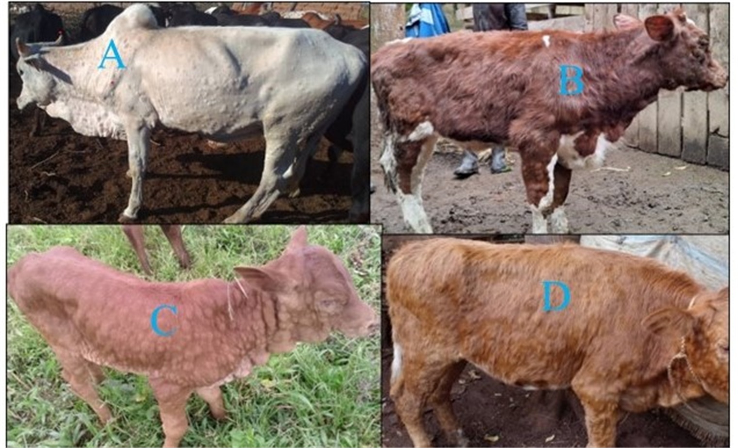
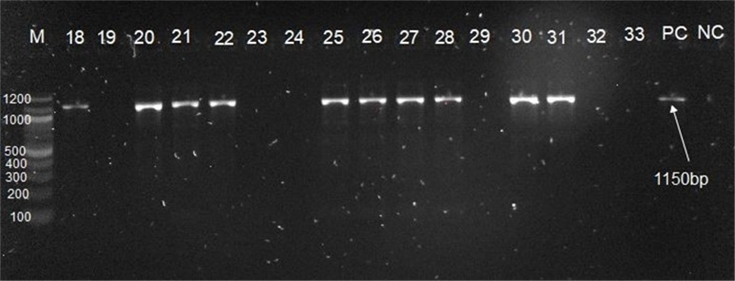
Preparation and storage of samplesThe procedure for preparing skin biopsy samples for analysis was performed in a Class II Biosafety Cabinet. The skin biopsy samples were chopped into small pieces using a sterile scalpel blade and then ground with a sterile mortar and pestle in PBS containing antibiotics to create a suspension of skin biopsy samples. The skin biopsy suspensions were then stored at −80°C for DNA extraction. DNA extractionDNA extraction was performed on skin biopsy suspensions and blood samples using the DNeasy Blood and Tissue Kit (Qiagen, Germany) according to the manufacturer’s instructions. A commercially available live attenuated LSDV Neethling vaccine (Lumpy Skin Disease Vaccine, Live, Neethling strain, Batch/Lot No. 7007) was used as a positive control, and nuclease-free water was used as a negative control. The DNA was eluted in 100 μl of elution buffer, its concentration was measured using a Nanodrop, and it was stored at −20°C. P32 gene amplification for detection of LSDV in cattleThe presence of LSDV was detected by PCR amplification of the P32 gene using OneTaq® Quick-Load® 2X Master Mix with Standard Buffer (New England Biolabs, Inc., USA), following the manufacturer’s instructions. The PCR amplification targeting the P32 gene was carried out using specific primers (forward 5′-ATGGCAGATATCCCATTATATGTTA-3′ and reverse 5′-GACGATAATCTAATTACATATG-3′), producing an amplicon size of 587 bp as described previously (Ireland and Binepal, 1998). The annealing temperature for P32 primers was optimized using gradient PCR across a range of 45°C to 55°C with DNA template volumes of 1, 3, and 5 µl. Each condition was run in two replicates, and the optimal annealing temperature of 51.3°C with 3 µl of DNA template was selected based on the best amplification consistency. The PCR master mix reactions were prepared at a final volume of 25 µl, with each reaction containing 12.5 µl master mix, 0.5 µl of each primer, 8.5 µl nuclease-free water, and 3 µl DNA template. The amplification was performed on a Bio-Rad thermal cycler machine under thermal conditions, including an initial denaturation at 94°C for 3 minutes for 1 cycle, followed by 34 cycles of denaturation for 30 seconds at 94°C, primer annealing for 45 seconds at 51.3°C, extension for 1 minute at 68°C, and final extension at 68°C for 5 minutes and a final hold at 4°C. All PCR reactions were performed in duplicate under sterile conditions using sterile consumables and separate workspaces to prevent cross-contamination. Each PCR run included a negative control (nuclease-free water) and a positive control (Neethling strain, live attenuated LSDV vaccine). The PCR products were then separated using 1.5% gel agarose, a 100 bp DNA ladder, and Tris-Borate-EDTA (TBE) buffer, stained with ethidium bromide, and visualized under a UV transilluminator to confirm LSDV-positive samples with a band size of 587 bp. GPCR gene amplification for genetic analysisAll samples positive for the P32 gene were amplified, targeting the GPCR gene (~1,150 bp) for genetic analysis, as described by Le Le Goff et al. (2009). The GPCR gene was amplified using OneTaq® Quick-Load® 2X Master Mix with Standard Buffer (New England Biolabs, Inc., USA), according to the manufacturer’s instructions, with specific primers: forward 5′-TTAAGTAAAGCATAACTCCAACAAAAATG-3′ and reverse 5′-TTTTTTTATTTTTTATCCAATGCTAATACT-3′ (Tulman et al., 2001). The annealing temperature for GPCR primers was optimized using gradient PCR over a temperature range of 48°C to 65°C with DNA template volumes of 1, 3, and 5 µl. Each setup was run in duplicate, and the optimal annealing temperature of 54.6°C with 3 µl of DNA template was chosen due to its consistent amplification. The PCR master mix reactions were prepared in a final volume of 25 µl, whereby one reaction contained 12.5 µl master mix, 0.5 µl of each primer, 8.5 µl nuclease-free water, and 3 µl DNA template. The GPCR gene amplification was performed on a Bio-Rad thermal cycler machine under thermal conditions, including an initial denaturation at 94°C for 3 minutes for 1 cycle, followed by 34 cycles of denaturation for 30 seconds at 94°C, primer annealing for 45 seconds at 54.6°C, extension for 1 minute at 68°C, and final extension at 68°C for 5 minutes and a final hold at 4°C. Each PCR reaction was performed in duplicate under sterile conditions using sterile consumables and separate workspaces to prevent cross-contamination. A negative control (nuclease-free water) and a positive control were included for every PCR reaction. The PCR products were separated using 1% gel agarose, a 100 bp DNA ladder, and TBE buffer, then stained with ethidium bromide and visualized under a UV transilluminator to confirm LSDV-positive samples with a band size of ~1,150 bp. Nucleotide sequencingThe PCR products positive for GPCR gene amplification following agarose gel electrophoresis were sequenced. The 20 GPCR gene-positive skin biopsy samples were sent to Macrogen (Amsterdam, the Netherlands) for purification and sequencing. The PCR products were purified using Macrogen standard proprietary EZ-Seq purification technology and sequenced directly using a BigDye Terminator Cycle Sequencing Kit (Applied Biosystems, USA) with a genetic analyzer (ABI 3730xl System from Applied Biosystems) to obtain nucleotide sequences. The forward and reverse primers used for GPCR gene amplification were also used for DNA sequencing. Sequencing depth for all samples was based on both direction (forward and reverse) coverage, with the resulting reads covering the entire ~1,150 bp region of the GPCR gene to generate a reliable consensus sequence. Partial gene sequencing analysisThe raw DNA sequence data were received from Macrogen and assessed using Chromas v2.6.6 (2018) to evaluate chromatogram quality and obtain Phred scores. The raw DNA sequence data were then edited and assembled to obtain consensus sequences using De Novo Assembly in Geneious Prime software version 2025.1. Then, the consensus sequences were visually inspected against chromatograms to verify their accuracy. For similarity and local alignment, the consensus sequences were subjected to the Basic Local Alignment Search Tool through the National Center for Biotechnology Information to determine the identity of the obtained sequences with other published GPCR gene sequences of LSDVs available in GenBank. After a BLAST search, LSDV reference sequences were selected based on the isolates’ nucleotide similarity and origin. Multiple sequence alignment was performed using the Multiple Sequence Comparison by Log-Expectation algorithm with the codon option in Molecular Evolutionary Genetics Analysis version 12 (MEGA 12) software to facilitate comparison. The percentage of sequence identity and genetic distance among sequences were calculated using Geneious Prime software version 2025.1 and MEGA version 12 software, respectively. The LSDV reference sequences from published vaccines and sequences from African and non-African countries were retrieved from GenBank for comparison with Tanzania field isolates. The retrieved nucleotide sequences were also translated into amino acid sequences using the ExPASy Translate tool, followed by multiple sequence alignment to check for LSDV-specific signatures of the amino acids. Phylogenetic analysis was performed using MEGA version 12 software to determine the genetic relatedness of Tanzanian LSDV field isolates with other isolates from African and non-African countries, as well as vaccine isolates. Therefore, the phylogenetic tree was created using the maximum likelihood method in MEGA version 12 software, with the Tamura 3-parameter model for nucleotide sequences and the Jones–Taylor–Thornton (JTT) model for amino acid sequences (Kumar et al., 2024). The phylogenetic tree was estimated using the bootstrap analysis method with 1,000 replicates and was thus annotated and visualized using the interactive tree of life (iTOL) tool (Letunic and Bork, 2024). Ethical approvalEthical approval was obtained from the Kibong'oto-Nelson Mandela-Cedha Health Research Ethical Committee (approval number: KNCHREC00009/09/2023). Official communication was also conducted through livestock extension officers. ResultsInvestigation of LSD outbreakThirty-three cattle suspected of being infected with LSDV were sampled from nine districts of Lake, Central, Northern, and Western zones, in Tanzania between 2023 and 2024. The sampled animals exhibited various clinical signs, with skin nodules being the most prominent, followed by a drop in milk production, loss of appetite, reluctance to move, and swelling of the limbs (Fig. 2). The animals were all unvaccinated against LSDV, and their ages ranged from 3 to 48 months. According to farmer reports, the sampled animals were unvaccinated due to limited awareness of vaccination benefits, fear about vaccinating vulnerable animals, and economic constraints related to herd size and vaccine dose packaging. LSDV was detected by PCR in 21 (64%) of 33 skin biopsy samples based on the P32 gene out of 33 samples (Table 2). Twenty-one PCR-positive samples for the P32 gene were used for a second PCR targeting the GPCR gene to determine genetic variability. From the second PCR, LSDV was confirmed by PCR in 20 (61%) skin biopsy samples, which were then sequenced using GPCR primers (Table 2 and Fig. 3). Moreover, the PCR results from blood samples revealed that 3 (9.1%) of 33 blood samples were positive for both the P32 and GPCR genes, indicating the detection rate within the collected samples. Among the 20 PCR-positive animals based on the GPCR gene, 18 were female and 2 were male, and their breed distribution included 13 Ayrshire-cross, 3 Jersey-cross, 3 Friesian-cross, and 1 Boran. Most animals were managed under a zero-grazing system (n=11), followed by a semi-intensive system (n=8) and pastoral system (n=1). Table 2. PCR results for skin biopsies samples collected from nine districts affected by LSD during 2023–2024 in Tanzania.
Fig. 2. Representative blood and skin biopsy samples of cattle with nodular skin presenting lumpy skin disease. Letters indicate sample locations: A=Bahi district; B=Meru district; C=Sengerema district; D=Babati district.
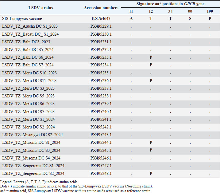
Fig. 3. Agarose gel electrophoresis showing amplification of the LSDV GPCR gene. Lane M, 100-bp DNA ladder; Lanes 18–33, tested samples with the numbers representing their ID; Lane PC, commercially available positive control vaccine (PC/VAC); Lane NC, negative control (RNase-free water); the expected band size is 1,150 bp. Sequence analysis of GPCRThe GPCR gene for 20 samples was successfully sequenced, and the resulting sequences were deposited in the GenBank database under accession numbers PX495229.1 to PX495248.1 (Table 3). All sequences had Phred scores above Q28, and the chromatograms showed clear peaks between nucleotide positions 20 and 1,130, indicating high-quality sequences. The Phred scores above Q28 and a representative chromatogram of the high-quality sequences are provided in Supplementary Figures 1 and 2. The percentage identity between the 20 Tanzania field isolates and vaccine strains ranged from 95% to 97%, with one isolate showing 95% identity, four isolates showing 96% identity, and 15 isolates showing 97% identity. Among Tanzania field isolates, sequence identity ranged from 98% to 100%, with one isolate at 98%, five at 99%, and fourteen at 100%. The overall mean genetic distance was 0.01. The overall genetic distance of 0.01 in a phylogenetic tree accounted for 1%, implying that, on average, 1% of aligned nucleotide positions between two sequences have undergone at least one mutation from the last shared common ancestor. Table 3. Signature amino acid positions in the GPCR gene identifying LSDV, their substitutions in some Tanzanian field isolates and GenBank accession numbers.
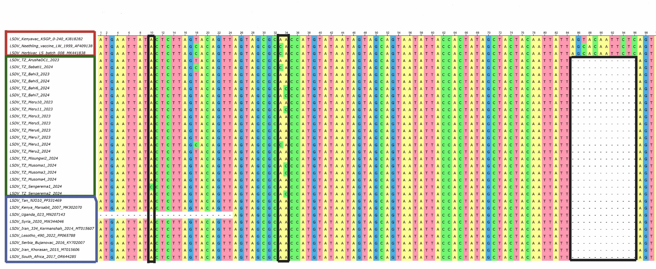
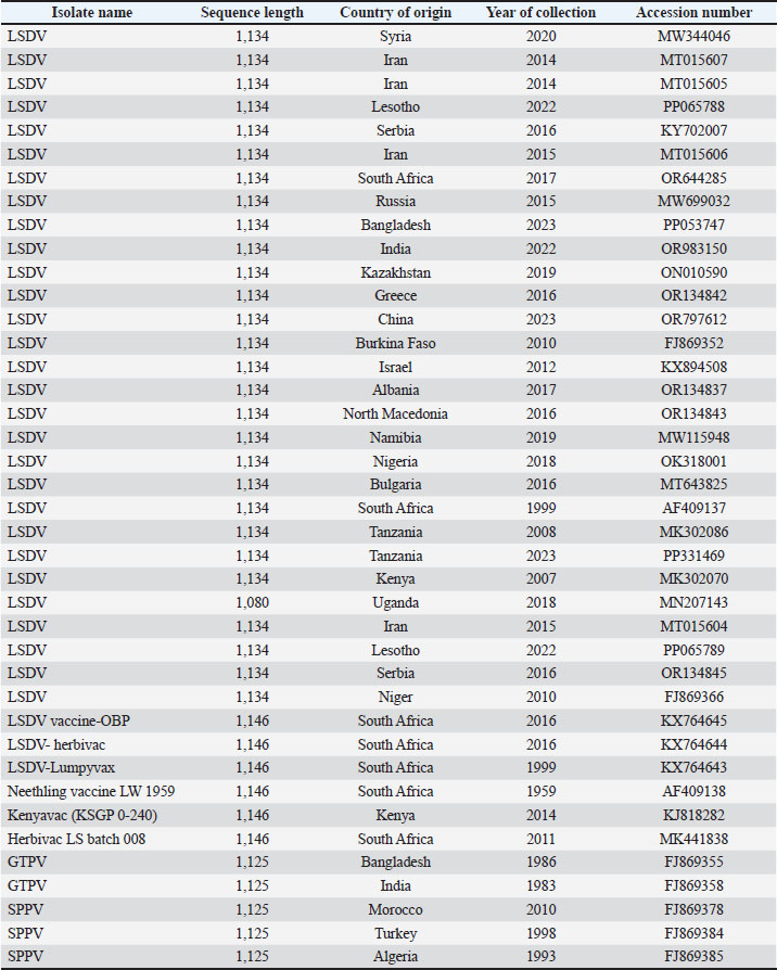
The nucleotide sequences of the GPCR gene from 20 Tanzanian samples were aligned with reference and vaccine strains to assess genetic relatedness. Details of the reference and vaccine strains and their GenBank accession numbers are provided in Supplementary Table S2. Nucleotide sequence analysis of the GPCR gene revealed a 12-nucleotide deletion at positions 85–96 in Tanzania and reference strains, while unique Adenine to Cytosine (A→C) substitutions at positions 10 and 34 were observed in some Tanzania strains but were absent in vaccine and reference strains (Fig. 4). Translation of these sequences into amino acids revealed signature residues at positions A11, T12, T34, S99, and P199, with a T→P substitution at position 12, which was observed in wild-type viruses from Bahi, Musoma, Sengerema, and Meru but absent in vaccine strains (Table 3). Together, these nucleotide and amino acid variations in the GPCR gene indicate that LSDV strains circulating in Tanzania are genetically distinct from vaccine strains.
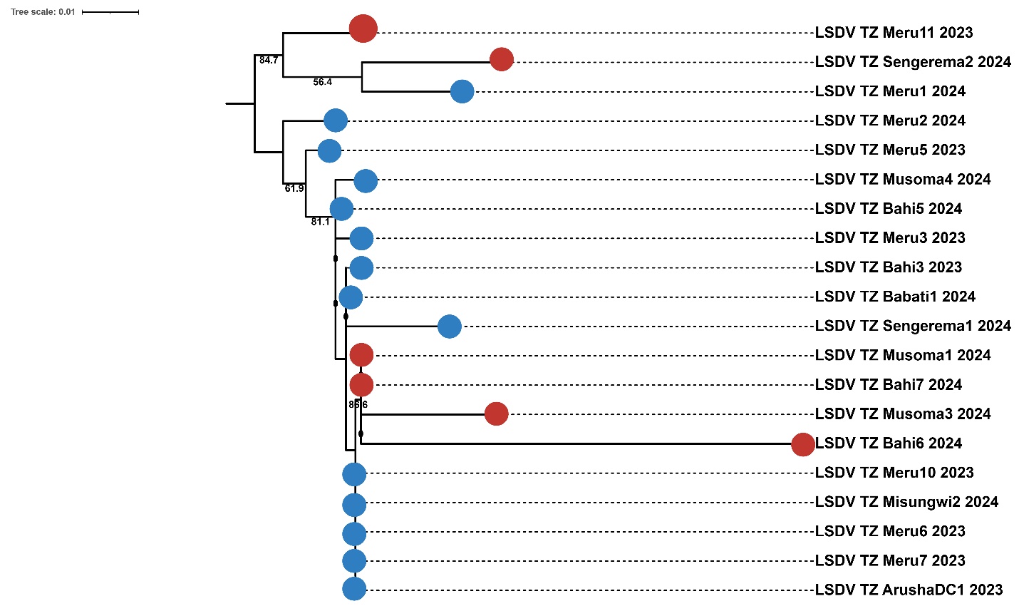
Fig. 4. Multiple sequence alignment showing a unique A→C substitution and a 12-nucleotide deletion (marked with a black rectangle). Phylogenetic analysis of GPCRPhylogenetic analysis of Tanzania isolates revealed clustering of Tanzanian isolates from different geographical locations (Fig. 5), indicating a lack of clear geographical segregation. The phylogenetic tree also showed that isolates with amino acid substitutions clustered with those without substitutions, with no distinct grouping based on this feature. From the phylogenetic tree, isolates from the Meru district clustered with those from several other districts.
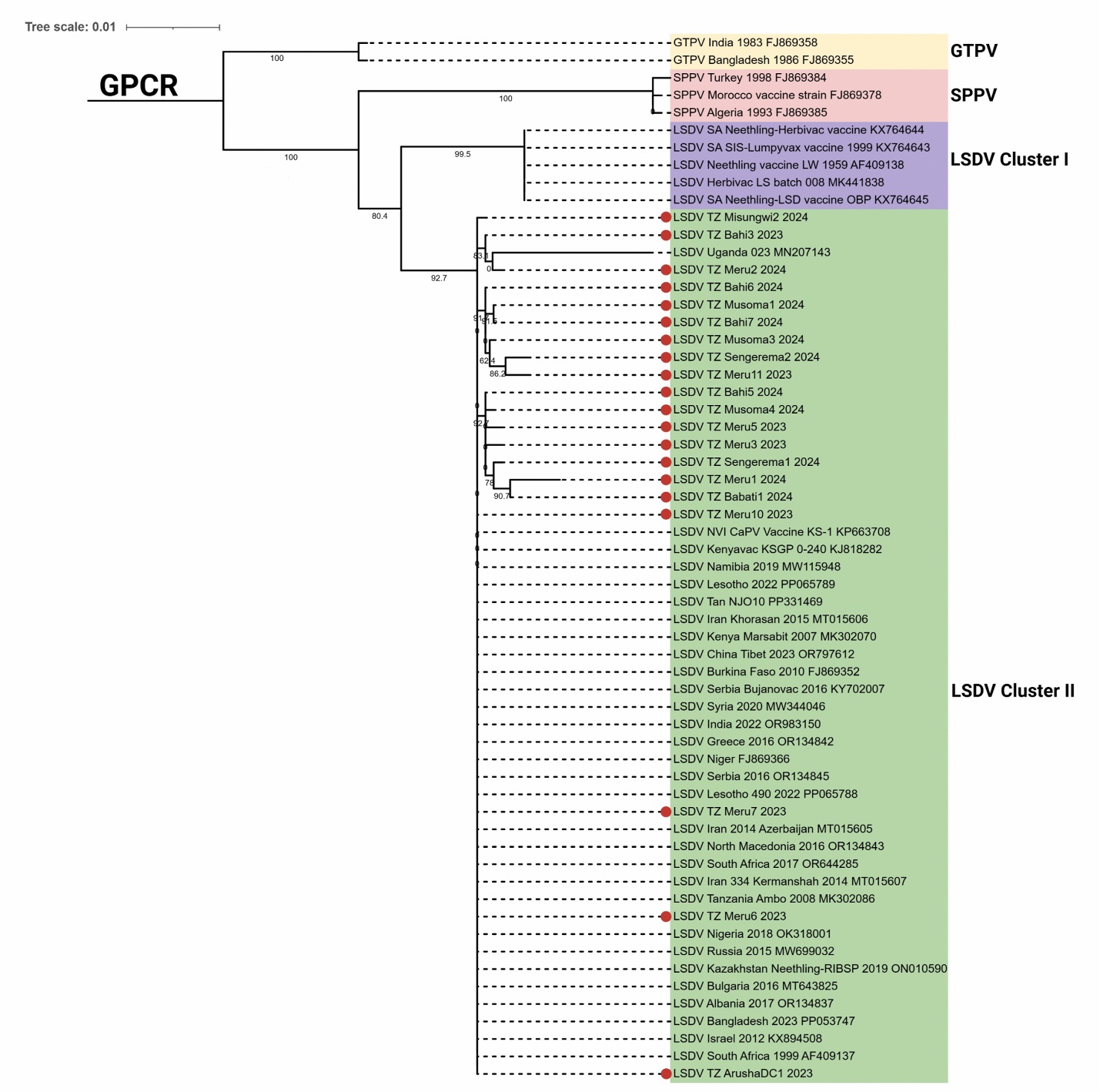
Fig. 5. Phylogenetic tree of the GPCR gene showing the relationships among Tanzania field strains based on amino acid sequences. The red circles indicate isolates with the amino acid substitution (T→P), while blue circles indicate isolates retaining T at the 12th position. The tree was generated using the maximum likelihood method and the JTT substitution model for amino acids, with 1,000 bootstrap replicates on MEGA version 12. The phylogenetic analysis of Tanzania isolates with reference strains revealed two subgroups of LSDV vaccine strains and field isolates (Fig. 6). The SPPV and GTPV, used as outgroup sequences, clustered in a separate clade (Fig. 6). Tanzania field isolates clustered closely with two reference vaccine strains, KSGP 0-240 (KJ818282) and KS-1 (KP663708), suggesting close genetic relatedness, but were genetically different from other reference vaccine strains. Although the Tanzania field isolates clustered closely with the vaccine strains KSGP 0-240 (KJ818282) and KS-1 (KP663708), they also grouped with reference strains from multiple regions, including Africa, Asia, Europe, and Eurasia (Fig. 6). These findings suggest that Tanzanian field isolates likely represent circulating wild-type viruses.
Fig. 6. Phylogenetic analysis of the GPCR gene of the Tanzania isolates (marked with a red circle) and a reference sequences, including field and vaccine strains from GenBank. The evolutionary distances were computed using the Tamura 3-parameter model with 1,000 bootstrap replicates on MEGA version 12 and visualized and annotated on iTOL. DiscussionThe detection of LSDV in skin and blood samples, along with GPCR-based analysis of genetic variability, provides a basis for understanding the epidemiology and molecular characteristics of LSDV. The countrywide molecular characterization of circulating LSDV strains in Tanzania has not been conducted, and information remains limited in several regions with high cattle populations, including Dodoma, Mwanza, Mara, Arusha, Manyara, and Kigoma. In this study, we investigated previously unexplored districts within these regions and identified the virus in all nine sampled districts. These findings provide novel insights into the genetic diversity of LSDV in the country. The clinical signs from the sampled animals, such as the appearance of skin nodules, fever, and reduced milk production, were consistent with a previous study (Babiuk et al., 2008), which reported similar signs. In this study, 64% of skin samples tested positive for LSDV based on the P32 gene, consistent with a previous study reporting high detection rate of 69.2% in skin tissue (Gupta et al., 2025). The high detection of LSDV in skin tissue suggests it is the most reliable specimen for LSDV detection, likely due to its higher viral load and the virus’s persistence in tissue for up to 92 days upon PCR detection (Tuppurainen et al., 2005). The low detection of LSDV in blood samples (9.4%) is consistent with previous reports indicating short viral persistence in blood (4–11 days), suggesting that blood samples are less reliable than skin tissue for LSDV detection (Tuppurainen et al., 2005; Zeynalova et al., 2016). The higher prevalence of LSD among female and crossbred cattle, which are predominantly managed under zero-grazing systems, may be due to their increased susceptibility to LSDV and higher likelihood of disease reporting. This finding is consistent with previous studies (Abera et al., 2015; Ochwo et al., 2019; Selim et al., 2021). The 12-nucleotide deletion identified in Tanzania field isolates represents an in-frame deletion that removes four amino acids without causing a frameshift; however, it contributes to genetic variation between field and vaccine strains. Although in-frame deletions could affect protein folding and potentially alter protein structure and function, their functional impact was not assessed in this study. Variations in the GPCR gene, including 12-nucleotide deletions, have been widely used as molecular markers to differentiate field strains from vaccine strains (El‐Tholoth and El‐Kenawy, 2016; Abdelsalam et al., 2023; Bich et al., 2024). Additionally, similar deletions have been reported in Ugandan wild-type strains (Ochwo et al., 2020). The unique A→C nucleotide substitutions together with the T→P amino acid substitution in some isolates highlight ongoing viral evolution and possible region-specific variation. Despite these changes, most sequences retained the characteristic LSDV signature residues at A11, T12, T34, S99, and P199, supporting their identity as LSDV (Le Goff et al., 2009). The presence of these unique signatures is consistent with reports from Uganda and Egypt (El‐Tholoth and El‐Kenawy, 2016; Ochwo et al., 2020), and the observed substitutions may contribute to strain-specific variability (Yadav et al., 2024; Uddin et al., 2025). In contrast, Ethiopian field isolates displayed an F→P substitution at a different GPCR position resulting from a C→T nucleotide mutation (Chibssa et al., 2021), suggesting that LSDV genetic diversity across regions is due to separate mutation events. Although the functional impact of the observed substitution T→P is unknown and not investigated in this study, these changes could potentially affect the ability of the virus to infect the host and suppress the host immune response, which are known functions of the GPCR protein. Generally, the nucleotide and amino acid variations indicate that circulating LSDV strains in Tanzania are genetically distinct from vaccine strains, reflecting regional evolution. Additional studies, including full genome sequencing, antigenic characterization and vaccine challenge experiments, are needed to determine whether these mutations affect vaccine efficacy. The clustering of isolates from Tanzania indicates that closely related LSDV strains are detected in different parts of the country. Additionally, the clustering of field isolates with and without amino acid substitution (T→P) suggests that this change alone is not a strong determinant of evolutionary lineage. From the phylogenetic tree, isolates from the Meru district clustered with several other districts because more cases were reported from the area. The higher number of cases reported from the Meru district may be associated with its proximity to the Tanzania Veterinary Laboratory Agency and the high proportion of crossbred cattle with exotic blood, which facilitates easy reporting of disease for intervention. Although the Tanzania field isolates clustered closely with the vaccine strains KSGP 0-240 (KJ818282) and KS-1 (KP663708), they also grouped with isolates from other countries, indicating close genetic relatedness. This suggests that Tanzania field isolates likely represent circulating wild-type viruses rather than vaccine-derived strains, which is consistent with the unvaccinated status of the sampled animals. In line with previous studies, these viruses may have been introduced into Tanzania through cross-border animal movement (Ochwo et al., 2020; Bianchini et al., 2023; Makoga et al., 2024). Active livestock markets characterize the northern part of Tanzania, particularly in the Meru district, which is near the Longido district. These markets facilitate the movement of animals across the Tanzania-Kenya border, which can lead to animal congregation and may increase the risk of disease transmission. These observations are consistent with previous studies describing livestock markets and cross-border movement as risk factors enhancing the introduction of LSDV to naive areas (Motta et al., 2019; Bianchini et al., 2023). An earlier study reported the clustering of field isolates with vaccine strains, which documented the occurrence of mixed features between the LSDV Neethling vaccine and field isolates (Chibssa et al., 2021). The distinct clustering of Tanzania field isolates and other vaccine strains in the phylogenetic tree highlights that they are genetically different, in agreement with previous studies (Ochwo et al., 2020; Makoga et al., 2024). Thus, these findings underscore ongoing viral evolution, suggest regional transmission, and confirm the circulation of wild-type viruses, emphasizing control measures to guide effective control strategies, including monitoring emerging variants and regulating animal movement. This study is limited by sequencing only 20 GPCR-positive biopsy samples; however, these represent all confirmed positive cases from the outbreak and provide robust insights into circulating LSDV variants in Tanzania. The study also relies on a single gene (GPCR) for viral characterization. The GPCR gene has been widely used in previous studies and is a well-established and highly informative marker for LSDV characterization and phylogenetic analysis (Abdallah et al., 2018; Ochwo et al., 2020; Seerintra et al., 2022; Abdelsalam et al., 2023; Bich et al., 2024; Tran et al., 2024). Previous comparative analyses of single-gene markers have shown that GPCR tends to recover a greater number of distinct clades than markers such as RPO30 and P32, supporting its utility in LSDV studies (Breman et al., 2023). Although the inclusion of additional genes or whole-genome sequencing would provide a comprehensive view of viral diversity and evolution, GPCR remains a reliable region for distinguishing circulating strains. Therefore, this study provides an overview of circulating LSDV strains in cattle-dense regions of Tanzania using GPCR gene analysis and contributes valuable information to guide effective LSD control and management strategies. ConclusionThis study provides a comprehensive molecular characterization of LSDV in regions with high cattle population in Tanzania. The observed genetic relatedness of Tanzania field isolates to reference sequences from other countries possibly resulted from international border movement, which underscores the need for control measures, including restrictions on animal movement. The genetic variation of Tanzania strains compared with reference vaccines confirms that the circulating viruses are predominantly wild-type and reflect ongoing viral evolution. Together, these findings enhance the understanding of LSDV genetic diversity in Tanzania and support the development of effective control strategies. AcknowledgmentThe authors acknowledge the Zonal Veterinary Centres in charge (ZVCs), District Veterinary Officers (DVOs) and Livestock Officers for their excellent cooperation throughout the period of sample collection. Conflict of interestThe authors declare no conflicts of interest. FundingThis study was funded by the Ministry of Education, Science and Technology of the United Republic of Tanzania through the Higher Education for Economic Transformation (HEET) project. Authors' contributionsPaskalina Badi Niima: Original draft writing, reviewing and editing, methodology, formal analysis, data curation, conceptualization, and investigation. Alexanda Danford Mzula: Supervision, writing, reviewing, and editing. Philemon Nyangi Wambura: Supervision, methodology, writing, reviewing, and editing. Gabriel Mkilema Shirima: Supervision, methodology, writing, reviewing, and editing. Data availabilityThe data used in this study will be made available on request. ReferencesAbdallah, F.M., El Damaty, H.M. and Kotb, G.F. 2018. Sporadic cases of lumpy skin disease among cattle in Sharkia province, Egypt: genetic characterization of lumpy skin disease virus isolates and pathological findings. Vet. World. 11(8), 1150–1158. Abdelsalam, A., El-Habbaa, A., Abo Hatab, E. and Sharawy, S. 2023. Molecular detection and phylogenetic analysis of recent isolate of lumpy skin disease virus from clinical cases in Menofia governorate between 2018 to 2020 based on GPCR gene. Benha. Vet. Med. J. 44(2), 84–87. Abera, Z., Degefu, H., Gari, G. and Kidane, M. 2015. Sero-prevalence of lumpy skin disease in selected districts of West Wollega zone, Ethiopia. BMC. Vet. Res. 11(1), 135. Abutarbush, S.M., Hananeh, W.M., Ramadan, W., Al Sheyab, O.M., Alnajjar, A.R., Al Zoubi, I.G., Knowles, N.J., Bachanek-Bankowska, K. and Tuppurainen, E.S.M. 2016. Adverse reactions to field vaccination against lumpy skin disease in Jordan. Transboundary. Emerg. Dis. 63(2), e213–e219. Aleksandr, K., Pavel, P., Olga, B., Svetlana, K., Vladimir, R., Yana, P. and Alexander, S. 2020. Emergence of a new lumpy skin disease virus variant in Kurgan Oblast, Russia, in 2018. Arch. Virol. 165(6), 1343–1356. Annandale, C.H., Holm, D.E., Ebersohn, K. and Venter, E.H. 2014. Seminal transmission of lumpy skin disease virus in heifers. Transboundary Emerg. Dis. 61(5), 443–448. Ayelet, G., Haftu, R., Jemberie, S., Belay, A., Gelaye, E., Sibhat, B., Skjerve, E. and Asmare, K. 2014. Lumpy skin disease in cattle in central Ethiopia: outbreak investigation and isolation and molecular detection of the virus. Rev. Sci. Tech. 33(3), 877–887. Babiuk, S., Bowden, T.R., Parkyn, G., Dalman, B., Manning, L., Neufeld, J., Embury-Hyatt, C., Copps, J. and Boyle, D.B. 2008. Quantification of lumpy skin disease virus following experimental infection in cattle. Transboundary Emerg. Dis. 55(7), 299–307. Bedeković, T., Šimić, I., Krešić, N. and Lojkić, I. 2018. Detection of lumpy skin disease virus in skin lesions, blood, nasal swabs and milk following preventive vaccination. Transboundary Emerg. Dis. 65(2), 491–496. Bianchini, J., Simons, X., Humblet, M.F. and Saegerman, C. 2023. Lumpy skin disease: a systematic review of mode of transmission, risk of emergence and risk entry pathway. Viruses 15(8), 1622. Bich, T., Trung, L., Hieu, T., Huyen, V., Hieu, T., Giang, H. and Chien, N. 2024. Characterization and molecular identification of the lumpy skin disease virus in cattle in the Mekong Delta of Vietnam. Open. Vet. J. 14(8), 1877–1895. Breman, F.C., Haegeman, A., Krešić, N., Philips, W. and De Regge, N. 2023. Lumpy skin disease virus genome sequence analysis: putative spatio-temporal epidemiology, single gene versus whole genome phylogeny and genomic evolution. Viruses 15(7), 1471. Chibssa, T.R., Sombo, M., Lichoti, J.K., Adam, T.I.B., Liu, Y., Elraouf, Y.A., Grabherr, R., Settypalli, T.B.K., Berguido, F.J., Loitsch, A., Sahle, M., Cattoli, G., Diallo, A. and Lamien, C.E. 2021. Molecular analysis of East African lumpy skin disease viruses reveals a mixed isolate with features of both vaccine and field isolates. Microorganisms 9(6), 1142. Davies, F.G. 1991. Lumpy skin disease, an African capripox virus disease of cattle. Br. Vet. J. 147(6), 489–503. El-Tholoth, M. and El-Kenawy, A.A. 2016. G-protein-coupled chemokine receptor gene in lumpy skin disease virus isolates from cattle and water buffalo (Bubalus bubalis) in Egypt. Transboundary Emerg. Dis. 63(6), e288–e295. Farah Gumbe, A.A. 2018. Review on lumpy skin disease and its economic impacts in Ethiopia. J. Dairy Vet. Anim. Res. 7(2), 39–46. Ghiglieri, G., Pittalis, D., Cerri, G. and Oggiano, G. 2012. Hydrogeology and hydrogeochemistry of an alkaline volcanic area: the NE Mt. Meru slope (East African Rift–Northern Tanzania). Hydrol. Earth Syst. Sci. 16(2), 529–541. Gupta, T., Patial, V., Bali, D., Angaria, S., Sharma, M. and Chahota, R. 2020. A review: lumpy skin disease and its emergence in India. Vet. Res. Commun. 44(3), 111–118. Gupta, V., Pravalika, A., Pandey, M.K., Mareddy, V., Jain, A.K., Singh, A., Nayak, A., Tripathi, S. and Rajoriya, S. 2025. Molecular detection, seroprevalence and biochemical analysis of lumpy skin disease virus. Viruses 17(3), 293. Haegeman, A., De Leeuw, I., Mostin, L., Campe, W.V., Aerts, L., Venter, E., Tuppurainen, E., Saegerman, C. and De Clercq, K. 2021. Comparative evaluation of lumpy skin disease virus-based live attenuated vaccines. Vaccines 9(5), 473. Ireland, D.C. and Binepal, Y.S. 1998. Improved detection of capripoxvirus in biopsy samples by PCR. J. Virol. Methods. 74(1), 1–7. Kumar, S., Stecher, G., Suleski, M., Sanderford, M., Sharma, S. and Tamura, K. 2024. MEGA12: molecular evolutionary genetic analysis version 12 for adaptive and green computing. Mol. Biol. Evol. 41(12), 263. Le Goff, C., Lamien, C.E., Fakhfakh, E., Chadeyras, A., Aba-Adulugba, E., Libeau, G., Tuppurainen, E., Wallace, D.B., Adam, T., Silber, R., Gulyaz, V., Madani, H., Caufour, P., Hammami, S., Diallo, A. and Albina, E. 2009. Capripoxvirus G-protein-coupled chemokine receptor: a host-range gene suitable for virus animal origin discrimination. J. Gen. Virol. 90(8), 1967–1977. Letunic, I. and Bork, P. 2024. Interactive tree of life (iTOL) v6: recent updates to the phylogenetic tree display and annotation tool. Nucleic. Acids. Res. 52(W1), W78–W82. Lubinga, J.C., Clift, S.J., Tuppurainen, E.S.M., Stoltsz, W.H., Babiuk, S., Coetzer, J.A.W. and Venter, E.H. 2014. Demonstration of lumpy skin disease virus infection in Amblyomma hebraeum and Rhipicephalus appendiculatus ticks using immunohistochemistry. Ticks. Tick-borne. Dis. 5(2), 113–120. Makoga, F.T., Chang’A, J.S., Meki, I.K., Mayenga, C., Settypalli, T.B.K., Bitanyi, S., Magidanga, B., Peter, E., Chengula, A., Cattoli, G. and Lamien, C.E. 2024. Detection and molecular characterization of lumpy skin disease and bovine papular stomatitis viruses in lumpy skin disease-suspected outbreaks in Tanzania. Virol. J. 21(1), 276. Maleko, D.D., Mbassa, G.N., Maanga, W.F. and Sisya, E.S. 2012. Impacts of wildlife-livestock interactions in and around Arusha National Park, Tanzania. Curr. Res. J. Biol. Sci. 4(4), 471–476. Molla, W., De Jong, M.C.M., Gari, G. and Frankena, K. 2017. Economic impact of lumpy skin disease and cost effectiveness of vaccination for the control of outbreaks in Ethiopia. Prev. Vet. Med. 147, 100–107. Motta, P., Porphyre, T., Handel, I.G., Hamman, S.M., Ngu Ngwa, V., Tanya, V.N., Morgan, K.L. and Bronsvoort, B.M.D.C. 2019. Characterizing livestock markets, primary diseases, and key management practices along the livestock supply chain in Cameroon. Front. Vet. Sci. 6, 101. Mulatu, E. and Feyisa, A. 2018. Review: lumpy skin disease. J. Vet. Sci. Technol. 9(535), 1–8. Mwanziva, C.E., Kitau, J., Tungu, P.K., Mweya, C.N., Mkali, H., Ndege, C.M., Sanga, A., Mtabho, C., Lukwaro, C., Myamba, J., Abdulalazizi, S., Magesa, S.M., Chilongola, J., Shekalaghe, S. and Mosha, F.W. 2011. Transmission intensity and malaria vector population structure in Magugu, Babati District in northern Tanzania. Tanzania. J. Health. Res. 13(1), 54–61. Ochwo, S., Vanderwaal, K., Munsey, A., Nkamwesiga, J., Ndekezi, C., Auma, E. and Mwiine, F.N. 2019. Seroprevalence and risk factors for lumpy skin disease virus seropositivity in cattle in Uganda. BMC Vet. Res. 15(1), 236. Ochwo, S., Vanderwaal, K., Ndekezi, C., Nkamwesiga, J., Munsey, A., Witto, S.G., Nantima, N., Mayanja, F., Okurut, A.R.A., Atuhaire, D.K. and Mwiine, F.N. 2020. Molecular detection and phylogenetic analysis of lumpy skin disease virus from outbreaks in Uganda 2017–2018. BMC. Vet. Res. 16(1), 66. Seerintra, T., Saraphol, B., Wankaew, S. and Piratae, S. 2022. Molecular identification and characterization of lumpy skin disease virus emergence from cattle in the northeastern part of Thailand. J. Vet. Sci. 23(5), 73. Selim, A., Manaa, E. and Khater, H. 2021. Seroprevalence and risk factors for lumpy skin disease in cattle in Northern Egypt. Trop. Anim. Health. Prod. 53(3), 350. Sharawi, S.S.A. and Abd El-rahim, I.H.A. 2011. The utility of polymerase chain reaction for diagnosis of lumpy skin disease in cattle and water buffaloes in Egypt. Revue Sci. Technique-OIE. 30(3), 821–830. Snow, M. 2011. The contribution of molecular epidemiology to the understanding and control of viral diseases of salmonid aquaculture. Vet. Res. 42(1), 56. Sprygin, A., Pestova, Y., Prutnikov, P. and Kononov, A. 2018. Detection of vaccine‐like lumpy skin disease virus in cattle and Musca domestica L. flies in an outbreak of lumpy skin disease in Russia in 2017. Transboundary Emerg. Dis. 65(5), 1137–1144. Tran, H., Truong, A., Tran, A., Chu, N., Nguyen, V. and Dang, H. 2024. A new variant of lumpy skin disease virus circulating in Vietnam based on sequencing analysis of GPCR gene. Open Vet. J. 14(7), 1701–1707. Tulman, E.R., Afonso, C.L., Lu, Z., Zsak, L., Kutish, G.F. and Rock, D.L. 2001. Genome of lumpy skin disease virus. J. Virol. 75(15), 7122–7130. Tuppurainen, E., Dietze, K., Wolff, J., Bergmann, H., Beltran-Alcrudo, D., Fahrion, A., Lamien, C.E., Busch, F., Sauter-Louis, C., Conraths, F.J., De Clercq, K., Hoffmann, B. and Knauf, S. 2021. Review: vaccines and vaccination against lumpy skin disease. Vaccines 9(10), 1136. Tuppurainen, E.S.M., Venter, E.H. and Coetzer, J.A.W. 2005. The detection of lumpy skin disease virus in samples of experimentally infected cattle using different diagnostic techniques. Onderstepoort J. Vet. Res. 72(2), 153–164. Uddin, M.B., Begum, R., Paul, J.K., Rahman, M.M., Cho, H.S. and Hossain, M.M. 2025. Genome-wide characterization of lumpy skin disease virus isolate from Bangladesh. Sci. Rep. 15(1), 36147. Yadav, P., Kumar, A., Nath, S.S., Devasurmutt, Y., Shashidhar, G., Joshi, M., Puvar, A., Sharma, S., Raval, J., Pandit, R., Chavda, P., Nagaraj, S., Revanaiah, Y., Patil, D., Raval, S.K., Raval, J., Kanani, A., Thakar, F., Kumar, N., Reddy, G.B.M., Joshi, C., Gulati, B.R. and Tatu, U. 2024. Unravelling the genomic origins of lumpy skin disease virus in recent outbreaks. BMC. Genomics. 25(1), 196. Zeynalova, S., Asadov, K., Guliyev, F., Vatani, M. and Aliyev, V. 2016. Epizootology and molecular diagnosis of lumpy skin disease among livestock in Azerbaijan. Front. Microbiol. 7, 1022. Supplementary MaterialSupplementary Table S1. Individual animal characteristics recorded during the 2023–2024 LSDV outbreaks.
Supplementary Table S2. Comprehensive information for the selected reference strains and vaccine strains.
Supplementary Fig. S1. Representative sequencing chromatogram of GPCR showing clear peaks across nucleotide positions 20–1,130.
Supplementary Fig. S2. Representative Phred quality scores of GPCR gene sequences, with all reads showing the values above Q28. | ||
| How to Cite this Article |
| Pubmed Style Niima PB, Mzula AD, Wambura PN, Shirima GM. Molecular characterization and phylogenetic analysis of lumpy skin disease virus detected in Tanzania between 2023 and 2024. Open Vet. J.. 2026; 16(6): 3384-3400. doi:10.5455/OVJ.2026.v16.i6.10 Web Style Niima PB, Mzula AD, Wambura PN, Shirima GM. Molecular characterization and phylogenetic analysis of lumpy skin disease virus detected in Tanzania between 2023 and 2024. https://www.openveterinaryjournal.com/?mno=285478 [Access: June 26, 2026]. doi:10.5455/OVJ.2026.v16.i6.10 AMA (American Medical Association) Style Niima PB, Mzula AD, Wambura PN, Shirima GM. Molecular characterization and phylogenetic analysis of lumpy skin disease virus detected in Tanzania between 2023 and 2024. Open Vet. J.. 2026; 16(6): 3384-3400. doi:10.5455/OVJ.2026.v16.i6.10 Vancouver/ICMJE Style Niima PB, Mzula AD, Wambura PN, Shirima GM. Molecular characterization and phylogenetic analysis of lumpy skin disease virus detected in Tanzania between 2023 and 2024. Open Vet. J.. (2026), [cited June 26, 2026]; 16(6): 3384-3400. doi:10.5455/OVJ.2026.v16.i6.10 Harvard Style Niima, P. B., Mzula, . A. D., Wambura, . P. N. & Shirima, . G. M. (2026) Molecular characterization and phylogenetic analysis of lumpy skin disease virus detected in Tanzania between 2023 and 2024. Open Vet. J., 16 (6), 3384-3400. doi:10.5455/OVJ.2026.v16.i6.10 Turabian Style Niima, Paskalina Badi, Alexanda Danford Mzula, Philemon Nyangi Wambura, and Gabriel Mkilema Shirima. 2026. Molecular characterization and phylogenetic analysis of lumpy skin disease virus detected in Tanzania between 2023 and 2024. Open Veterinary Journal, 16 (6), 3384-3400. doi:10.5455/OVJ.2026.v16.i6.10 Chicago Style Niima, Paskalina Badi, Alexanda Danford Mzula, Philemon Nyangi Wambura, and Gabriel Mkilema Shirima. "Molecular characterization and phylogenetic analysis of lumpy skin disease virus detected in Tanzania between 2023 and 2024." Open Veterinary Journal 16 (2026), 3384-3400. doi:10.5455/OVJ.2026.v16.i6.10 MLA (The Modern Language Association) Style Niima, Paskalina Badi, Alexanda Danford Mzula, Philemon Nyangi Wambura, and Gabriel Mkilema Shirima. "Molecular characterization and phylogenetic analysis of lumpy skin disease virus detected in Tanzania between 2023 and 2024." Open Veterinary Journal 16.6 (2026), 3384-3400. Print. doi:10.5455/OVJ.2026.v16.i6.10 APA (American Psychological Association) Style Niima, P. B., Mzula, . A. D., Wambura, . P. N. & Shirima, . G. M. (2026) Molecular characterization and phylogenetic analysis of lumpy skin disease virus detected in Tanzania between 2023 and 2024. Open Veterinary Journal, 16 (6), 3384-3400. doi:10.5455/OVJ.2026.v16.i6.10 |


