Open Veterinary Journal, (2026), Vol. 16(6): 3932-3941
Research Article
10.5455/OVJ.2026.v16.i6.62
Neuroprotective potential of intranasal quercetin in the early phase of ischemic stroke: Evidence from a rat model
Ika Satya Perdhana1,2, Ekowati Handharyani1, Huda Shalahuddin Darusman1, Setyo Widi Nugroho3,
Hera Maheshwari1* and Sjaikhurrizal El Muttaqien4
1Department of Anatomy, Physiology and Pharmacology, The School of Veterinary Medicine and Biomedical Sciences (SVMBS), IPB University, Bogor, Indonesia
2Medical Faculty, Gunadarma University, Depok, Indonesia
3Medical Faculty, University of Indonesia, Jakarta, Indonesia
4Research Center for Vaccine and Drugs, Research and Innovation Agency (BRIN), Tangerang, Indonesia
*Corresponding Author: Hera Maheshwari. Department of Anatomy, Physiology and Pharmacology, The School of Veterinary Medicine and Biomedical Sciences (SVMBS), IPB University, Bogor, Indonesia. Email: hera_maheshwari [at] apps.ipb.ac.id
Submitted: 08/09/2025 Revised: 05/05/2026 Accepted: 19/05/2026 Published: 20/06/2026
© 2025 Open Veterinary Journal
This is an Open Access article distributed under the terms of the Creative Commons Attribution-Non Commercial-No Derivatives License (http://creativecommons.org/licenses/by-nc-nd/4.0/), which permits non-commercial re-use, distribution, and reproduction in any medium, provided the original work is properly cited, and is not altered, transformed, or built upon in any way.
ABSTRACT
Background: Ischemic stroke remains a major cause of morbidity and mortality worldwide, highlighting its significance as a critical public health concern. Oxidative stress plays a pivotal role in neuronal injury, and quercetin, a natural flavonoid with potent antioxidant properties, has shown promise as a neuroprotective agent. Intranasal administration provides a direct nose-to-brain delivery route, potentially enhancing brain delivery efficiency.
Aim: This study aimed to evaluate the potential neuroprotective effects of intranasally administered quercetin-loaded bovine serum albumin–polydopamine nanoparticles (NPs) in a rat model of ischemic stroke.
Methods: A total of 24 male Sprague–Dawley rats were divided into six groups using the Federer formula. Ischemic stroke was induced by middle cerebral artery occlusion for 60 minutes. Quercetin NPs were administered intranasally at 45 minutes post-induction. Neurological outcomes were assessed using the Bederson score at 24 and 48 hours post-induction. Brain tissue samples were collected for histopathological evaluation, malondialdehyde (MDA) quantification, and superoxide dismutase (SOD) activity measurement.
Results: Rats receiving intranasal quercetin NPs (Groups 3–5) showed improved neurological scores at 48 hours compared with the untreated ischemic groups, which exhibited persistent deficits. Histopathological analysis revealed reduced neuronal necrosis in the treated groups. The biochemical analysis indicated a trend toward lower MDA levels and higher SOD activity in the quercetin-treated rats.
Conclusion: Intranasal quercetin NPs have a potential neuroprotective effect, possibly associated with modulation of oxidative stress. However, the results should be considered preliminary, and further studies with larger sample sizes and more comprehensive outcome measures are required to confirm these findings.
Keywords: Early-phase neuroprotection, Intranasal delivery, Ischemic stroke model, Quercetin nanoparticles.
Introduction
Stroke remains a major public health and medical problem (Karenberg, 2020). Stroke is recognized as the second most common factor responsible for premature death and chronic disability globally (Kuriakose & Xiao, 2020). Ischemic stroke remains a leading cause of mortality and long-term disability, accounting for 65.3% of all deaths worldwide (Feigin et al., 2025). According to the updated World Health Organization ICD-11, cerebral ischemic stroke is an acute focal neurological dysfunction caused by focal infarction at a single or multiple sites of the brain confirmed by symptoms for more than 24 hours or neuroimaging (Groff et al., 2024).
Currently, the recommended therapy for ischemic stroke is tPA. It restores tissue perfusion, aiming to restore oxygen and nutrient delivery to ischemic brain tissue, but has a narrow therapeutic time range, which is only 4.5 hours from ischemic stroke onset (Barthels and Das, 2021). This short window poses a major obstacle, especially because many patients and communities remain unaware of the urgent need for timely stroke management (Fitriani et al., 2024).
Several factors influence the limited success rate of ischemic stroke therapy, one of which is the delay in accessing treatment. Therefore, timely intervention is critical, and the development of early, bridging therapies is essential to slow stroke progression before definitive treatment becomes available. Such early interventions must be based on the underlying mechanisms and subsequent pathological processes of ischemia. Despite advances in acute management, effective neuroprotective strategies targeting mechanisms of secondary brain injury remain limited, highlighting the need for novel therapeutic approaches.
The pathophysiology of ischemic stroke is complex and leads to neuronal death and neurological function loss (Dewi & Fitraneti, 2024). Neuronal injury primarily results from oxygen depletion in ischemic areas (Jiang et al., 2017), which disrupts mitochondrial electron transport and cellular redox homeostasis, leading to increased generation of reactive oxygen species (ROS) accompanied by the release of pro-inflammatory mediators, ultimately triggering cellular damage and death (Soares et al., 2019).
The increase in ROS can be counteracted by endogenous and exogenous antioxidants. Exogenous antioxidants are widely available from natural sources. One of the most abundant antioxidants is quercetin, which is commonly found in onions, vegetables, and fruits. Quercetin is readily accessible and has been used for its protective role against cellular damage (Yang et al., 2020). Quercetin reduces oxidative stress and delays the progression of ischemic stroke (Guo et al., 2022). It exerts multimodal potential neuroprotective effects by scavenging ROS, suppressing inflammatory signaling, and modulating platelet activation; thus, it counteracts the mechanisms underlying ischemic brain injury (Zhang et al., 2022). Conversely, quercetin protects blood–brain barrier (BBB) integrity during focal cerebral ischemia and reduces neuronal apoptosis (Guo et al., 2022). Therefore, enhancing quercetin delivery to the brain during the acute phase of ischemic stroke may attenuate oxidative damage, as evidenced by malondialdehyde (MDA) and superoxide dismutase (SOD) levels modulation. Despite extensive evidence supporting quercetin’s antioxidant and neuroprotective properties, its translation into ischemic stroke therapy remains hampered by its limited bioavailability.
Bioavailability plays a crucial role in the effectiveness of pharmacological therapy and is affected by various factors, including the mode of administration and hepatic metabolism (Stielow et al., 2023). There is a type of drug administration that can reduce hepatic metabolism and shorten drug delivery to the brain as the target organ. It offers a unique nose-to-brain pathway that bypasses the restrictive BBB, which limits the penetration of most therapeutic agents into the brain following oral or intravenous administration. The nasal cavity provides direct anatomical connections to the central nervous system (CNS) via the olfactory and trigeminal nerve pathways. This pathway enables therapeutic agents to rapidly reach the brain regions without first entering the systemic circulation. This mechanism allows drugs to enter the brain within minutes while avoiding the selective endothelial tight junctions of the BBB that exclude most molecules from the CNS (Selvaraj et al., 2018). These mechanisms also enhance drug bioavailability and reduce systemic exposure with all related side effects. This provides a clear mechanistic rationale for investigating the use of intranasal quercetin delivery as a targeted early intervention strategy (Nguyen and Duong, 2025).
The potential of intranasal delivery to enable rapid quercetin nose-to-brain transport during the acute phase of ischemic stroke has not been adequately investigated. Intranasal administration offers a non-invasive nose-to-brain pathway that may enable early neuroprotection during the acute phase of ischemic injury. However, small dosing volumes and rapid mucociliary clearance limit intranasal administration, which may reduce drug residence time and therapeutic efficacy (Tamaddon et al., 2021). To address these challenges, nanoparticle (NP)-based formulations have been proposed as enabling platforms to enhance nasal residence and improve drug transport to the brain (Erdő et al., 2018). Therefore, the present study aimed to evaluate the neuroprotective potential of intranasally administered quercetin-loaded NPs in an experimental model during the early phase of ischemic stroke, with a particular focus on early oxidative stress modulation and neuronal preservation. This study introduces a novel therapeutic paradigm by combining NP-based formulation with intranasal delivery to maximize brain targeting of quercetin during the critical early window of ischemic injury.
Although various antioxidant compounds have been investigated in experimental ischemic stroke, most studies have focused on systemic administration or long-term outcomes. This study addresses a distinct gap by evaluating the potential neuroprotective effects of intranasal NP quercetin in the early phase of ischemia in a rat model. By targeting this early window, the study explores the potential of quercetin to slow the progression of ischemia-induced brain damage rather than to reverse established injury and examined its association with brain tissue-based oxidative stress and histopathological outcomes. These findings provide new evidence supporting the use of intranasal nanocarrier strategies for quercetin delivery in cerebral ischemia.
Materials and Methods
Materials
The study was conducted from December 2023 to July 2025 at the Veterinary Teaching Hospital of IPB University, the Pathology Laboratory of SVMBS IPB University, and the National Research and Innovation Agency. The instruments used in this study included surgical tools and equipment for the preparation and examination of histopathological slides. The nylon filament used for middle cerebral artery (MCA) occlusion is a 6.0 nylon filament, which is modified by heating the tip of a monofilament to create a rounded end. This modification follows that of the previous researcher, (Ramli et al., 2017). This modification allows researchers to conduct ischemic stroke studies without relying on costly imported materials. Pure quercetin was obtained from Haihang Corporation, China.
Animals and experimental design
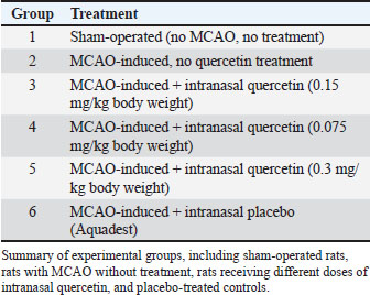
A total of 24 adult male Sprague–Dawley rats weighing 200–250 g were used in this study. The sample size was determined based on the Federer formula for experimental animal studies and ethical considerations following the 3R principles (Replacement, Reduction, and Refinement). A minimum of four animals per group was selected to allow valid statistical evaluation while minimizing animal use in this exploratory middle cerebral artery occlusion (MCAO) study. All rats were housed at the Veterinary Teaching Hospital, IPB University, and allowed to acclimate for 2 weeks under controlled conditions (22°C–24°C), with unrestricted access to food and water. This exploratory in vivo study did not implement formal randomization and investigator blinding. All surgical procedures, treatments, and outcome assessments were conducted using standardized protocols and performed by the same trained investigator to minimize variability. The animals were then assigned into six experimental groups: Group 1: sham-operated; Group 2: MCAO without treatment; Group 3: MCAO + quercetin (0.075 mg/kg); Group 4: MCAO + quercetin (0.15 mg/kg); Group 5: MCAO + quercetin (0.3 mg/kg); and Group 6: MCAO + placebo (Aquadest), as shown in Table 1. The sham-operated group underwent all surgical procedures identically, including anesthesia, neck incision, and vessel exposure, but without insertion of a filament to induce MCAO.
Table 1. Treatment groups.
Induction of ischemic stroke
A modified MCAO method was used to induce transient focal ischemia. Rats were administered intraperitoneal anesthesia with ketamine (70 mg/kg BW) and xylazine (4 mg/kg BW). A ~2 cm midline cervical incision was made to access the internal carotid artery (ICA). These modifications provide an accessible alternative to the standard MCAO method (Makkiyah and Sadewo, 2019). Similarly, Ramli et al. (2017) modified the occluder used in MCAO by heating the tip of a monofilament to create a rounded end, replacing the commercially available blunted monofilament, which is relatively expensive (Ramli et al., 2017). This modification allows researchers to conduct ischemic stroke studies without relying on costly imported materials.
A blunted 6.0 nylon filament was modified and attached to an intravenous catheter that was inserted and advanced within the ICA until it obstructed the MCA, producing ischemia in the distal territory. The filament was carefully removed after 60 minutes of occlusion to restore blood flow (Perdhana et al., 2025). No formal exclusion criteria were predefined for unsuccessful occlusion. However, all procedures were performed by the same experienced operator following a standardized MCAO protocol, and all animals exhibited neurological deficits that were scored with the Bederson score, indicating that the model was reliable.
Rats in group 3–5 that underwent ischemia induction received intranasal quercetin administration through the ipsilateral nostril as the ischemic site (left side) each for the same volume, 40 µl. To model acute-phase ischemic stroke therapy, quercetin administration was done 45 minutes after induction. All rats were evaluated neurologically using the Bederson score at 1, 24, and 48 hours after ischemic induction, and then terminated. The Bederson score represents a composite neurological score ranging from 0 to 3, calculated as the sum of three binary parameters (forelimb flexion, resistance to lateral push, and circling behavior), where each abnormal response is scored as 1. The Bederson score is calculated as follows: Thus, a total score of three indicates the simultaneous presence of all three neurological deficits.
A previous study showed that infarct volume and the number of necrotic neurons peaked at 24 hours and started to decrease at 48 hours after ischemic induction (Ramli et al., 2017). These findings provided the rationale for performing termination at 48 hours after ischemic induction.
Preparation of quercetin NPs
Quercetin NPs were developed using bovine serum albumin (BSA) and polydopamine (PDA) as stabilizing agents. Quercetin powder was dissolved in ethanol (15 mg/ml), and BSA was dissolved in a 4-(2-hydroxyethyl)-1-piperazineethanesulfonic acid (HEPES) buffer (20 mg/ml). The quercetin solution was added dropwise to the BSA solution, followed by the addition of ethylcarbodiimide (1 mg) as a crosslinking agent. The mixture was stirred for 3 hours at room temperature for 3 hours.
Subsequently, 500 l of dopamine hydrochloride (0.5 mg/ml in HEPES buffer) was added (500 µl) and stirred overnight to form a PDA coating on the NPs. The resulting quercetin–BSA–PDA NPs were purified by three cycles of centrifugation at 10,000 rpm, washed with distilled water, and freeze-dried for storage (Li et al., 2023). NPs were prepared using a standardized protocol with identical material concentrations and preparation conditions for all batches.
Statistical analysis
Data from histopathological analysis (percentage of necrotic brain cells) are expressed as mean ± standard deviation (SD). Statistical comparisons among groups for histopathological data (percentage of necrotic cells) were performed using one-way analysis of variance followed by Tukey’s post hoc test. A value of p < 0.05 was considered statistically significant. Only biologically relevant pairwise comparisons are reported in this manuscript. Statistical analyses were conducted using the Statistical Package for the Social Sciences version 16.0. Neurological deficit scores (Bederson score) were presented descriptively without statistical analysis because of their ordinal nature and limited sample size. The behavioral outcomes assessed using the Bederson score were analyzed descriptively due to the ordinal nature of the data and the limited sample size (n=4 per group).
Ethical approval
The Animal Ethics Committee of the School of Veterinary Medicine and Biomedical Sciences approved this study ethically (Approval No. 140/KEH/SKE/XII/2023), and all animal procedures adhered to institutional and national guidelines.
Results
Physicochemical characterization of quercetin NPs
Dynamic light scattering analysis demonstrated a consistent particle size distribution and acceptable polydispersity indices (PDIs) across formulations. Alb NPs exhibited a narrow size distribution with a low PDI, indicating high particle homogeneity (Table 2). The particle size of surface functionalization with PDA (Alb-PDA NPs) increased, consistent with the formation of a PDA coating layer, while maintaining a relatively narrow PDI.
Table 2. Physicochemical characteristics of the prepared NPs.
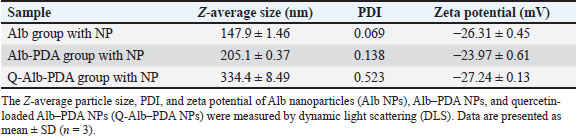
All NP formulations exhibited zeta potential values below −20 mV, indicating sufficient electrostatic repulsion and acceptable colloidal stability for biological applications.
Clinical observations
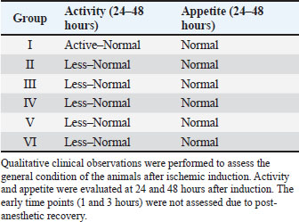
Clinical assessment was conducted by evaluating the activity and appetite of the rats, as shown in Table 4. Behavioral observations at 1 hour post-induction were not interpreted due to reduced spontaneous activity after anesthetic administration, due to the ischemic induction. None of the rats consumed food or water during the study period. By 24 hours post-surgery, rats in groups 1, 3, 4, and 5 had resumed normal activity, whereas those in groups 2 and 6 remained less active. Although food and water intake were observed in all groups at 24 hours, it remained below pre-surgical levels. At 48 hours, all rats displayed normal activity and feeding behavior, which were comparable to baseline.
Table 4. Clinical observations of experimental rats following ischemic induction.
Neurological function was assessed using the Bederson score (Table 3) (Makkiyah & Sadewo, 2019). Neurological function was analyzed descriptively due to its ordinal nature and limited sample size. The Bederson score was assessed using a composite scoring system based on forelimb flexion, lateral push resistance, and circling activity (Table 5). Each abnormal neurological response was assigned a score of 1, whereas normal responses were assigned a score of 0. The total Bederson score (range, 0–3) reflects the severity of neurological deficits, with higher scores indicating more severe impairment. This precludes reliable neurological evaluation; therefore, these observations were not included in the comparative analyses.
Table 3. Bederson scale.
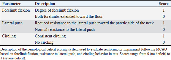
Table 5. Bederson neurological scores 24 and 48 hours after ischemic induction.
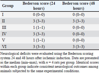
Neurological function assessment was not feasible 1 hour post-surgery due to inactivity. At 24 hours, Group 1 rats (sham-operated) scored 0 on the Bederson scale, indicating intact neurological function. In contrast, groups 2 and 6 (untreated and placebo, respectively) exhibited severe deficits (score 3). Groups 3 and 5, which were treated with 0.15 and 0.3 mg/kg BW quercetin, indicated mild impairment (score 1), whereas Group 4 (0.075 mg/kg BW) suggested severe deficits (score 3). At 48 hours, neurological recovery was observed in Groups 3–5, all of which had scored 0 on the Bederson scale. Groups 2 and 6 continued to exhibit severe deficits (score 3), and Group 1 consistently showed no impairment throughout the observation period.
Macroscopic and histopathological findings
All animals were sacrificed at 48 hours after induction, and their brain tissue was harvested for both macroscopic and microscopic analyses. Macroscopic examination revealed widened cerebral vessels, particularly in Groups 2 and 6, which had not received Quercetin NPs. This indicated post-ischemic vascular congestion and endothelial dysfunction. In contrast, rats treated with quercetin (Groups 3–5) exhibited less prominent vascular dilation, with 0.15 and 0.3 mg/kg doses suggesting the most apparent protective effect (Fig. 1).
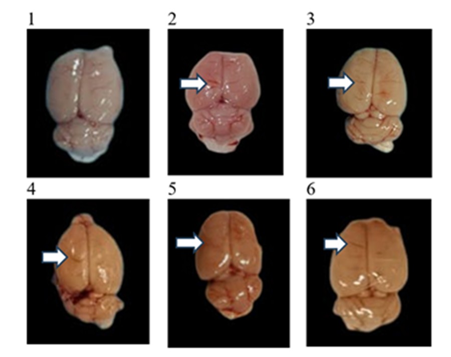
Fig. 1. Macroscopic appearance of rat brain sections at 48 hours after ischemic induction. Arrows indicate widened cerebral vessels. Marked vascular dilation is observed in the untreated (Group 2) and placebo (Group 6) groups, whereas quercetin-treated groups (Groups 3–5) show less pronounced vessel widening.
Following macroscopic examination, the brain tissue was divided into two parts for histopathological preparation and spectrophotometric analysis. Histopathological slides were processed and stained with hematoxylin and eosin (H&E), and the results are presented in Figure 2.
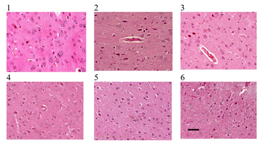
Fig. 2. Histopathological changes in rat brain tissue after MCAO and intranasal quercetin treatment. Representative H&E-stained brain sections from the (1) Sham group, (2) MCAO-induced group without treatment, (3) MCAO + quercetin 0.15 mg/kg BW, (4) MCAO + quercetin 0.075 mg/kg BW, (5) MCAO + quercetin 0.3 mg/kg BW, and (6) MCAO + placebo group. Necrotic neurons are characterized by pyknotic nuclei and eosinophilic cytoplasm. Table 6 presents the quantitative analysis of necrotic neurons. Scale bar=50 µm.
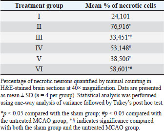
Histopathological examination using H&E staining revealed neuronal necrosis in Groups 2–6, which induced ischemic, whereas group 1 (shade preserved neuronal morphology. Table 6 presents the quantitative analysis of necrotic neurons.
Table 6. Quantification of necrotic neurons in rat brain tissue after treatment with MCAO and intranasal quercetin.
Biochemical analysis
The other portion of the brain tissue was subjected to spectrophotometric analysis for MDA level and SOD activity. The levels of MDA and SOD activity in brain tissue across experimental groups are presented in Figure 3. MDA levels were elevated in the ischemic groups compared with the sham group (Group 1), with higher values observed in the untreated (Group 2) and placebo (Group 6) groups. Among the quercetin-treated groups, MDA levels varied across doses, with relatively lower values noted in the 0.15 mg/kg (Group 3) and 0.3 mg/kg (Group 5) groups than in the untreated group. SOD activity suggested a decreasing trend in ischemic animals relative to the sham group, with reduced values in the untreated (Group 2) and placebo (Group 6) groups.
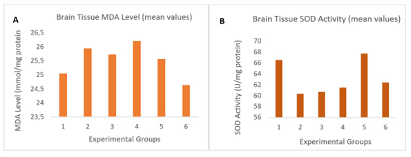
Fig. 3. Oxidative stress markers in brain tissue. Mean values of (A) MDA levels and (B) SOD activity in brain tissue across experimental groups. Data are presented as group means due to the unavailability of individual animal-level measurements.
Discussion
This study was preceded by the development of quercetin-loaded NPs for intranasal administration. These NPs were formulated using BSA and PDA, resulting in a mean particle size of approximately 334.4 nm with a PDI of 0.523. The PDI reflects the degree of size distribution heterogeneity within a NPs system, with values between 0.05 and 0.7 indicating varying dispersion levels (Danaei et al., 2018). The relatively higher PDI observed in the present formulation may be attributed to the incorporation of quercetin and the formation of the PDA coating, which can increase the heterogeneity of protein-based NP systems.
Zeta potential analysis indicated that the NPs possessed sufficient surface charge to maintain moderate colloidal stability, as values greater than +20 mV or less than −20 mV are generally associated with stable dispersions due to electrostatic repulsion (Thilagam and Gnanamani, 2020). These characteristics suggest that the formulated NPs are suitable for in vivo application. However, comprehensive physicochemical characterization was not extensively explored in this study, and further analyses are required to better understand the relationship between NP properties and biological performance.
Ischemic stroke was induced using the MCAO model, followed by intranasal administration of quercetin NPs 45 minutes after occlusion. This time point was selected to evaluate the intervention’s potential as an early therapeutic strategy during the acute phase of ischemic injury.
Clinical observations indicated a transient reduction in motor activity at 24 hours post-induction in the ischemic groups, followed by recovery at 48 hours. Although these observations were qualitative, they suggest a temporary functional impairment associated with ischemia. Neurological assessment using the Bederson score demonstrated that compared with untreated ischemic groups, animals receiving intranasal quercetin NPs exhibited milder neurological deficits at 24 hours and improved outcomes at 48 hours. These findings suggest that the administration of intranasal quercetin may be associated with improved neurological outcomes following ischemic injury.
Gross examination of brain tissue revealed vascular changes ipsilateral to MCA occlusion, which may reflect leptomeningeal collateral dilation as a compensatory response to reduced cerebral perfusion (Liu et al., 2014; Epp et al., 2023). Histopathological analysis further demonstrated a lower proportion of necrotic neurons in quercetin-treated groups than in untreated ischemic controls. These observations indicate that intranasal quercetin administration may be associated with a potential reduction in neuronal damage associated with quercetin NP administration. However, the absence of infarct volume measurement limits the strength of conclusions regarding the extent of neuroprotection.
Quercetin has antioxidant and neuroprotective properties (Lin et al., 2021). In the present study, the lower degree of neuronal necrosis observed in the treated groups may reflect a protective effect; however, this interpretation should be made cautiously due to the absence of quantitative infarct volume assessment. Interestingly, the lower dose (0.15 mg/kg BW) appeared to be associated with a more favorable histopathological outcome than the higher dose (0.3 mg/kg BW). One possible explanation is the dual redox behavior of quercetin, which may exhibit pro-oxidant effects under certain conditions. However, this remains speculative because direct measurements of ROS and related pathways were not performed in this study.
Biochemical analysis showed a tendency toward increased MDA levels in ischemic groups, indicating enhanced lipid peroxidation following cerebral ischemia. This finding is consistent with the known pathophysiology of ischemic stroke, in which reduced cerebral blood flow disrupts cellular metabolism and promotes oxidative stress (Salaudeen et al., 2024). MDA is a well-established marker of lipid peroxidation that reflects oxidative damage to cell membranes (Jadoon and Malik, 2017).
In addition, SOD activity tended to be higher in quercetin-treated groups than in untreated ischemic controls. SOD plays a critical role in endogenous antioxidant defense by catalyzing superoxide radical dismutation. The observed increase in SOD activity may indicate an enhancement of antioxidant capacity; however, the variability of SOD responses in acute stroke models remains widely reported. These findings should be interpreted with caution because the assessment of oxidative stress was limited to MDA and SOD quantification without direct ROS quantification.
Taken together, the biochemical and histopathological findings suggest that the observed effects of intranasal quercetin NPs may be partly associated with oxidative stress modulation. Nevertheless, the absence of direct ROS measurement and broader antioxidant pathway analysis limits mechanistic interpretation.
Overall, the findings of this study suggest that intranasally administered quercetin NPs have a potential neuroprotective effect during the acute phase of ischemic stroke. The intranasal route offers a non-invasive approach that may facilitate rapid nose-to-brain delivery, potentially enabling early intervention during ischemic injury. This strategy may represent a potential supportive approach to mitigate early oxidative damage; however, further studies are required to confirm its therapeutic relevance.
This study has several limitations. First, the relatively small sample size (n=4 per group), determined using the Federer formula, may reduce statistical power and increase variability. Although this approach is commonly applied in exploratory animal studies and aligns with the principle of reduction within the 3Rs framework, future studies should consider larger sample sizes with formal power calculations.
Second, the absence of formal randomization and blinding during group allocation and outcome assessment may introduce bias. Although standardized experimental procedures and controlled conditions were applied to minimize variability, future studies should incorporate these methodological approaches to improve rigor.
Third, the evaluation of ischemic brain injury did not include quantitative infarct volume measurement, such as 2,3,5-triphenyltetrazolium chloride staining or neuroimaging. While histopathological analysis provides important information regarding cellular damage, infarct volume quantification would allow a more comprehensive assessment of neuroprotective effects.
Fourth, the oxidative stress assessment was limited to MDA and SOD. Although these are commonly used biomarkers, the direct measurement of ROS, for example, using 2',7'-dichlorofluorescin diacetate, and the evaluation of additional antioxidant pathways would provide deeper mechanistic insight.
Finally, the use of a single sex, a single strain (Sprague Dawley), a short observation period, and limited behavioral assessment may restrict the generalizability of the findings. In addition, the lack of pharmacokinetic and biodistribution data further limits the interpretation of NP delivery efficiency.
Taken together, these limitations indicate that the findings should be interpreted cautiously and warrant further validation in more comprehensive experimental designs. From a translational perspective, extrapolation from rodent models to humans requires careful consideration. Differences in nasal anatomy, mucociliary clearance, and metabolic processes may influence drug absorption and brain delivery efficiency. Nevertheless, the intranasal route remains a clinically attractive, non-invasive strategy for CNS drug delivery, supporting the relevance of the present findings as a proof-of-concept for early antioxidant intervention in ischemic stroke.
Conclusion
This study suggests a potential neuroprotective effect of intranasally administered quercetin-loaded BSA–PDA NPs in an experimental MCAO model. This intervention was associated with improved neurological outcomes, reduced neuronal necrosis, and favorable trends in oxidative stress markers during the ischemic stroke acute phase.
However, these findings should be considered preliminary and hypothesis-generating. Further studies incorporating larger sample sizes, infarct volume assessment, and comprehensive mechanistic analyses are required to confirm and extend these observations.
Acknowledgments
The authors would like to sincerely thank IPB University, Gunadarma University, and the National Research and Innovation Agency for their assistance. We also acknowledge the financial support from BIMA, the Directorate of Research and Community Service, Indonesian Ministry of Research, Technology, and Higher Education, which made this research possible. Special thanks to our colleagues and academic advisors for their valuable insights and constructive suggestions throughout the research process.
Conflict of interest
The authors declare no conflict of interest.
Funding
This research was funded by the Indonesian Ministry of Higher Education, Science, and Technology (BIMA).
Authors' contributions
- Perdhana, I.S. conceived and designed the study, performed the experiments, analyzed the data, and drafted the manuscript.
- Maheshwari, H. contributed to the study design and revised the manuscript.
- Handharjani, E. supervised the study, contributed to the study design, and revised the manuscript.
- Darusman, H.S. and Widi Nugroho, S. provided scientific advice and critically reviewed the manuscript.
- All authors have read and approved the final version of the manuscript.
Data availability
Data supporting the results of this research are restricted for confidentiality reasons and may be provided by the corresponding author upon request.
References
Barthels, D. and Das, H. 2020. Current advances in ischemic stroke research and therapies. Biochim. Biophys. Acta. Mol. Basis Dis. 1866(4), 165260; doi:10.1016/j.bbadis.2018.09.012
Danaei, M., Dehghankhold, M., Ataei, S., Hasanzadeh Davarani, F., Javanmard, R., Dokhani, A., Khorasani, S. and Mozafari, M.R. 2018. Impact of particle size and polydispersity index on the clinical applications of lipidic nanocarrier systems. Pharmaceutics 10(2), 1–17; doi:10.3390/pharmaceutics10020057
Dewi, L. and Fitraneti, E. 2024. Stroke iskemik. Sci. J. 3(6), 365–374; doi:10.56260/sciena.v3i6.173
Epp, R., Glück, C., Binder, N.F., El Amki, M., Weber, B., Wegener, S., Jenny, P. and Schmid, F. 2023. The role of leptomeningeal collaterals in redistributing blood flow during stroke. PLoS Comput. Biol. 19(10), 1–33; doi:10.1371/journal.pcbi.1011496
Erdő, F., Bors, L.A., Farkas, D., Bajza, A. and Gizurarson, S. 2018. Evaluation of intranasal delivery route of drug administration for brain targeting. Brain. Res. Bull. 143, 155–170; doi:10.1016/j.brainresbull.2018.10.009
Feigin, V.L., Brainin, M., Norrving, B., Martins, S.O., Pandian, J., Lindsay, P., Grupper, F.M. and Rautalin, I. 2025. World stroke organization: global stroke fact sheet 2025. Int. J. Stroke. 20(2), 132–144; doi:10.1177/17474930241308142
Fitriani, E., Kriasa, I.M., Adam, N.M., Herawati, T. and Mulyatsih, E. 2024. Analysis of factors associated to pre-hospital delay in therapeutic window administration of intravenous thrombolysis for acute ischaemic stroke. Depok, Indonesia: Universitas Indonesia Library. Available via https://lontar.ui.ac.id/detail?id=9999920540362&lokasi=lokal
Groff, H., Yousfani, S., Pantoja-Ruiz, C., Douiri, A., Bhalla, A., Wolfe, C. and Marshall, I.J. 2024. A systematic review of the incidence and outcomes of ICD-11 defined stroke. J. Stroke. Cerebrovascular. Dis. 33(8), 107784; doi:10.1016/j.jstrokecerebrovasdis.2024.107784
Guo, C., Wang, W.J., Liao, Y.C., Zhao, C., Yin, Y., Yao, M.N., Ding, Y. and Wang, J.W. 2022. Effect and mechanisms of quercetin for experimental focal cerebral ischemia: a systematic review and meta-analysis. Oxid. Med. Cell. Longev. 2022, 9749461; doi:10.1155/2022/9749461
Jadoon, S. and Malik, A. 2017. A review article on the formation, mechanism and biochemistry of Mda and Mda As a biomarker of oxidative stress. Int. J. Adv. Res. 5(12), 811–818; doi:10.21474/ijar01/6024
Jiang, L.J., Zhang, S.M., Li, C.W., Tang, J.Y., Che, F.Y. and Lu, Y.C. 2017. Roles of the Nrf2/HO-1 pathway in the anti-oxidative stress response to ischemia-reperfusion brain injury in rats. Eur. Rev. For. Med. Pharmacol. Sci. 21(7), 1532–1540.
Karenberg, A. 2020. Historic review: select chapters of a history of stroke. Karenberg Neurol. Res. Pract. 2(1), 34; doi:10.1186/s42466-020-00082-0
Kuriakose, D. and Xiao, Z. 2020. Pathophysiology and treatment of stroke: present status and future perspectives. Int. J. Mol. Sci. 21(20), 1–24; doi: 10.3390/ijms21207609
Li, H., He, E., Wang, Y., Fu, J., Liu, T., Gou, R., Shi, S. and Gu, C. 2023. Albumin-stabilized polydopamine nanoparticles for chemo-photothermal synergistic therapy of melanoma. J. Drug. Del. Sci. Technol. 87, 104759, doi:10.1016/j.jddst.2023.104759
Lin, M.C., Liu, C.C., Liao, C.S. and Ro, J.H. 2021. Neuroprotective effect of quercetin during cerebral ischemic injury involves regulation of essential elements, transition metals, cu/zn ratio, and antioxidant activity. Molecules 26(20), 26; doi:10.3390/molecules26206128
Liu, J., Wang, Y., Akamatsu, Y., Lee, C.C., Stetler, R.A., Lawton, M.T. and Yang, G.Y. 2014. Vascular remodeling after ischemic stroke: mechanisms and therapeutic potentials. Prog. Neurobiol. 115(C), 138–156; doi:10.1016/j.pneurobio.2013.11.004
Makkiyah, F.A. and Sadewo, W. 2019. Technical report: simple method of animal stroke model of luminal occlusion of middle cerebral artery in Indonesia. Surg. Neurol. Int. 10(143), 10; doi:10.25259/SNI_62_2019
Nguyen, L.T. and Duong, V. 2025. Nose-to-brain drug delivery. Encyclopedia 5(91), 1–15; doi:10.3390/encyclopedia5030091
Perdhana, I.S., Handharyani, E., Darusman, H.S. and Nugroho, S.W. 2025. Method modification of developing ischemic stroke animal models using intravenous catheters in rats. Acta Vet. Indones. 3(1), 67–76.
Ramli, Y., Alwahdy, A.S., Kurniawan, M., Juliandi, B., Wuyung, P.E. and Susanto, Y.D.B. 2017. Permanent flame-blunted monofilament of middle cerebral artery occlusion technique for ischemia stroke induction in animal models. Med. J. Indonesia 26(3), 183–189; doi:10.13181/mji.v26i3.1645
Salaudeen, M.A., Bello, N., Danraka, R.N. and Ammani, M.L. 2024. Understanding the pathophysiology of ischemic stroke: the basis of current therapies and opportunity for new ones. Biomolecules 14(3), 1–23; doi:10.3390/biom14030305
Selvaraj, K., Gowthamarajan, K. and Karri, V.V.S.R. 2018. Nose to brain transport pathways an overview: potential of nanostructured lipid carriers in nose to brain targeting. Artif. Cells. Nanomed. Biotechnol. 46(8), 2088–2095; doi:10.1080/21691401.2017.1420073
Soares, R.O.S., Losada, D.M., Jordani, M.C., Évora, P. and Castro-E-Silva, O. 2019. Ischemia/reperfusion injury revisited: an overview of the latest pharmacological strategies. Int. J. Mol. Sci. 20(20), 5034; doi:10.3390/ijms20205034
Stielow, M., Witczyńska, A., Kubryń, N., Fijałkowski, Ł., Nowaczyk, J. and Nowaczyk, A. 2023. The bioavailability of drugs—the current state of knowledge. Molecules 28(24), 28; doi:10.3390/molecules28248038
Tamaddon, L., Mohamadi, N. and Bavarsad, N. 2021. Preparation and characterization of mucoadhesive loratadine nanoliposomes for intranasal administration. Turkish J. Pharm. Sci. 18(4), 492–497; doi:10.4274/tjps.galenos.2020.33254
Thilagam, R. and Gnanamani, A. 2020. Preparation, characterization and stability assessment of keratin and albumin functionalized gold nanoparticles for biomedical applications. Appl. Nanosci. (Switzerland). 10(6), 1879–1892; doi:10.1007/s13204-020-01250-z
Yang, D., Wang, T., Long, M. and Li, P. 2020. Quercetin: its main pharmacological activity and potential application in clinical medicine. Oxid. Med. Cell. Longev. 2020, 8825387; doi:10.1155/2020/8825387
Zhang, L., Ma, J., Yang, F., Li, S., Ma, W., Chang, X. and Yang, L. 2022. Neuroprotective effects of quercetin on ischemic stroke: a literature review. Front. Pharmacol. 13, article number 854249; doi:10.3389/fphar.2022.854249








