


| Research Article | ||
Open Vet. J.. 2026; 16(2): 1162-1170 Open Veterinary Journal, (2026), Vol. 16(2): 1162-1170 Research Article Comparative in vitro cytotoxicity of aqueous extracts of Brassica oleracea and Flammulina velutipes on prostate cancer cellsAlaa M. Dh. Al-Haidari1, Roaa M. H. Shoker2* and Israa Ahmed Ali11Department of Biology, College of Science, University of Baghdad, Baghdad, Iraq 2Department of Biology, College of Science, University of Wasit, Wasit, Iraq *Corresponding Author: Roaa M. H. Shoker. Department of Biology, College of Science,University of Wasit, Wasit, Iraq. Email: rmhasan [at] uowasit.edu.iq Submitted: 05/09/2025 Revised: 22/12/2025 Accepted: 08/01/2026 Published: 28/02/2026 © 2026 Open Veterinary Journal
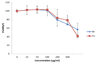
ABSTRACTBackground: Prostate cancer is one of the most common cancers in men. Current treatments include surgery, radiotherapy, and chemotherapy. These options improve survival but often cause serious side effects. Therefore, researchers are looking for natural products that may provide safer anticancer effects. Broccoli (Brassica oleracea) and mushrooms (Flammulina velutipes) contain many bioactive compounds. These include phenols, flavonoids, and polysaccharides. Such compounds are known for their antioxidant and anticancer activities. Aim: This study aimed to compare the cytotoxic effects of aqueous extracts from broccoli and mushroom. The cytotoxic activity of aqueous extracts of broccoli and mushroom was evaluated on human prostate cancer cells and normal human foreskin fibroblast-1 (HFF-1) cells. This study used the total aqueous extract of broccoli without separating specific phytochemicals. No quantification of a specific ingredient was included because the focus was on the overall cytotoxic effect. Methods: Extracts were prepared by drying, grinding, and dissolving the plant material in distilled water. They were then tested at concentrations between 25 and 800 μg/ml. The 3-(4,5-dimethylthiazol-2-yl)-2,5-diphenyltetrazolium bromide (MTT) assay was used to measure cell viability after 24 hours of treatment. Normal HFF-1 levels were also tested for safety. Morphological changes were observed using SEM. Results: Both extracts reduced cancer cell viability in a dose-dependent manner. At 800 μg/ml, broccoli extract reduced viability to 56.75%, whereas mushroom extract reduced viability to 41.09%. The IC50 values were 856.33 μg/ml for broccoli and 713.37 μg/ml for mushroom. SEM analysis revealed more pronounced cell shrinkage and surface damage with mushroom extract. Normal HFF-1 cells remained largely unaffected, but broccoli showed slightly more toxicity than mushroom. The differences between the two extracts were significant (p ≤ 0.05). The IC50 values were 856.3 µg/ml for broccoli and 713.4 µg/ml for mushroom extract. Normal HFF-1 cells remained largely unaffected, indicating that mushroom extract has a better safety profile and higher selectivity. Conclusion: Mushroom extract was more effective than broccoli extract in reducing prostate cancer cell viability. Keywords: Anticancer activity, Brassica oleracea, Cytotoxicity, Flammulina velutipes, Prostate cancer. IntroductionProstate cancer is one of the most common cancers among men worldwide and is considered the second leading cause of cancer-related death (Rawla, 2019; Sung et al., 2020). Its incidence increases with age, with the average age of diagnosis around 66 years (Cheng et al., 2023). Despite advances in surgery, radiotherapy, and chemotherapy, many patients experience side effects that reduce their quality of life. Due to these challenges, attention has turned toward alternative treatments, especially those derived from natural sources. Many plants and fungal extracts contain compounds that may slow cancer progression or reduce tumor survival (Kallifatidis et al., 2016). Broccoli is a widely consumed cruciferous vegetable known for its health-promoting properties. It is rich in bioactive compounds, including phenols, flavonoids, terpenes, vitamins, and glucosinolates (Li et al., 2021). These compounds are associated with antioxidant and anticancer activities. When broccoli is cut, chewed, or boiled, the enzyme myrosinase converts glucoraphanin into sulforaphane, a phytochemical with strong cancer-preventive potential (Kaiser et al., 2021). Sulforaphane can act through several mechanisms. It increases detoxifying enzyme activity, blocks phase I carcinogen-activating enzymes, and promotes apoptosis in cancer cells. It also inhibits histone deacetylases and modulates cell cycle regulators, which help stop abnormal cell proliferation (Amjad et al., 2015; Baladia et al., 2024). Sulforaphane is an important dietary compound for cancer prevention. Several studies have shown that sulforaphane protects against cancers of the skin, lung, prostate, colon, and stomach. It works by inducing detoxifying enzymes and stopping tumor growth (Amjad et al., 2015; Brooks et al., 2021). Mushrooms also contain many bioactive compounds. Flammulina velutipes (golden needle mushroom or enoki) is edible and medicinal. Its fruiting bodies and stipes carry polysaccharides, proteins, phenols, and anticancer enzymes (Chen et al., 2023). Flammulina velutipes extracts can reduce prostate cancer cell survival. They lower the expression of the androgen receptor and prostate-specific antigen (PSA). They also activate apoptosis through caspase pathways (Devere White et al., 2002; Maruyama and Ikekawa, 2007). These results suggest that F. velutipes may act as a natural anticancer agent. Although several studies have identified and quantified bioactive compounds, such as sulforaphane, phenolics, and polysaccharides, in broccoli and mushroom extracts (Li et al., 2021; Chen et al., 2023), their cytotoxic potentials have not been directly compared under identical experimental conditions. This study adds novelty by offering a direct comparative evaluation of their anticancer and safety profiles using the same assay system, highlighting their relative selectivity toward PCa cells. Although numerous studies have reported the anticancer activity of broccoli and mushroom extracts individually, comparative evidence evaluating their selective effects on PCa cells using identical conditions is limited. Both broccoli and mushroom extracts have been studied for their role in reducing oxidative stress and blocking cancer cell growth. Cruciferous vegetables, such as broccoli, show strong antioxidant and anticancer activity, even in leaves and stalk residues (Gaafar et al., 2020). Mushrooms, such as F. velutipes, also contain high levels of polysaccharides. These compounds exert strong antioxidant and immune-modulating effects (Ferreira et al., 2010). Mushroom polysaccharides slow tumor growth by affecting immune responses and directly reducing cell proliferation (Zhao et al., 2019). This indicates that both plants and mushrooms provide natural anticancer compounds. The comparison between broccoli and mushroom extract is justified because the two species have distinct yet complementary biological makeups. The species of broccoli contain glucosinolates and isothiocyanates, whereas the mushroom species F. velutipes has been shown to have anticancer activity and contains polysaccharides and phenolic compounds. The comparison of two unrelated natural sources within the context of the same condition also allows the determination of the two classes of dietary bioactive compounds to have greater selective cytotoxicity. This provides more insight into the comparison of two closely related species. This study aimed to prepare aqueous extracts of broccoli and mushroom, test their cytotoxic effects on prostate cancer cell lines using the MTT assay, compare their IC50 values, observe cell morphology under scanning electron microscopy, and test both extracts on normal HFF-1 cells for safety. The comparison was done to determine which extract was more effective in lowering the viability of prostate cancer cells. These findings may help guide future strategies that use dietary plants and mushrooms as supportive anticancer agents. The aims of this study were: To prepare aqueous extracts of broccoli and mushroom. To test their cytotoxic effects on prostate cancer (PC-3) cell lines using the MTT assay. To compare their IC50 values. To observe morphological changes in treated cells under a scanning electron microscope. To test the safety and selectivity of both extracts on normal HFF-1 cells. Natural extracts from edible plants and mushrooms are increasingly being studied as safer alternatives to conventional chemotherapy. By comparing Brassica oleracea and F. velutipes, which are both common dietary sources, this research seeks to identify potential natural agents that could complement existing prostate cancer treatments while minimizing toxicity. This study aimed to evaluate the comparative cytotoxic potential of aqueous extracts from broccoli and mushroom against PC-3 cells. The objectives of this study were to prepare and standardize the extracts, assess their inhibitory effects on the PC-3 prostate cancer cell line using the MTT assay, determine the IC50 values, and observe treatment-related morphological changes using scanning electron microscopy. This study also compared the effects of the extract on normal HFF-1 cells to evaluate its safety and selectivity. The cytotoxic activity of aqueous extracts of broccoli and mushroom was evaluated on human PC-3 cells and normal HFF-1 cells. Although this study used human prostate cancer cells, the mechanisms explored—oxidative stress, detoxifying enzymes, and selective cytotoxicity—are shared across mammalian systems. Therefore, the findings provide a foundation for later application in veterinary oncology and toxicology, where natural plant and mushroom extracts are being studied as alternative or supportive treatments for animal cancers. Materials and MethodsCollection, drying, and grinding of the samplesFresh broccoli was purchased from a local market in Baghdad. The dried fruiting bodies of the mushrooms were obtained from the Organic Agriculture Section of the Ministry of Agriculture, Iraq. The broccoli was washed several times with tap water to remove soil and dirt. The edible parts were cut into small pieces and spread on trays. The samples were dried in an electric oven at 40 °C until crisp. The mushroom samples were already dried, but were given an additional low-temperature drying step. This preserved the active compounds and stopped the microbial growth. The dried broccoli and mushroom were ground separately into fine powders using an electric grinder. The powders were sieved for uniform size and stored in airtight glass containers. The containers were labeled and kept in a dry place at room temperature. This storage preserves phytochemicals and stops oxidation. The fine powder form also improved extraction by allowing maximum release of bioactive components. Preparation of the aqueous extractsFifty grams of broccoli powder and fifty grams of mushroom powder were suspended in 250 ml of boiled distilled water. The mixtures were placed on a shaker for 24 hours to allow thorough mixing. Suspensions were first filtered through a muslin cloth to remove residues. The samples were then filtered again with Whatman filter paper. Clear filtrates were collected in clean glass dishes. After filtration, the aqueous extracts were evaporated at 40°C until completely dry. The dried residue was scraped and ground into a fine powder before redissolving to prepare the stock solution. This low-temperature drying step preserved heat-sensitive compounds that might degrade under high temperatures. The dried extracts were carefully scraped, weighed, and dissolved in sterile distilled water to prepare stock solutions. Six working concentrations (25, 50, 100, 200, 400, and 800 μg/ml) were prepared from each stock by diluting with distilled water. The solutions were transferred to sterile tubes and stored at 4°C until required for experiments. These steps were repeated for both broccoli and mushroom powders to ensure consistency in extraction. The method ensured that the bioactive compounds, such as polysaccharides, phenols, and flavonoids, were preserved and available for biological testing. Cell cultureHuman prostate cancer cells and normal HFF-1 cells were obtained from the American Type Culture Collection. PC-3 (ATCC® CRL-1435™) was used as the cancer model. Before use, the viability of the cells was tested using trypan blue exclusion dye, which distinguishes living cells from dead ones. Cells were stored in vapor-phase liquid nitrogen at −196°C until they were required. The culture medium used was RPMI-1640 (Capricorn Scientific, Germany). The medium was enriched with 10% FBS. The medium also contained 2 ml of penicillin (100x) and 1 ml of amphotericin-B (250 μg/mi). Before use, it was sterilized with a 0.22 μm filter to prevent contamination. Cells were cultured in T-25 flasks at 37°C in a humidified incubator with 5% CO2. Subculturing was performed daily to keep the cells healthy. For each experiment, the cells were seeded into plates and left for 24 hours to attach. Healthy fibroblasts were used as controls. This step ensured that safety was tested and that anticancer effects could be separated from general toxicity. These measures kept the results accurate and free from errors caused by poor or contaminated cultures. Cells were used between passages 5 and 15 to maintain stable growth characteristics. The average doubling time under culture conditions was 24–30 hours. Cytotoxicity assay (MTT assay)The MTT assay was used to measure the cytotoxicity of the broccoli and mushroom extracts. Approximately 7,000 cells were seeded into each well of a 96-well plate. Cells were allowed to attach for 24 hours. Different concentrations of the extracts were added, and untreated wells were used as controls. Incubation was continued for 24 hours at 37°C. All experiments were performed in triplicate (three independent biological replicates), and each treatment concentration was tested in three technical repeats. Untreated cells were used as a control and represented 100% viability. To calculate the percentage of viable cells, all treated groups were compared with this control. This ensured that any reduction in cell survival was due to exposure to the extract and not to experimental handling. After incubation, the wells were gently rinsed once with PBS to remove the residual extract without disturbing the attached cells. This step prevents color interference with the MTT reaction. The medium was discarded after incubation. The wells were gently rinsed with PBS. To assess viability, 20 μl of serum-free medium and 20 μl of MTT solution (5 mg/ml in PBS) were added to each well. Plates were kept in the dark for 3 h. During this time, metabolically active cells reduced MTT to purple formazan crystals. Extract-only blanks (without cells) were run in parallel to correct for any background absorbance or direct reduction of MTT by the extracts. For SEM analysis, treated and control cells were fixed with 2.5% glutaraldehyde for 2 hours and then washed in PBS. The samples were dehydrated using graded ethanol (50%–100%) and dried under vacuum. They were mounted on aluminum stubs, coated with gold using a sputter coater, and examined under a field-emission scanning electron microscope (TESCAN VEGA3, Czech Republic) at 15 kV and 1 × 10⁻⁴ Pa pressure. These steps produced clear surface images showing treatment-related changes. Cell viability values were normalized to the untreated control, defined as 100%. Minor increases above 100% indicate small fluctuations or metabolic stimulation at low extract concentrations, which are commonly observed in MTT assays. After incubation, 50 μl of dimethyl sulfoxide (DMSO) was added to dissolve the crystals. The absorbance was measured using a microplate reader at 570 nm. Cell viability was calculated using the following formula: Viability %=[(A test A blank)/(A control A blank)] × 100. The inhibitory concentration (IC50), defined as the extract dose that killed 50% of cells, was obtained from the dose-response curve using GraphPad Prism software. Statistical analysisThe Statistical Package for the Social Sciences (version 2019) was used for statistical analysis. T Data were analyzed using one-way analysis of variance with LSD post hoc comparisons. Normality and variance homogeneity were tested using Shapiro–Wilk and Levene’s tests. Differences were considered significant at p ≤ 0.05. Ninety-five percent confidence intervals were calculated for IC50 values. In this study, extract type (broccoli or mushroom) and concentration were treated as independent variables, while cell viability percentage was the dependent variable analyzed using one-way ANOVA followed by the least significant difference test. Ethical approvalNot needed for this study. ResultsEffects of broccoli and mushroom extracts on prostate cancer cellsThe cytotoxic effect of aqueous extracts of broccoli and mushroom on prostate cancer cells was evaluated using the MTT assay. Both extracts reduced cancer cell viability in a concentration-dependent manner. At lower concentrations ranging from 25 to 100 μg/ml, no significant difference was observed compared with the control, and cell viability remained close to 100%. A clear inhibitory effect was observed when the concentrations were increased to 200 μg/ml. At 200 μg/ml, viability decreased to 79.41% and 83.86% with broccoli and mushroom extracts, respectively, showing a significant reduction compared with the control group. As the concentration reached 400 μg/ml, the inhibition became more obvious, with 68.83% viability in broccoli-treated cells and 78.37% in mushroom-treated cells. The strongest effect was recorded at the highest concentration, 800 μg/ml, where broccoli extract reduced viability to 56.75%, while mushroom extract reduced viability to 41.09%. The IC50 values calculated were 856.33 μg/ml for broccoli extract and 713.37 μg/ml for mushroom extract, confirming that mushroom extract was more potent. The SEM findings supported the MTT results. SEM analysis revealed that mushroom-treated cells at 800 μg/ml showed extensive shrinkage and surface damage compared with broccoli-treated cells, which displayed less damage under the same conditions. These findings confirmed that mushroom extract induced stronger cytotoxicity against PCa cells than broccoli extract (Table 1; Figs. 1 and 2). High-resolution scanning electron microscopy images (Fig. 2) clearly show progressive membrane damage and cell shrinkage with increasing extract concentration. The effects were more pronounced in mushroom-treated cells than in untreated cells, confirming the MTT data.
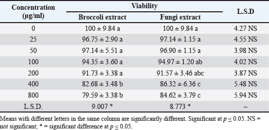
Fig. 1. The viability percentage of prostate cancer cell line after treatment with different concentrations of broccoli plant extract and mushroom extract. Br=broccoli extract; Fu=mushroom extract. Table 1. The viability of aqueous extracts of broccoli and mushroom against the prostate cancer cell line after 24 hours incubation.
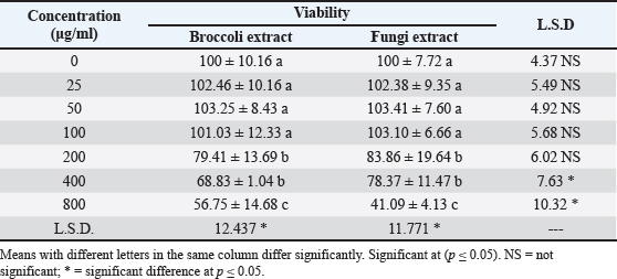
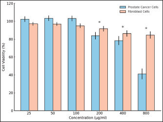
Fig. 2. Scanning electron micrographs showing the effects of aqueous extracts of broccoli and mushroom (Flammulina velutipes) on PC-3 cells. The images reveal a progressive reduction in cell number and size with increasing extract concentration, indicating dose-dependent cytotoxicity. Morphological alterations, such as surface shrinkage and membrane damage, were more pronounced in mushroom-treated cells. (A) Untreated control; (B, D, F, H, J, and L) broccoli extract at 25, 50, 100, 200, 400, and 800 µg/ml; (C, E, G, I, K, and M) mushroom extract at the same concentrations. Scale bar=10 µm. Even if the magnification of the acquisition was 10 µm, the magnification and scale layers were absent in the final image due to automated software exporting. Appropriate blocks will ensure that scale bars and magnification indicators are embedded in each micrograph to meet the FE-SEM guidelines for reporting the imaging. The calculated IC50 values for HFF-1 cells were 798.2 µg/ml for broccoli extract and 841.8 µg/ml for mushroom extract, giving SIs of 0.93 and 1.18, respectively. Effects of broccoli and mushroom extracts on HFF-1 levelsThe safety of the broccoli and mushroom extracts was evaluated using normal HFF-1. Both extracts showed minimal toxicity to normal cells compared with their effect on cancer cells. At low concentrations from 25 to 100 μg/ml, viability was maintained above 94% in all treatments. At 200 μg/ml, broccoli extract reduced viability to 91.73%, whereas mushroom extract maintained viability at 91.57%. These results indicate that both extracts exerted limited cytotoxic effects at low and moderate concentrations. At higher concentrations, the differences between broccoli and mushroom became clearer. At 400 μg/ml, broccoli extract reduced viability to 82.68%, whereas mushroom extract retained viability at 86.32%. At 800 μg/ml, the effect on fibroblast cells was more pronounced, but the cells remained more resistant than cancer cells. Broccoli- and mushroom-treated fibroblast cells showed 79.59% and 84.62%, respectively. SEM analysis revealed that both extracts caused mild surface changes, but these changes were less severe than those observed in cancer cells. SEM measurements confirmed that fibroblast cells remained relatively intact, supporting the extracts’ selective action. These findings suggest that mushroom extract is not only more effective against cancer cells but also less harmful to normal cells, making it a safer option (Table 2; Fig. 3). The cytotoxic activity of broccoli and mushroom extracts in prostate cancer and normal HFF-1 cells was compared. Figure 4 shows the effect of the broccoli extract. Cell viability decreased gradually with increasing concentration in both cell types. At concentrations up to 100 µg/ml, no significant difference was observed, and viability remained above 95%. At 200 µg/ml, a marked reduction in prostate cancer cells was observed, while fibroblasts were less affected. At 400 and 800 µg/ml, broccoli extract strongly inhibited prostate cancer cells, reducing their viability to 68.8% and 56.7%, respectively. In contrast, fibroblast cells showed only mild reduction, maintaining 82.7% and 79.6% viability at the same doses. The difference between cancer and normal cells at higher concentrations was statistically significant (p ≤ 0.05). Figure 5 presents the response to F. velutipes (mushroom) extract. The extract also reduced cancer cell viability in a dose-dependent pattern but produced a stronger effect than broccoli at high doses. At 200 µg/ml, viability dropped to 83.9%, and at 800 µg/ml, to 41.1%. However, fibroblast cells remained relatively stable, with 86.3% and 84.6% viability at the same doses. The comparative bar graphs clearly show that the mushroom extract was more cytotoxic toward PCa cells while maintaining better safety for normal cells.
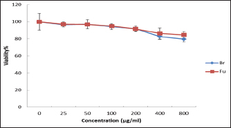
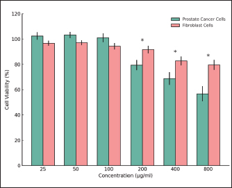
Fig. 3. The viability percentage of HFF-1 fibroblast cells after 24 h of incubation with different concentrations of broccoli and mushroom extracts. Br=broccoli extract; Fu=mushroom extract.
Fig. 4. Comparison of the viability of prostate cancer and normal HFF-1 cells after 24 h treatment with different concentrations of broccoli extract Asterisks (*) indicate significant differences (p ≤ 0.05) between cancer and normal cells treated with the same concentration. Table 2. The viability, SD, and SEM of the aqueous extracts of broccoli and mushroom against the HFF-1 (fibroblast) cell line after 24 hours of incubation.
Fig. 5. Comparison of the viability of prostate cancer and normal HFF-1 cells after 24 hours treatment with different mushroom extract concentrations Asterisks (*) denote significant differences (p ≤ 0.05) between cancer and normal cells treated with the same concentration. DiscussionEffects of broccoli and mushroom extracts on prostate cancer cellsThe results showed that both broccoli and mushroom extracts inhibited the growth of PCa cells in a concentration-dependent manner, with mushroom extract showing greater potency. This observation supports earlier findings that broccoli contains sulforaphane, a compound capable of inducing detoxifying enzymes and preventing tumor growth (Brooks et al., 2021; Kaiser et al., 2021). Türker et al. (2019) confirmed that broccoli extract suppressed the proliferation of Du145 prostate cancer cells, whereas Abdulah et al. (2009) found that selenium-enriched broccoli sprouts enhanced chemosensitivity in LNCaP cells. The present study agrees with these reports but highlights that the mushroom extract produced stronger cytotoxicity at the same concentration. At 400 µg/ml, broccoli extract may show stronger inhibition because sulforaphane and related glucosinolates reach effective intracellular levels that trigger detoxifying enzymes and oxidative stress in cancer cells. At higher doses, the mushroom extract activity plateaus, while it continues to release high levels of polysaccharides and phenolic compounds that enhance apoptosis. This explains why mushroom extract becomes more effective at 800 µg/ml (Maruyama and Ikekawa, 2007; Chen et al., 2023). The more potent effect of mushroom extract is supported by several studies. Devere White et al. (2002)demonstrated that mushroom mycelium extract inhibited prostate cancer cell growth. Maruyama and Ikekawa (2007) reported that prolamin isolated from F. velutipes exerted immunomodulatory and antitumor effects. Chen et al. (2023) showed that mushroom stipe extracts activated caspase-dependent apoptosis and reduced the survival of prostate cancer cells. These outcomes are consistent with our finding that the IC50 value of mushroom extract was lower than that of broccoli, confirming a higher cytotoxic potential. Together, the present data and earlier studies indicate that mushroom extract may serve as a stronger anticancer agent than broccoli against prostate cancer. This preliminary study focused on cell viability and morphology as the first indicators of cytotoxicity. To better define the underlying mechanisms, future studies will explore specific biochemical markers such as caspase activation, ROS generation, and apoptosis-related gene expression. The surface shrinkage and membrane blebbing observed in the SEM images are typical features of apoptosis. These findings support previous studies showing that F. velutipes extracts can activate caspase-dependent apoptotic pathways in PCa cells (Devere White et al., 2002; Chen et al., 2023). Thus, the observed morphological alterations may reflect early apoptotic events induced by the extracts. Effects of broccoli and mushroom extracts on HFF-1 levelsIn this study, both broccoli and mushroom extracts had limited effects on normal fibroblast cells. Viability remained above 90% at low to moderate concentrations, indicating low toxicity. However, at high concentrations, broccoli extract reduced fibroblast viability more than mushroom extract. These findings confirm earlier reports that cruciferous vegetables contain bioactive compounds that can show mild cytotoxic effects in normal cells (Gaafar et al., 2020). Sulforaphane from broccoli has been described as selective in targeting cancer cells while sparing normal cells (Amjad et al., 2015). Our results partly agree, but also suggest that mushroom extract may be safer than broccoli in this context. The safety profile of mushroom extract aligns with that of earlier work. Zhao et al. (2019) reported that F. velutipes polysaccharides enhanced immune responses in mice without causing harmful side effects. confirmed that mushroom caps contain compounds with strong antioxidant and anticancer activity, yet minimal toxicity to healthy cells. Ferreira et al. (2010) also described mushroom polysaccharides as selective anticancer agents that act through immunomodulation. These earlier reports agree with our results, where mushroom extract showed stronger anticancer activity with less toxicity to normal fibroblast cells, making it a more favorable candidate than broccoli extract. HFF-1 cells were selected to represent normal healthy cells because they are widely used as a standard non-tumor model in cytotoxicity studies. They exhibit stable growth, uniform morphology, and high sensitivity to toxic compounds. Their use allows direct comparison with earlier research that tested plant or mushroom extracts on normal fibroblasts (Türker et al., 2019; Karbon and Alhammer, 2020). Although epithelial cells from the same tissue could also be used, fibroblasts provide a general measure of safety across cell types. HFF-1 cells were used to evaluate the safety of extracts. Fibroblasts are frequently used in cytotoxicity testing because they are easy to maintain and reflect normal cell metabolism (Freshney, 2000; Türker et al., 2019). Their use ensures consistency with other in vitro screening studies. The comparative analysis in Figures 4 and 5 shows that both broccoli and mushroom extracts inhibited prostate cancer cell growth while sparing normal fibroblast cells. This selective effect indicates that their bioactive compounds mainly act on transformed cells. The stronger cytotoxicity of F. velutipes may be related to its higher polysaccharide and phenolic compound content, which triggers apoptosis and oxidative stress in tumor cells. This observation agrees with previous reports that mushroom polysaccharides can induce programmed cell death and reduce proliferation in several cancer models (Maruyama and Ikekawa, 2007; Chen et al., 2023). Broccoli extract also reduced cancer cell survival, particularly at 400 µg/ml, which may correspond to the optimal intracellular sulforaphane level. This compound activates detoxifying enzymes and blocks the metabolism of phase I carcinogens (Amjad et al., 2015; Kaiser et al., 2021). However, at higher concentrations, its effect reached a plateau, whereas mushroom extract continued to increase cytotoxicity, probably due to the combined action of multiple compounds. The fibroblast response confirmed that both extracts are relatively safe, with limited structural damage observed under SEM. Together, these findings indicate that F. velutipes possesses stronger cytotoxic and selective activity than broccoli against PCa cells. The data suggest that mushroom extracts may provide a broader therapeutic window and could serve as promising natural sources for complementary cancer treatment. The MTT assay at 24 hours was used as a primary screening step. To confirm these findings, future work will extend testing to longer exposures and additional viability and apoptosis assays. The higher SI value of the mushroom extract suggests better selectivity and safety than that of the broccoli extract, which is consistent with its lower toxicity in normal fibroblast cells. A limitation of this study is the absence of a standard cytotoxic drug and detailed extract characterization. A positive control, such as doxorubicin or cisplatin, was not included because the main goal was to compare the two natural extracts rather than benchmark them against a chemotherapeutic agent. Future studies will include a reference compound such as doxorubicin and quantify key components such as sulforaphane, phenolics, and polysaccharides. Although both extracts reduced cancer cell viability, their IC50 values were relatively high, indicating weak potency compared with standard chemotherapeutic agents. Therefore, their role may be supportive or preventive rather than therapeutic. These extracts could serve as natural dietary supplements with mild anticancer potential rather than primary treatment options. The IC50 values observed in this study were relatively high, suggesting that crude aqueous extracts have limited potency in direct anticancer action. These concentrations may not reflect physiological relevance but provide important proof-of-concept data. Future studies should focus on isolating active components, such as sulforaphane and mushroom polysaccharides, which are expected to act at lower concentrations. Among the two extracts tested, F. velutipes showed stronger cytotoxicity toward PCa cells while maintaining a higher safety level in normal fibroblast cells. This dual effect highlights its potential as a safe and selective natural anticancer candidate for further research. Some concentrations showed large standard deviations, which may indicate some biological variability or a possible lack of biological replicates. In theory, including additional independent experiments should reduce variability in the estimates, and subsequently, the estimates of viability may acquire stronger confidence. The variability of some treatment groups could point to a simple biological variability in fibroblast responsiveness and perhaps a lack of replicates. Greater sequential replication is needed to reduce the overall dispersion. ConclusionThis study demonstrated that both broccoli and mushroom aqueous extracts reduced the growth of PCa cells in vitro, with mushroom extract showing stronger cytotoxic activity and a lower IC50 value. SEM analysis confirmed greater damage in cancer cells treated with mushroom than broccoli, while both extracts had minimal effects on normal fibroblast cells. These findings suggest that F. velutipes has higher anticancer potential and better safety than broccoli, supporting its use as a natural and affordable dietary source for prostate cancer prevention or complementary therapy. These results also suggest the potential of using such extracts in veterinary contexts, as many molecular pathways of cancer development are conserved between humans and animals. Further studies using veterinary cell lines or animal models are recommended to confirm these effects. AcknowledgmentThe authors thank the College of Biology in the University of Wasit and University of Baghdad for their support in this study. FundingThe authors have self-funded the study. No external funding source is available. Authors’ contributionsAll authors have participated in the study. Conflict of interestThe authors have no conflicts of interest to declare. Data availabilityData are available when requested by the corresponding author. ReferencesAbdulah, R., Faried, A., Kobayashi, K., Yamazaki, C., Suradji, E.W., Ito, K., Suzuki, K., Murakami, M., Kuwano, H. and Koyama, H. 2009. Selenium enrichment of broccoli sprout extract increases chemosensitivity and apoptosis of LNCaP prostate cancer cells. BMC. Cancer. 9, 414; doi:10.1186/1471-240-9-414 Amjad, A.I., Parikh, R.A., Appleman, L.J., Hahm, E.R., Singh, K. and Singh, S.V. 2015. Broccoli-derived sulforaphane and chemoprevention of prostate cancer: from bench to bedside. Curr. Pharmacol. Rep. 1, 382–390; doi:10.1007/s40495-015-0034-x Baladia, E., Moñino, M., Pleguezuelos, E., Russolillo, G. and Garnacho-Castaño, M.V. 2024. Broccoli consumption and risk of cancer: an updated systematic review and meta-analysis of observational studies. Nutrients 16, 1583; doi:10.3390/nu16111583 Brooks, J.D., Paton, V.G. and Vidanes, G. 2021. Potent induction of phase 2 enzymes in human prostate cells by sulforaphane. Cancer Epidemiol. Biomark. Prev. 10, 949–954. Chen, C.C., Tang, C.T., Lai, C.Y., Han, C.K., Cheng, Y.H., Lin, Y.F., Lin, C.T. and Huang, Y.L. 2023. In vitro assessment of the antioxidant and anticancer properties of Flammulina velutipes stipe extracts. Anticancer Res. 43(7), 3057–3067; doi:10.21873/anticanres.16477 Cheng, B., Li, L., Wu, Y., Luo, T., Tang, C., Wang, Q., Zhou, Q., Wu, J., Lai, Y., Zhu, D., Du, T. and Huang, H. 2023. The key cellular senescence related molecule RRM2 regulates prostate cancer progression and resistance to docetaxel treatment. Cell. &. Bioscience. 13(211), M2–20; doi:10.1186/s13578-023-01157-6 Devere White, R.W., Hackman, R.M., Soares, S.E., Beckett, L.A. and Sun, B. 2002. Effects of a mushroom mycelium extract on the treatment of prostate cancer. Urology 60(4), 640–644; doi:10.1016/S0090-4295(02)01856-3 Ferreira, I., A. Vaz, J., Vasconcelos, M.H. and Martins, A. 2010. Compounds from wild mushrooms with antitumor potential. Anti-Cancer Agents MedChem. 10(5), 424–436; doi:10.2174/1871520611009050424 Freshney. 2000. Culture of animal cells: a manual for basic technique. 4th ed., NJ: Wiley-Liss. Gaafar, A.A., Salama, Z.A. and El-Baz, F.K. 2020. A comparative study on the active constituents, antioxidant capacity and anti-cancer activity of cruciferous vegetable residues. Baghdad. Sci. J. 17(3), 743–753; doi:10.21123/bsj.2020.17.3.0743 Kaiser, A.E., Baniasadi, M., Giansiracusa, D., Giansiracusa, M., Garcia, M., Fryda, Z., Wong, T.L. and Bishayee, A. 2021. Sulforaphane: a broccoli bioactive phytocompound with cancer preventive potential. Cancers 13, 4796. Kallifatidis, G., Hoy, J.J. and Lokeshwar, B.L. 2016. Bioactive natural products for chemoprevention and treatment of castration-resistant prostate cancer. Seminars. Cancer. Biol. 40–41, 160–169; doi:10.1016/j.semcancer.2016.06.003 Karbon, M.H. and Alhammer, A.H. 2020. Cytotoxic effect of aqueous-ethanol extract of Typha domingensis Pers. (Pollen) against human breast cancer cells in vitro. Systematic. Rev. Pharm. 11(10), 1158–1161. Li, H., Xia, Y., Liu, H.Y., Guo, H., He, X.Q., Liu, Y., Wu, D.T., Mai, Y.H., Li, H.B., Zou, L. and Gan, R.Y. 2021. Nutritional values, beneficial effects, and food applications of broccoli (Brassica oleracea var. italica Plenck). Trends Food Sci. &. Technol. 119, 288–308; doi:10.1016/j.tifs.2021.12.015 Maruyama, H. and Ikekawa, T. 2007. Immunomodulation and antitumor activity of a mushroom product, proflamin, isolated from Flammulina velutipes (W. Curt.: fr.) singer (Agaricomycetideae). Int. J. MedMushrooms. 9(2), 109–122; doi:10.1615/IntJMedMushr.v9.i2.20 Rawla, P. 2019. Epidemiology of prostate cancer. World J. Oncol. 10(2), 63. Sung, H., Ferlay, J., Siegel, R., Laversanne, M., Soerjomataram, I., Jemal, A. and Bray, F. 2020. Globocan estimates of incidence and mortality worldwide for 36 cancers in 185 countries. CA. A Cancer J. For Clinicians 71(3), 209–249; doi:10.3322/caac.21660 Türker, N.P., Bağcı, U. and Onsekizoglu Bagci, P. 2019. Investigation of the anticancer and proliferative effect of broccoli extract on Du145 prostate cancer and MEF healthy fibroblast cell lines. Int. J. Innov. Approaches Agricult. Res. 3(4), 550–556. Zhao, R., Hu, Q., Ma, G., Su, A., Xie, M., Li, X., Chen, G. and Zhao, L. 2019. Effects of Flammulina velutipes polysaccharide on immune response and intestinal microbiota in mice. J. Funct. Foods 56, 255–264; doi:10.1016/j.jff.2019.03.031 | ||
| How to Cite this Article |
| Pubmed Style Al-haidari AMD, Shoker RMH, Ali IA. Comparative in vitro cytotoxicity of aqueous extracts of Brassica oleracea and Flammulina velutipes on prostate cancer cells. Open Vet. J.. 2026; 16(2): 1162-1170. doi:10.5455/OVJ.2026.v16.i2.34 Web Style Al-haidari AMD, Shoker RMH, Ali IA. Comparative in vitro cytotoxicity of aqueous extracts of Brassica oleracea and Flammulina velutipes on prostate cancer cells. https://www.openveterinaryjournal.com/?mno=282125 [Access: February 27, 2026]. doi:10.5455/OVJ.2026.v16.i2.34 AMA (American Medical Association) Style Al-haidari AMD, Shoker RMH, Ali IA. Comparative in vitro cytotoxicity of aqueous extracts of Brassica oleracea and Flammulina velutipes on prostate cancer cells. Open Vet. J.. 2026; 16(2): 1162-1170. doi:10.5455/OVJ.2026.v16.i2.34 Vancouver/ICMJE Style Al-haidari AMD, Shoker RMH, Ali IA. Comparative in vitro cytotoxicity of aqueous extracts of Brassica oleracea and Flammulina velutipes on prostate cancer cells. Open Vet. J.. (2026), [cited February 27, 2026]; 16(2): 1162-1170. doi:10.5455/OVJ.2026.v16.i2.34 Harvard Style Al-haidari, A. M. D., Shoker, . R. M. H. & Ali, . I. A. (2026) Comparative in vitro cytotoxicity of aqueous extracts of Brassica oleracea and Flammulina velutipes on prostate cancer cells. Open Vet. J., 16 (2), 1162-1170. doi:10.5455/OVJ.2026.v16.i2.34 Turabian Style Al-haidari, Alaa M. Dh., Roaa M. H. Shoker, and Israa Ahmed Ali. 2026. Comparative in vitro cytotoxicity of aqueous extracts of Brassica oleracea and Flammulina velutipes on prostate cancer cells. Open Veterinary Journal, 16 (2), 1162-1170. doi:10.5455/OVJ.2026.v16.i2.34 Chicago Style Al-haidari, Alaa M. Dh., Roaa M. H. Shoker, and Israa Ahmed Ali. "Comparative in vitro cytotoxicity of aqueous extracts of Brassica oleracea and Flammulina velutipes on prostate cancer cells." Open Veterinary Journal 16 (2026), 1162-1170. doi:10.5455/OVJ.2026.v16.i2.34 MLA (The Modern Language Association) Style Al-haidari, Alaa M. Dh., Roaa M. H. Shoker, and Israa Ahmed Ali. "Comparative in vitro cytotoxicity of aqueous extracts of Brassica oleracea and Flammulina velutipes on prostate cancer cells." Open Veterinary Journal 16.2 (2026), 1162-1170. Print. doi:10.5455/OVJ.2026.v16.i2.34 APA (American Psychological Association) Style Al-haidari, A. M. D., Shoker, . R. M. H. & Ali, . I. A. (2026) Comparative in vitro cytotoxicity of aqueous extracts of Brassica oleracea and Flammulina velutipes on prostate cancer cells. Open Veterinary Journal, 16 (2), 1162-1170. doi:10.5455/OVJ.2026.v16.i2.34 |

