





| Case Report | ||
Open Vet. J.. 2026; 16(6): 3569-3574 Open Veterinary Journal, (2026), Vol. 16(6): 3569-3574 Case Report Racemose hemangioma of the bronchial artery in a dogToshihide Furuike1, Shunsuke Shimamura2*, Shougo Hirata3, Shunichi Watanabe2 and Hiroyuki Tani41Fleur Animal Hospital Veterinary Cardiovascular Center Shiga, Moriyama, Japan 2Department of Veterinary Medicine, Laboratory of Small Animal Clinical Medicine, Graduate School of Veterinary Sciences, Osaka Metropolitan University, Osaka, Japan 3Veterinary Medical Center, Graduate School of Veterinary Sciences, Osaka Metropolitan University, Osaka Japan 4Laboratory of Internal Medicine, Graduate School of Veterinary Sciences, Osaka Metropolitan University, Osaka, Japan *Corresponding Author: Shunsuke Shimamura. Department of Veterinary Medicine, Laboratory of Small Animal Clinical Medicine, Graduate School of Veterinary Sciences, Osaka Metropolitan University, Osaka, Japan. Email: shimamur [at] omu.ac.jp Submitted: 02/02/2026 Revised: 05/05/2026 Accepted: 15/05/2026 Published: 05/06/2026 © 2025 Open Veterinary Journal
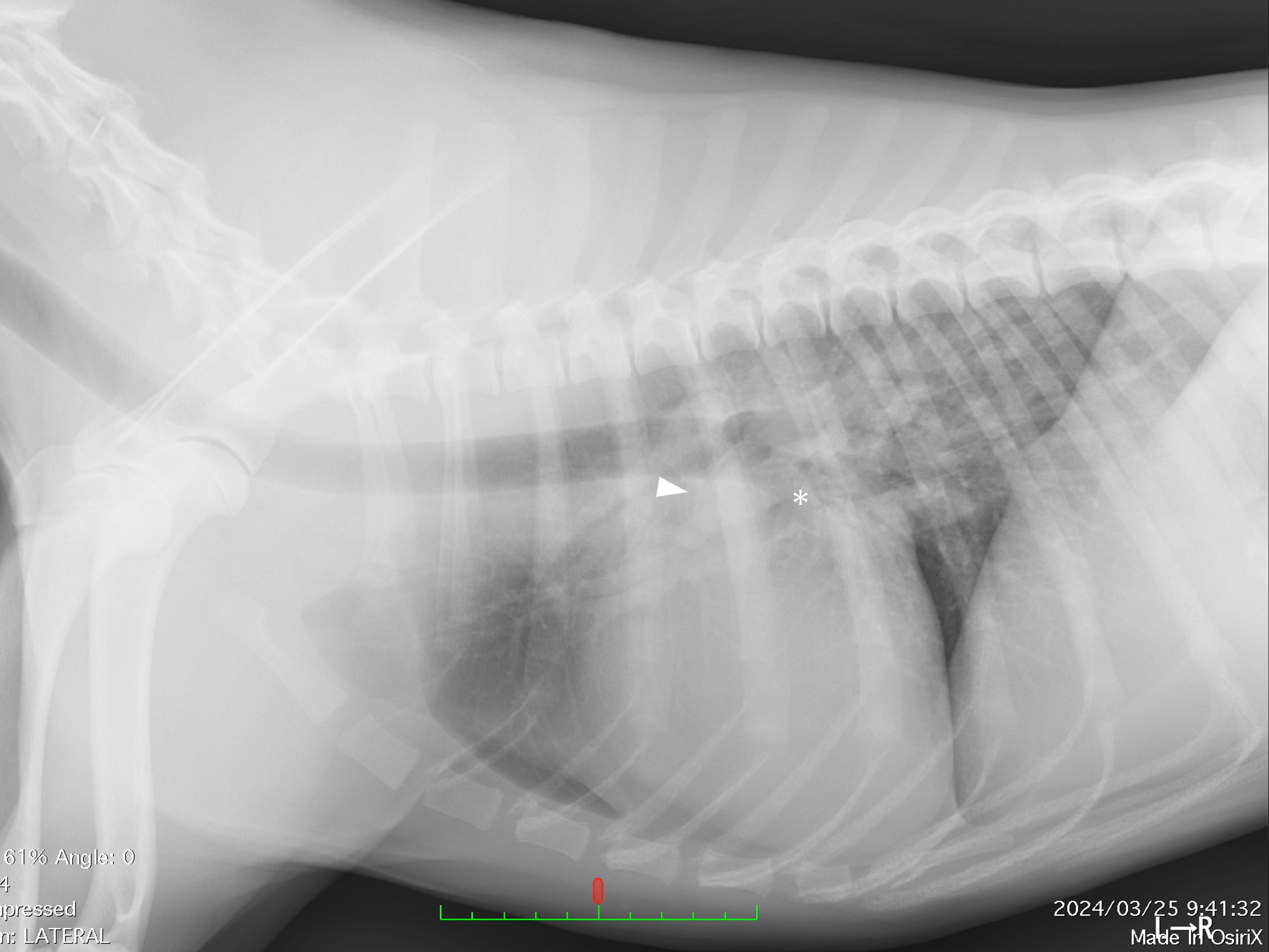
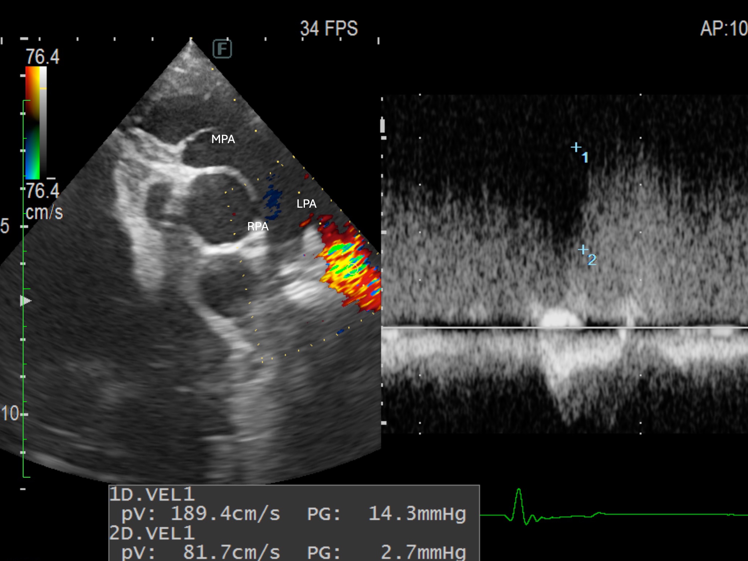
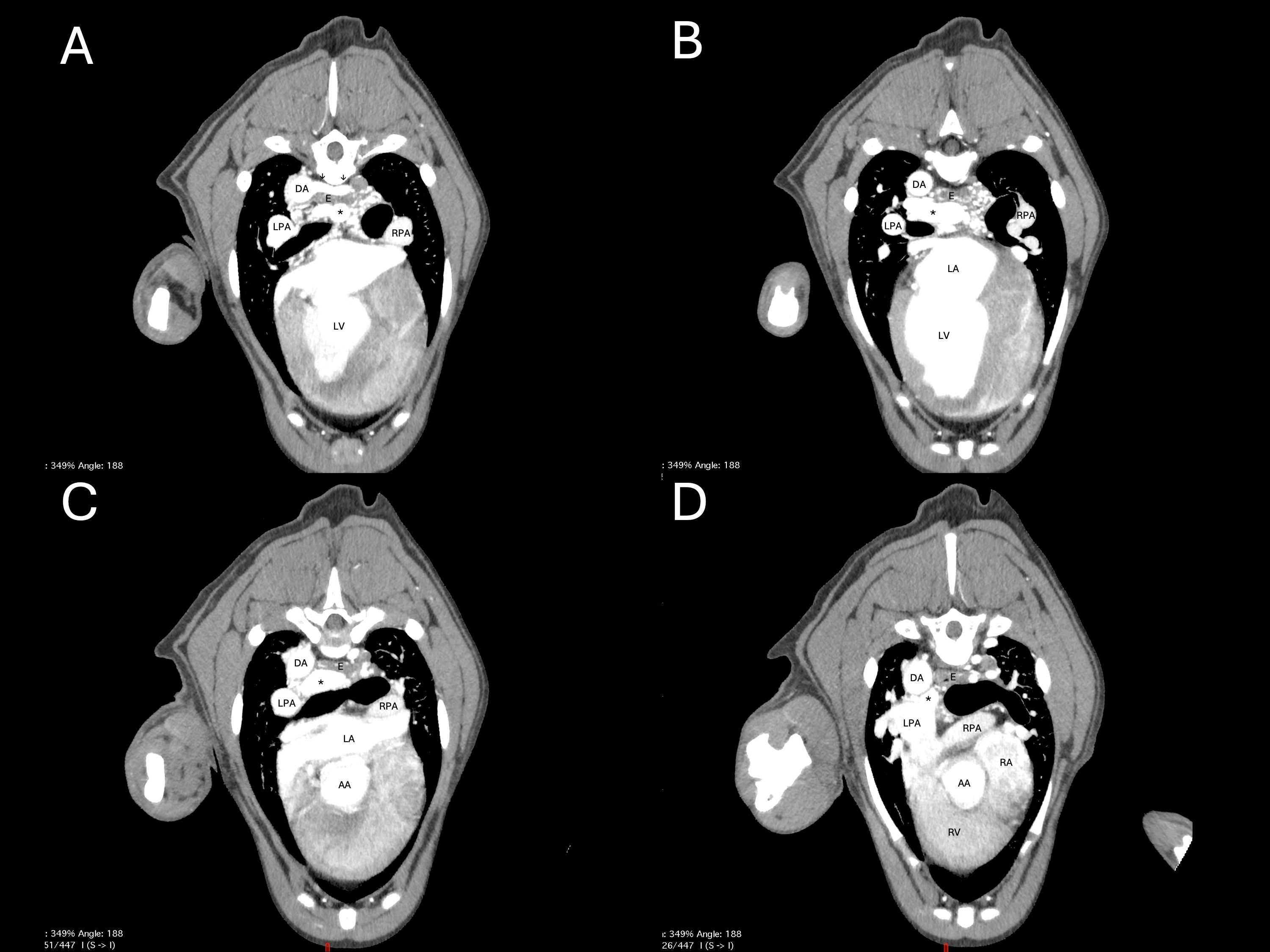
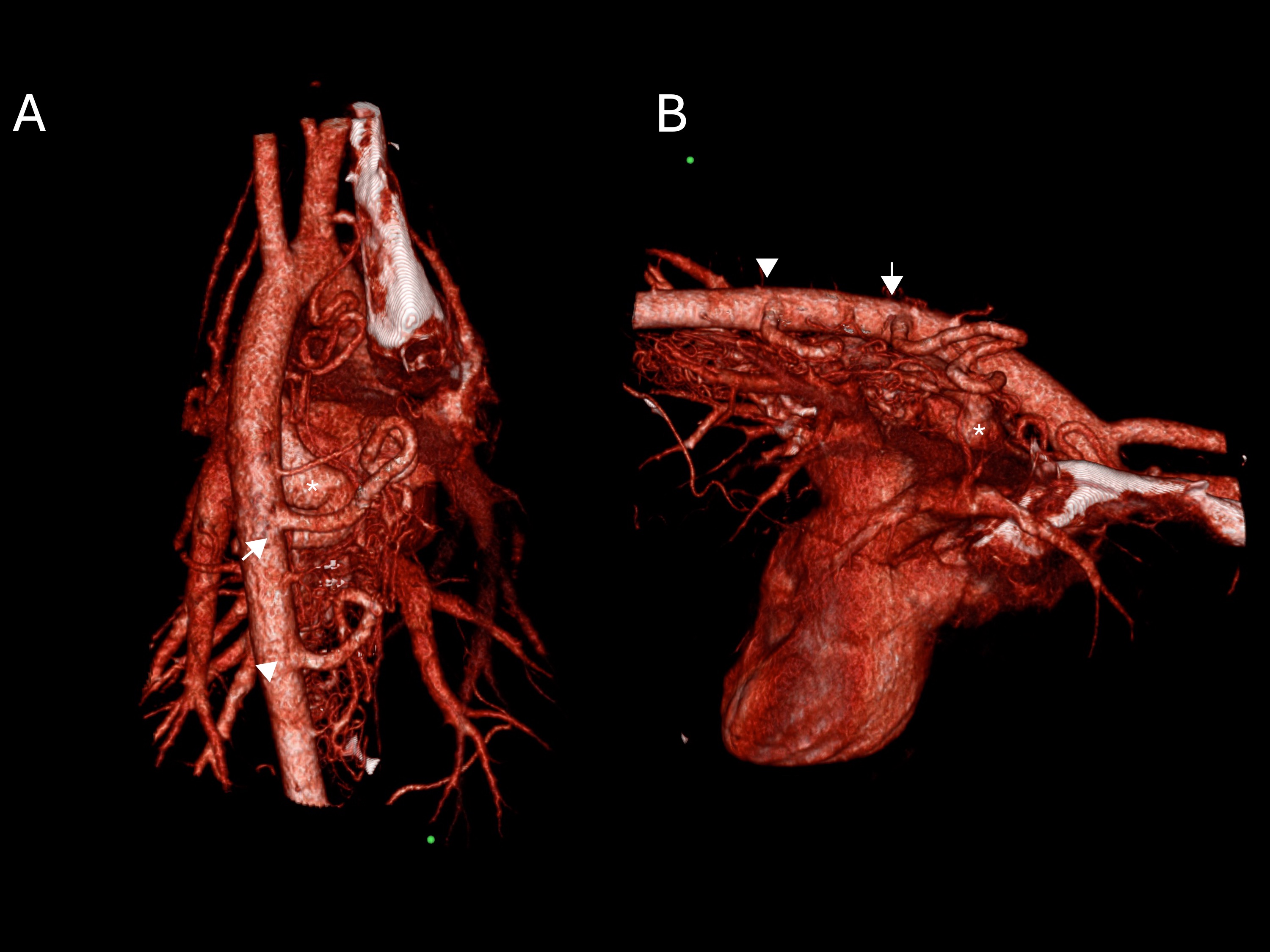
ABSTRACTBackground: Racemose hemangioma of the bronchial artery is a condition in which the bronchial artery is significantly curved, dilated, or tortuous, either congenitally or acquired, and abnormal anastomosis with the pulmonary artery and vein is sometimes observed. However, asymptomatic cases are occasionally incidentally diagnosed. Diagnosis was made using selective angiography and computed tomography (CT). This study reports the incidental diagnosis of racemose hemangioma of the bronchial artery in asymptomatic dogs and the results of the subsequent observations. Case Description: A 1-year-old Labrador Retriever with asymptomatic cardiac enlargement underwent an ultrasound examination, during which abnormal blood flow into the pulmonary artery was observed. A CT scan revealed a racemose hemangioma of the bronchial artery with a shunt to the pulmonary artery. The dog is alive and showed no clinical symptoms 352 days after diagnosis. Conclusion: This is the first report to describe a rare case of racemose hemangioma of the bronchial artery and to demonstrate the usefulness of CT in diagnosing this condition. Keywords: Bronchopulmonary artery fistula, Canine, Racemose hemangioma of the bronchial artery. IntroductionRacemose hemangioma of the bronchial artery is a condition in which the bronchial artery is significantly curved, dilated, or tortuous, either congenitally or acquired, and sometimes shows an abnormal anastomosis with the pulmonary artery and vein. It is characterized by the following: The first report of a racemose hemangioma of the bronchial artery was published in 1976 by v Babo et al. (1976) in Germany, who described it as a hyperplastic change in the bronchial artery (v Babo et al., 1976). In 1980, Cain and Spanel histologically examined angiomatoid vascular convolutions of the bronchial artery and proposed the term “primary angioma arterio-venosum racemosum” for cases without inflammation (Cain and Spanel, 1980). However, this disease has various names. The term “racemose hemangioma of the bronchial artery” is mainly used in the field of medicine in Japan (Takano et al., 2020). This condition is referred to as hypervascularity, bronchial artery dilation and tortuosity, arteriovenous malformations, and bronchial artery varices in the United States and Europe (Soda et al., 1995; Revel et al., 2002; Remy-Jardin et al., 2004). A case of anastomosis between the bronchial and pulmonary arteries in a horse has been reported as a “bronchopulmonary artery fistula” (Sano et al., 2021). The main symptom of this disease in humans is hemoptysis (Tanaka et al., 2003). Bronchoscopy often reveals aneurysms covered by the normal mucosa. Rupture of these aneurysms is thought to cause hemoptysis (Takano et al., 2020). In veterinary medicine, cases have been reported in horses in which the bronchial and pulmonary arteries were anastomosed, resulting in death due to respiratory distress caused by a pulmonary hemorrhage. This is the first report to describe racemose hemangioma of the bronchial artery in a dog. Case DetailsThe patient was a 1-year-old neutered male Labrador Retriever. Elevated cardiac biomarker levels (N-terminal pro-brain natriuretic peptide: 1,371 pg/ml, ref: <900 pg/ml; cardiac troponin I: 0.03 ng/ml, ref: <0.09 ng/ml) prompted a cardiac ultrasound examination, which revealed mild left ventricular enlargement and abnormal mosaic signals in the pulmonary artery. The dog was referred to the Osaka Metropolitan University Veterinary Clinical Center for further examination. The dog weighed 25.4 kg and had a heart rate of 120 bpm. The general condition of the dog was good, and no clinical signs were observed. No abnormalities were observed during the physical examination or blood tests. Thoracic radiography revealed an enlarged cardiac silhouette with a vertebral heart scale of 12.1v (ref: 8.5–10.5) and a soft tissue shadow at the cardiac base that resembled a tumor (Fig. 1). Echocardiography showed mild enlargement of the left ventricular internal dimension in diastole (LVIDDN: 1.96, ref: 1.14–1.61) and pulmonary artery dilation. Color flow revealed a continuous abnormal mosaic signal in the left pulmonary artery (Fig. 2). Blood flow velocity was measured using the Doppler method and was 1.89 m/s during systole and 0.81 m/s during diastole. These tests raised suspicion of a pulmonary artery shunt. A computed tomography (CT) scan was performed to clarify the anatomical structure of the arteriovenous shunt. A power injector was used to inject the contrast agent. Electrocardiographic gating was not performed. The time delay for angiography was planned as follows after the administration of 2 ml/kg of the contrast agent: arterial phase (5 seconds), venous phase (20 seconds), and equilibrium phase (180 seconds). Spiral angiography scans were performed for each phase. CT images obtained during the arterial phase revealed a dilated tracheoesophageal artery branching from the aorta and curving into complex small vessels along the esophagus (Fig. 3A). This complex branching of blood vessels formed a network of vascular clusters centered on the esophagus and trachea within the mediastinum (Fig. 3B). Additionally, a significantly dilated and tortuous vessel was observed between the esophagus and trachea, which was anastomosed with the left pulmonary artery. Blood inflow from the bronchial artery enhanced the left pulmonary artery more than the right pulmonary artery (Fig. 3C and D). Based on these findings, a racemose hemangioma of the bronchial artery was diagnosed. The sixth intercostal artery was markedly dilated and branched into the esophagus (Fig. 4). Follow-up observation without further treatment was decided as the course of action. The examination was conducted on day 352 after diagnosis. The patient remained stable, and no clinical signs were observed. Thoracic radiography revealed no changes in the cardiac shadows (VHS 11.3v) or in the shadows of the cardiac base compared to the initial examination. Slight pulmonary vessel dilation was observed. Echocardiography showed LVIDDN of 1.9, with no changes in the abnormal mosaic signals in the pulmonary artery.
Fig. 1. Right lateral radiograph of the dog showing displacement of the left primary bronchus (*). A soft tissue shadow (arrow) is observed on the dorsal side of the left superior lobar bronchus.
Fig. 2. Two-dimensional echocardiographic images showing the right parasternal short-axis view at the heart base. Continuous blood flow entering the left pulmonary artery was detected by color Doppler ultrasonography. The continuous wave Doppler measured flow velocities of up to 1.89 m/s and down to 0.81 m/s. MPA, main pulmonary artery; LPA, left pulmonary artery; RPA, right pulmonary artery.
Fig. 3. Transverse post-contrast images of the heart at the level of A) the branch of the bronchoesophageal artery (arrow) and descending aorta, D) Anastomosis between a hemangioma (*) and the pulmonary artery. B) and C) indicate hemangiomas between slices A) and D). DA, descending aorta; AA, ascending aorta; LPA, left pulmonary artery; RPA, right pulmonary artery; LA, left atrium; LV, left ventricle; E, esophagus.
Fig. 4. A 3-dimensional reconstruction of the heart was created from the CT images. A) Dorsal-ventral view and B) left lateral view. The bronchial esophageal artery, which branches from the aorta (white arrow), is divided into numerous small vessels. The dilated bronchial artery, which anastomoses with the left pulmonary artery, is indicated by an asterisk. The sixth intercostal artery (white arrowhead) is dilated and branches into the esophagus. DiscussionIn dogs, the tracheoesophageal artery originates from near the fifth intercostal artery on their right side. It runs alongside the esophagus and branches into the esophageal branch. It supplies blood to most of the esophagus in the thoracic cavity. The artery then enters the lung hilum. Tracheal arteries supply blood to the trachea, pulmonary airways, regional lymph nodes, and pulmonary arteries and veins (Bezuidenhout, 2013). In humans, this disease is characterized by dilation and bending of the bronchial arteries and shunting of the pulmonary artery or vein (Iwasaki et al., 2001). The tracheoesophageal artery, which branched off the aorta, was dilated and branched into small, complex, and tortuous vessels that supplied the esophagus with abundant blood. Subsequently, it branched into a severely dilated and complex tortuous vessel that shunted into the left pulmonary artery. This expanded area was identified as the tracheal artery, resulting in a racemose hemangioma of the bronchial artery. The clinical findings reported for this disease are similar to those previously observed in dogs with arteriovenous shunts (Fujii et al., 2009). Continuous murmurs were detected in these asymptomatic dogs, and blood flow into the pulmonary artery was detected using transthoracic echocardiography. Selective angiography revealed shunt flow from the aorta to the pulmonary artery; however, the anatomical structure of the shunt was unclear. The authors suggested that CT examination is necessary for detailed anatomical evaluation. In this case, a CT scan revealed a shunt from the dilated and tortuous bronchial artery to the left pulmonary artery. Shunting of the pulmonary artery or vein has been reported in many cases of bronchial artery racemose hemangiomas. However, the locations of the shunted pulmonary arteries varied. A heart murmur was not heard in this case, despite being heard in a previously reported case (Fujii et al., 2009), depending on the anatomical location of the shunt and the blood flow through the shunt. The etiology of racemose hemangiomas of the bronchial artery can be broadly categorized as primary, such as bronchial artery malformations or developmental abnormalities, or secondary, such as vascular abnormalities resulting from bronchi or lung inflammation (v Babo et al., 1976; Yon and Ravenel, 2010). The mechanism underlying the formation of secondary vascular malformations is thought to involve narrowing or obstruction of the pulmonary artery due to inflammatory lesions, such as bronchiectasis, pulmonary tuberculosis, or pneumonia. This leads to increased local oxygen demand and bronchial artery compensatory aneurysmal dilation (Ishikawa, 1977; Do et al., 2001). No narrowing or obstruction of the pulmonary artery was observed. No history or symptoms of respiratory disease were reported in the past or present. Therefore, congenital disorders were considered as possible diagnoses. As cardiovascular catheterization was not performed, the pulmonary artery pressure could not be measured. As there was no tricuspid regurgitation and the pulmonary regurgitation was mild, the pulmonary vascular resistance could not be estimated from the regurgitant flow velocity during echocardiography. Additionally, the fistula in the left pulmonary artery was difficult to visualize on ultrasonography. Therefore, the measured shunt flow velocity was lower than the actual value, making it unsuitable for estimating pulmonary artery pressure. However, the absence of right ventricular enlargement or ventricular septal displacement, in addition to the continuous shunt flow into the left pulmonary artery, suggested that pulmonary vascular resistance was not elevated. Therefore, secondary pulmonary hypertension was excluded from the diagnosis. In human medicine, racemose hemangioma of the bronchial artery is considered a preventive treatment because of the risk of rupture, even in cases in which the patient does not experience symptoms. However, no guidelines exist for determining treatment based on factors such as the degree of vascular dilation. Common human treatments include embolization, bronchial artery ligation, and surgical resection of the affected lung tissue (Tanaka et al., 2003; Sanno et al., 2009; Matsubara et al., 2015). In the present case, bronchial artery dilatation was observed. However, no such treatment is required. The main symptom of this disease in humans is hemoptysis (Tanaka et al., 2003). Bronchoscopy often reveals aneurysms covered by normal mucosa. The rupture of these aneurysms is thought to cause hemoptysis (Takano et al., 2020). Bronchoscopy was not performed in this case, and no signs of hemoptysis were observed. However, the risk of hemoptysis cannot be excluded. The esophageal artery showed strong staining on CT angiography. An aneurysm may form in the esophagus in dogs when the bronchial artery branches into the esophageal artery. Therefore, there is a risk of bleeding due to esophageal mucosa damage. Therefore, the dog was monitored for hemoptysis, gastrointestinal bleeding, and cardiac volume overload. No signs of respiratory symptoms or gastrointestinal bleeding were observed during the observation period, which lasted until day 352. No morphological changes due to cardiac volume overload were noted. ConclusionThis report describes a rare case of a dog with racemose bronchial artery hemangioma. If radiographic examination reveals dilated vascular shadows around the bronchi, this disease should be suspected, and a CT scan should be performed to confirm the diagnosis. Further research is needed to determine the prognosis of this disease in dogs, as it poses a risk of hemoptysis in humans. AcknowledgmentsNone. Conflict of interestThe authors declare no conflict of interest. FundingThis study received no specific grant. Authors’ contributionsF.T. found this case and was responsible for regular follow-up examinations. S.S., S.H., W.A., and H.T. were involved in diagnosing this case and participated in the follow-up. All authors have reviewed, discussed, and agreed upon their respective contributions to this manuscript before and during its submission. Data availabilityAll data supporting this study’s findings are available within the manuscript. ReferencesBezuidenhout, A. 2013. The heart and arteries. In Miller’s anatomy of the dog. Eds., de Lahunta, A., and Evans, H.E. 5th ed, St. Louis, MO: Elsevier, pp: 428–504. Cain, H. and Spanel, K. 1980. Etiology and morphogenesis of the socalled bronchial arterioma (author's transl). Klin. Wochenschr. 58, 347–357; doi:10.1016/j.klinwwr.1980.09.016 Do, K.H., Goo, J.M., Im, J.G., Kim, K.W., Chung, J.W. and Park, J.H. 2001. Systemic arterial supply to the lungs in adults: spiral CT findings. Radiographics 21, 387–402. Fujii, Y., Aoki, T., Takano, H., Ishikawa, R. and Wakao, Y. 2009. Arteriovenous shunts resembling patent ductus arteriosus in dogs: 3 cases. J. Vet. Cardiol. 11, 147–151. Ishikawa, T. 1977. Systemic artery—pulmonary artery communication in Takayasu's arteritis. AJR. Am. J. Roentgenol. 128, 389–393. Iwasaki, M., Kobayashi, H., Nomoto, T., Arai, T. and Kondoh, T. 2001. Primary racemose hemangioma of the bronchial artery. Intern. Med. 40, 650–653. Matsubara, Y., Morisaki, K., Matsuda, D., Aoyagi, Y., Tanaka, S., Okadome, J., Fukunaga, R., Matsumoto, T. and Maehara, Y. 2015. Thoracic endovascular aortic repair and coil embolization of the pulmonary artery for primary racemose hemangioma of the bronchial artery with a bronchial-pulmonary artery fistula. Vascular 23, 436–439. Remy-Jardin, M., Bouaziz, N., Dumont, P., Brillet, P.Y., Bruzzi, J. and Remy, J. 2004. Bronchial and nonbronchial systemic arteries at multi-detector row CT angiography: comparison with conventional angiography. Radiology 233, 741–749. Revel, M.P., Fournier, L.S., Hennebicque, A.S., Cuenod, C.A., Meyer, G., Reynaud, P. and Frija, G. 2002. Can CT replace bronchoscopy in the detection of the site and cause of bleeding in patients with large or massive hemoptysis?. AJR. Am. J. Roentgenol. 179, 1217–1224. Sanno, K., Hatanaka, N., Yamagishi, T., Nakazawa, I., Hirano, Y., Hosaka, K. and Kamemura, H. 2009. Selective gelfoam embolization of primary racemose hemangioma of the bronchial artery. Respirology 14, 609–611. Sano, Y., Kuwajima, H., Kanai, H., Baba, C., Azami, K. and Matsuda, K. 2021. A bronchopulmonary artery fistula in a horse. J. Vet. Med. Sci. 83, 1225–1228. Soda, H., Oka, M., Kohno, S., Watanabe, M., Takatani, H. and Hara, K. 1995. Arteriovenous malformation of the bronchial artery showing endobronchial protrusion. Intern. Med. 34, 797–800. Takano, R., Matsutani, T., Hagiwara, N., Nomura, T. and Yoshida, H. 2020. Racemose hemangioma of the bronchial artery mimicking esophageal submucosal tumor: a case report. Clin. J. Gastroenterol. 13, 1022–1027. Tanaka, K., Ihaya, A., Horiuci, T., Morioka, K., Kimura, T., Uesaka, T., Sasaki, M., Uchinami, M., Tsuda, T., Yamada, N., Li, W., Hirai, S., Tanabe, S., Okubo, Y. and Tanaka, S. 2003. Giant mediastinal bronchial artery aneurysm mimicking benign esophageal tumor: a case report and review of 26 cases from literature. J. Vasc. Surg. 38, 1125–1129. v Babo, H.A., Huzly, H.K., Deininger, and Barth, V. 1976. '[Angiomas and angioma-like changes of the bronchial arteries (author's transl) 1976. Rofo 124, 103–110. Yon, J.R. and Ravenel, J.G. 2010. Congenital bronchial artery-pulmonary artery fistula in an adult. J. Comput. Assist. Tomogr. 34, 418–420. | ||
| How to Cite this Article |
| Pubmed Style Furuike T, Shimamura S, Hirata S, Watanabe S, Tani H. Racemose hemangioma of the bronchial artery in a dog. Open Vet. J.. 2026; 16(6): 3569-3574. doi:10.5455/OVJ.2026.v16.i6.26 Web Style Furuike T, Shimamura S, Hirata S, Watanabe S, Tani H. Racemose hemangioma of the bronchial artery in a dog. https://www.openveterinaryjournal.com/?mno=281587 [Access: June 26, 2026]. doi:10.5455/OVJ.2026.v16.i6.26 AMA (American Medical Association) Style Furuike T, Shimamura S, Hirata S, Watanabe S, Tani H. Racemose hemangioma of the bronchial artery in a dog. Open Vet. J.. 2026; 16(6): 3569-3574. doi:10.5455/OVJ.2026.v16.i6.26 Vancouver/ICMJE Style Furuike T, Shimamura S, Hirata S, Watanabe S, Tani H. Racemose hemangioma of the bronchial artery in a dog. Open Vet. J.. (2026), [cited June 26, 2026]; 16(6): 3569-3574. doi:10.5455/OVJ.2026.v16.i6.26 Harvard Style Furuike, T., Shimamura, . S., Hirata, . S., Watanabe, . S. & Tani, . H. (2026) Racemose hemangioma of the bronchial artery in a dog. Open Vet. J., 16 (6), 3569-3574. doi:10.5455/OVJ.2026.v16.i6.26 Turabian Style Furuike, Toshihide, Shunsuke Shimamura, Shougo Hirata, Shunichi Watanabe, and Hiroyuki Tani. 2026. Racemose hemangioma of the bronchial artery in a dog. Open Veterinary Journal, 16 (6), 3569-3574. doi:10.5455/OVJ.2026.v16.i6.26 Chicago Style Furuike, Toshihide, Shunsuke Shimamura, Shougo Hirata, Shunichi Watanabe, and Hiroyuki Tani. "Racemose hemangioma of the bronchial artery in a dog." Open Veterinary Journal 16 (2026), 3569-3574. doi:10.5455/OVJ.2026.v16.i6.26 MLA (The Modern Language Association) Style Furuike, Toshihide, Shunsuke Shimamura, Shougo Hirata, Shunichi Watanabe, and Hiroyuki Tani. "Racemose hemangioma of the bronchial artery in a dog." Open Veterinary Journal 16.6 (2026), 3569-3574. Print. doi:10.5455/OVJ.2026.v16.i6.26 APA (American Psychological Association) Style Furuike, T., Shimamura, . S., Hirata, . S., Watanabe, . S. & Tani, . H. (2026) Racemose hemangioma of the bronchial artery in a dog. Open Veterinary Journal, 16 (6), 3569-3574. doi:10.5455/OVJ.2026.v16.i6.26 |


