


| Research Article | ||
Open Vet. J.. 2025; 15(12): 6245-6252 Open Veterinary Journal, (2025), Vol. 15(12): 6245-6252 Research Article NT-proBNP versus cardiac troponin-I (cTnI) for the early detection of cardiac diseases in dogs with other cardiographic findingsIntan Permatasari Hermawan1*, Palestin Palestin2, Ady Kurnianto1, Hana Cipka Pramuda Wardhani3 and Dian Ayu Kartika Sari41Laboratorium of Veterinary Internal Medicine, Faculty of Veterinary Medicine, Wijaya Kusuma Surabaya University, Surabaya, Indonesia 2Laboratorium of Clinical Pathology, Faculty of Veterinary Medicine, Wijaya Kusuma Surabaya University, Surabaya, Indonesia 3Laboratorium of Physiology, Faculty of Veterinary Medicine, Wijaya Kusuma Surabaya University, Surabaya, Indonesia 4Laboratorium of Biochemistry, Faculty of Veterinary Medicine, Wijaya Kusuma Surabaya University, Surabaya, Indonesia *Corresponding Author: Intan Permatasari Hermawan. Laboratorium of Veterinary Internal Medicine, Faculty of Veterinary Medicine, Wijaya Kusuma Surabaya University, Surabaya, Indonesia. Email: intanpermatasari [at] uwks.ac.id Submitted: 21/08/2025 Revised: 02/11/2025 Accepted: 10/11/2025 Published: 31/12/2025 © 2025 Open Veterinary Journal
AbstractBackground: Heart diseases are common in dogs, geriatric, overweight, to obese individuals, with metabolic problems, lack of nutrition, and breed problems. NT-proBNP and cardiac troponin-I (cTnI) are selected for the diagnosis of heart damage in early detection. Both of these biomarkers have another function. This study compared NT-proBNP with cTnI for the early detection of cardiac diseases in dogs. Aim: This study compared NT-proBNP with cTnI levels for the early detection of cardiac diseases in dogs. Methods: A total of five dogs of various breeds, ages, sexes, and body weights were included. All animals underwent a comprehensive diagnostic workup, including a thorough history and physical examination, cardiorespiratory auscultation, and laboratory diagnostics. These diagnostics include blood pressure measurement, thoracic radiography, and echocardiography, which are performed on every patient, and biomarker levels used a commercial kit rapid test (®Flash test). Results: Four dogs had a cardiac troponin I level of >0.1 ng/ml (abnormal), and 1 dog has a normal cTnI level, but four dogs had a normal NT-proBNP level ≤ 2 ng/ml, but 1 dog has an abnormal level. Every dog has laboratory results based on blood pressure, electrocardiography, X-ray, and echocardiography per case. Based on cardiographic results relevant with cTnI that more sensitive if asymptomatic patients rather than NT-proBNP Conclusion: cTnI is more sensitive than NT-proBNP, supported by other laboratory examinations to establish a more accurate diagnosis. However, a larger number of case samples is needed to draw a more accurate diagnosis. Keywords: NT-proBNP, cTn-I, Cardiography, Dogs. IntroductionCardiac diseases in dogs, such as Dilated Cardiomyopathy, Hypertrophic Cardiomyopathy, Myxomatous Mitral Valvular Disease (MMVD), and Congestive Heart Failure, are major concerns in veterinary cardiology. These conditions can be fatal, especially when diagnostic tools are limited. The gold standard for cardiac diagnosis includes blood pressure measurement, echocardiography, radiography, electrocardiography, and cardiac biomarkers, such as those detected by immunofluorescence techniques. According to the American College of Veterinary Internal Medicine consensus guidelines, the diagnosis and staging of MMVD are based on clinical signs, echocardiographic findings, and thoracic radiographs. In this context, biomarkers serve as indicators of physiological or pathological processes in the body. Two of the most studied cardiac biomarkers are N-terminal pro–B-type natriuretic peptide (NT-proBNP) and cardiac troponin-I (cTnI), both of which provide valuable insight into myocardial stress and damage. NT-proBNP is a marker of cardiac wall stress and has been associated with subclinical cardiac disease, helping in the diagnosis and prognosis of HF (Cushman et al., 2016) reported its potential in identifying silent cardiac conditions, while a recent study emphasized its role in evaluating the progression of heart failure (Nasab et al., 2023). Cardiac troponin-I is a highly specific and sensitive marker of myocardial injury that can be measured in plasma through immunoassay techniques. Demonstrated that in dogs with cardiomyopathy and mitral valve disease, plasma cTnI levels were positively correlated with left ventricular and atrial enlargement (Oyama and Sisson, 2004). In dogs with subaortic stenosis, cTnI showed a moderate correlation with ventricular wall thickness. Furthermore, dogs with CM and cTnI concentrations >0.20 ng/ml had a significantly shorter median survival time (112 days) than those with lower concentrations (<0.20 ng/ml; 357 days; p=0.006). Serum cTnI levels were significantly elevated in dogs with IE compared with those with preclinical MMVD or inflammatory myocardial disease. In clinically suspected cases, a serum cTnI concentration of >0.625 ng/ml supports the diagnosis of IE (Kilkenny et al., 2021) Despite the increasing use of cardiac biomarkers in veterinary cardiology, comparative research on the effectiveness of NT-proBNP versus cTnI for the early detection of cardiac diseases in dogs remains limited. Although both biomarkers are individually well-documented for diagnosing and monitoring heart disease, few studies have directly compared their diagnostic accuracy, sensitivity, specificity, and prognostic value in clinical settings. This study aimed to address this gap by evaluating NT-proBNP and cTnI levels in dogs with early clinical signs of cardiac disease, supported by echocardiographic and radiographic evidence. By analyzing each patient’s clinical presentation and differential diagnosis, this research seeks to determine which biomarker provides more reliable information for early and accurate detection, potentially improving patient outcomes and guiding treatment decisions in veterinary cardiology. Materials and MethodsThis study was conducted from January 2025 to March 2025 at the Teaching Animal Hospital WEKA, Faculty of Veterinary Medicine, Universitas Wijaya Kusuma Surabaya, Indonesia. A total of five dogs of various breeds, ages, sexes, and body weights were included. All animals underwent a comprehensive diagnostic workup, including a thorough history and physical examination, cardiorespiratory auscultation, and laboratory diagnostics. These diagnostics include blood pressure measurement, thoracic radiography, and echocardiography, which are performed on every patient. Radiographic examinationThoracic radiographs were obtained in the right lateral position to assess cardiac dimensions. The following parameters were evaluated: • Vertebral heart size (VHS) • Thoracic inlet heart size (THIS) • Tracheal diameter • Pulmonary patterns (e.g., pulmonary edema or vascular changes) EchocardiographyAn Edan Aclarix AX-3 ultrasound system with a microcurve probe was used for echocardiographic evaluations. Dogs were positioned in both the right and left lateral recumbencies to allow right and left parasternal views, respectively. Imaging included: • Brightness mode (B mode) • Motion mode (M-mode) • Color Flow Doppler The echocardiographic probe was aligned in long axis and short-axis planes to visualize cardiac structures and assess functional abnormalities. All diagnoses of cardiac abnormalities were made based on these imaging results. Biomarker analysisSerum samples were collected from all five dogs following diagnosis. The concentrations of NT-proBNP and cTnI were measured using the ®FLASH Test (immunofluorescence method). The interpretation criteria were as follows: • NT-proBNP: o ≤ 2 ng/ml=normal o 2–10 ng/ml=suspect o 10 ng/ml=abnormal • Cardiac Troponin-I (cTnI): o ≤ 0.1 ng/ml=normal o 0.1 ng/ml=abnormal test. A p-value < 0.05 was considered statistically significant. Ethical approvalAll dogs included in this study were enrolled following informed consent from their respective owners. The study protocol was reviewed and approved by the Ethics Committee of the Faculty of Veterinary Medicine, University of Wijaya Kusuma Surabaya (approval no. 132B-KKE) Date: 30 December, 2024. ResultsSymptoms in patients with cardiac problems can be seen in Table 1. Generally, geriatric dogs with heart disorders exhibit symptoms such as coughing, murmur, dyspnea, and exercise intolerance. These symptoms may occur as a result of compensatory mechanisms due to weakened cardiac function, which consequently affects the respiratory system. Table 1. Symptoms in dogs.
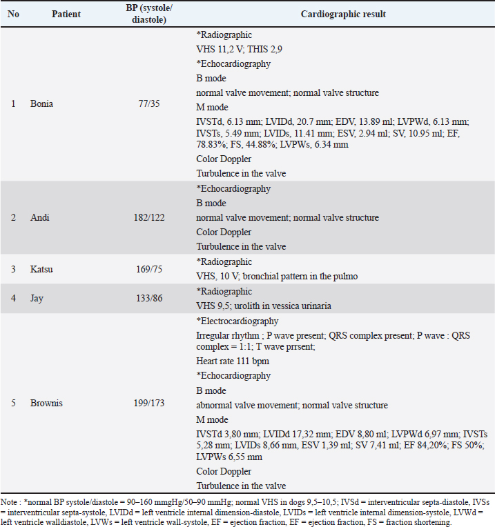
Table 2 presents the results of cardiac examinations from various cases, including radiography, echocardiography, and electrocardiography. The type of cardiac examination was selected based on the patient’s cardiac condition. In Table 2, radiographic examinations were performed on patients Bonia, Katsu, and Jay to measure the VHS. Echocardiographic examinations were conducted on patients Bonia, Andi, and Brownis to evaluate the valves and chambers using B-mode, M-mode, and color Doppler. Meanwhile, the electrocardiographic examination was performed only on Brownis, as this patient exhibited an abnormal heart rhythm. Table 2. Cardiographic result.
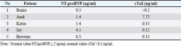
Table 3 shows the results of cardiac biomarkers, including NT-proBNP and cTnI, which are used to detect cardiac injury. Among the four patients, abnormalities in NT-proBNP were found in Jay (4.1 ng/ml), while abnormalities in cTnI were observed in Andi (7.77 ng/ml). Table 3. NT-proBNP and cTnI.
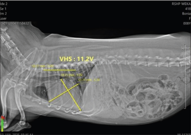
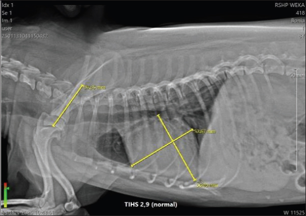
Figure 1 shows the VHS measurement of Bonia’s heart, with a value of 11.2 V, whereas the normal range in dogs is 9.5–10.5 V. This indicates that Bonia is experiencing cardiomegaly. Figure 2 shows the TIHS, a method used to detect MMVD. Under normal conditions, the TIHS value is less than 3 (Marbela et al., 2023); therefore, Bonia is not affected by MMVD.
Fig. 1. VHS 11,2 V of Bonia.
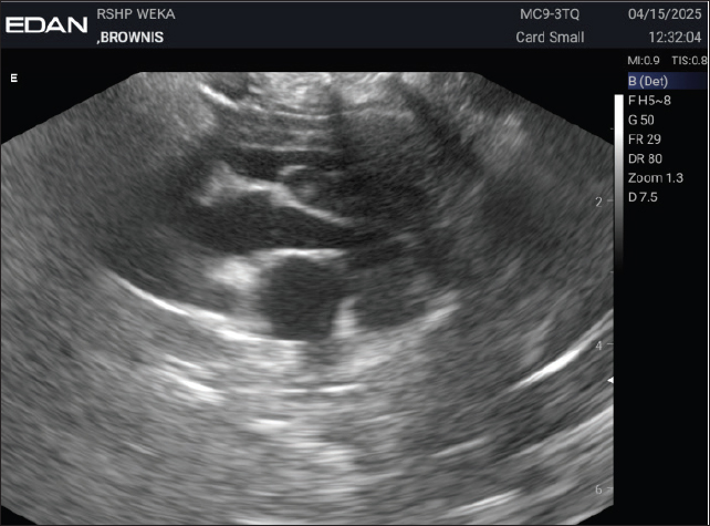
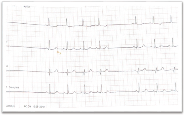
Fig. 2. Thoracic Inlet heart size (THIS) 2,9 of Bonia. Figures 3 and 4 show the results of the echocardiographic examination, while Figure 5 presents the electrocardiographic findings of the dog, Brownis. The motion mode (M-mode) evaluation (Fig. 3) was used to measure the left ventricular dimensions during systole and diastole, as well as systolic and diastolic function values. The M-mode results showed a decrease in interventricular septal thickness during diastole and systole (IVSTd=5.28 mm, IVSTs=3.80 mm) and a reduction in left ventricular internal dimension during systole (LVIDs=8.66 mm). Figure 4 represents the brightness mode (B-mode) evaluation, used to assess valve thickening and movement. The results showed abnormal valve movement with normal valve structure. Figure 5 presents the electrocardiographic examination performed at a paper speed of 50 mm/second using a 5-lead system, which revealed sinus arrhythmia.
Fig. 3. M-mode Echocardiography of Brownis.
Fig. 4. Echocardiography B mode of Brownis.
Fig. 5. ECG of Brownis. DiscussionTable 1 shows that each patient presented with different clinical signs and laboratory results. In Table 2, Bonia was reported with hypotension (77/35 mmHg) and a VHS value of 11.2 V, which falls into the cardiomegaly category. The THIS score was 2.9, indicating no evidence of MMVD because the normal THIS value for dogs is below 3 (Lam et al., 2021). Echocardiography in B-mode showed normal valve movement and valve structure, whereas M-mode measurements were within the normal range. However, color Doppler examination revealed turbulence, suggesting disturbed blood flow and valvular regurgitation. Color Doppler function is used to detect congenital or acquired cardiac diseases (Wess et al., 2021). According to the cardiac biomarkers presented in Table 3, Bonia had NT-proBNP levels of 0.5 ng/ml and cTnI levels <0.1 ng/ml. These values are within the normal range, indicating no myocardial damage or cardiac injury. Andi’s examination results (Table 2) showed hypertension (182/122 mmHg). Echocardiography in B-mode demonstrated normal valve movement and valve structure, while color Doppler revealed turbulence, indicating disturbed blood flow and valvular regurgitation. The cardiac biomarkers (Table 3) showed that NT-proBNP was within the normal range at 1.4 ng/ml, whereas cTnI was elevated at 7.77 ng/ml. This finding suggests that Andi experienced early myocardial cell injury, as evidenced by the abnormal cTnI value. Katsu radiographic evaluation revealed a VHS of 10 V (within normal limits), but a bronchial pattern was detected in the lungs, indicating a tendency for coughing and dyspnea. The normal VHS value in dogs is about 9,5–10,5 V based on each breed, and abnormality in the heart showed cardiac silhouette size in dogs (Costanza et al., 2023). Katsu has normal VHS, but the pulmonic imaging has a bronchial pattern, and this condition includes cardiorespiratory because cTnI showed abnormal. Blood pressure measurement showed systolic/diastolic values of 169/75 mmHg, with an elevated systolic value consistent with situational hypertension. Katsu cardiac biomarkers showed NT-proBNP at 1.4 ng/ml and cTnI at 0.13 ng/ml. Both values were within normal limits, indicating no evidence of myocardial damage. The X-ray examination of Jay revealed a VHS of 9.5 V, with the presence of uroliths in the bladder. Blood pressure was within the normal range at 133/86 mm Hg. However, NT-proBNP levels were elevated at 4.1 ng/ml, and cardiac troponin I was also abnormal at 0.12 ng/ml. In the case of brownies, electrocardiography (ECG) showed an irregular rhythm with the presence of P waves, QRS complexes, a 1:1 P wave to QRS complex ratio, T waves, and a heart rate of 111 bpm. B-mode echocardiography revealed abnormal valve movement with normal valve structure. M-mode measurements were as follows: IVSTd 3.80 mm (normal 5.2–6.5 mm), LVIDd 17.32 mm (normal 15.83–18.53 mm), EDV 8.80 ml, LVPWd 6.97 mm (normal 5.1–6.1 mm), IVSTs 5.28 mm (normal 6.95–8.22 mm), LVIDs 8.66 mm (normal 9.01–11.5 mm), ESV 1.39 ml, SV 7.41 ml, EF 84.20% (normal 25%–55%), FS 50% (normal 55%–85%), and LVPWs 6.55 mm (normal 6.6–8.12 mm) (Cerbu et al., 2023). Color Doppler examination demonstrated turbulence at the valve, indicating abnormal blood flow velocity, visualized as red and blue gradients on the Doppler image. NT-proBNP levels were within normal limits at 0.5 ng/ml, whereas cardiac troponin I was elevated at 0.13 ng/ml, indicating myocardial cell injury. The results of cardiac biomarker examinations in the five patients showed that cTnI was more sensitive than NT-proBNP. As presented in Table 3, compared with NT-proBNP, troponin levels could detect myocardial abnormalities at an earlier stage. NT-proBNP levels were elevated in patients with symptomatic heart disease compared to asymptomatic cases. Conversely, cTnI could already be detected at abnormal levels in asymptomatic patients. This finding is consistent with previous study which reported that NT-proBNP levels in the symptomatic group were significantly higher than those in the asymptomatic (p < 0.001) and control (p < 0.001) groups, while cTnI levels in the control group were significantly lower than those in both the asymptomatic (p=0.039) and symptomatic (p=0.001) groups (Chanmongkolpanit et al., 2024). These results confirm that cardiac troponin I is a more sensitive biomarker than NT-proBNP. Among the five dogs, Andi presented with hypertension (182/122 mmHg) accompanied by snoring, coughing, occasional dyspnea, heart murmur, and exercise intolerance. These findings were correlated with abnormal levels of both NT-proBNP (1.4 ng/ml) and cTnI (7.77 ng/ml). Echocardiography using color Doppler also revealed turbulence, supporting the presence of valvular regurgitation and blood flow disturbance. Hypertension imposes additional stress on the heart, particularly on the left ventricle, resulting in myocardial cell damage. This is in agreement with a study which demonstrated that serum troponin I concentrations in dogs with pulmonary hypertension averaged 0.21 ng/ml (range: 0.10–2.10 ng/ml; p < 0.001) (Guglielmini et al., 2010). Another cardiographic test, such as radiographic, echocardiographic, or electrocardiographic, is the gold standard for diagnosing cardiac problems. Cardiac biomarkers as one of the diagnostic tools to complete the diagnosis. It takes a veterinarian’s experience and expertise to diagnose heart disease. ConclusionIn conclusion, cTnI is more sensitive than NT-proBNP, which is supported by other laboratory examinations to establish a more accurate diagnosis. However, a larger number of case samples are needed to draw a more accurate diagnosis. AcknowledgmentsThe author would like to thank the Faculty of Veterinary Medicine, University of Wijaya Kusuma Surabaya, the Teaching Animal Hospital of Weka in Surabaya, and K and P Clinic Surabaya for their support of this research. Conflict of interestThe authors have no conflicts of interest to declare. This study received funding from the Faculty of Veterinary Medicine, University of Wijaya Kusuma Surabaya. These entities had no role in the study design, data collection and analysis, decision to publish, or manuscript preparation. FundingThis research was funded by LPPMLPPM University of Wijaya Kusuma Surabaya to support this research funding 2025. Authors' contributionsIPH was the conceptualization, research design, data curation, data analysis, interpretation, and discussion of the results. P was the practitioner to prepare the data. AK, HCPW, and DAKS were the research administrators. Data availabilityThe data used to support the findings of this study are available upon request from the corresponding author. ReferencesCerbu, M., Cerbu, C. and Papuc, I. 2023. M-mode echocardiography in canine veterinary practice: a comprehensive review of left ventricular measurements in 44 different dog breeds. Animals. (Basel). 13, 2986. Chanmongkolpanit, K., Riengvirodkij, N., Channgam, P., Kaenchan, P., Buayam, W., Janhirun, Y., Phonarknguen, R., Tansakul, M. and Sakcamduang, W. 2024. How accurate are NT-proBNP, ANP, and cTnI levels in diagnosing myxomatous mitral valve disease in dogs?. Open Vet. J. 14, 1426–1441. Costanza, D., Greco, A., Piantedosi, D., Bruzzese, D., Pasolini, M.P., Coluccia, P., Castiello, E., Baptista, C.S. and Meomartino, L. 2023. The heart to single vertebra ratio: a new objective method for radiographic assessment of cardiac silhouette size in dogs. Vet. Radiol. Ultrasound. 64, 378–384. Cushman, M., Callas, P.W., Mcclure, L.A., Unverzagt, F.W., Howard, V.J., Gillett, S.R., Thacker, E.L. and Wadley, V.G. 2016. N-terminal pro-b-type natriuretic peptide and risk of future cognitive impairment in the REGARDS cohort. J. Alzheimers. Dis. 54, 497–503. Guglielmini, C., Civitella, C., Diana, A., Di Tommaso, M., Cipone, M. and Luciani, A. 2010. Serum cardiac troponin I concentration in dogs with precapillary and postcapillary pulmonary hypertension. J. Vet. Intern. Med. 24, 145–152. Kilkenny, E., Watson, C., Dukes-McEwan, J., Bode, E.F., Hezzell, M.J., Payne, J.R. and Borgeat, K. 2021. Evaluation of serum cardiac troponin-I concentrations for infective endocarditis diagnosis in dogs. J. Vet. Intern. Med. 35, 2094–2101. Lam, C., Gavaghan, B.J. and Meyers, F.E. 2021. Radiographic quantification of left atrial size in dogs with myxomatous mitral valve disease. J. Vet. Intern. Med. 35, 747–754. Marbella Fernández, D., García, V., Santana, A.J. and Montoya-Alonso, J.A. 2023. The Thoracic Inlet Length as a Reference Point to Radiographically Assess Cardiac Enlargement in Dogs with Myxomatous Mitral Valve Disease. Animals. (Basel). 13, 2666. Nasab Mehrabi E., Toupchi-Khosroshahi V., Athari S, S. 2023. Relationship of atrial fibrillation and N terminal pro brain natriuretic peptide in heart failure patients. ESC Heart Fail. 6, 3250–3257. Oyama, M.A. and Sisson, D.D. 2004. Cardiac troponin-I concentration in dogs with cardiac disease. J. Vet. Intern. Med. 18, 831–839. Wess, G., Bauer, A. and Kopp, A. 2021. Echocardiographic reference intervals for volumetric measurements of the left ventricle using the Simpson’s method of discs in 1331 dogs. J. Vet. Intern. Med. 35, 724–728. | ||


| How to Cite this Article |
| Pubmed Style Hermawan IP, Palestin P, Kurnianto A, Wardhani HCP, Sari DAK. NT-proBNP versus cardiac troponin-I (cTnI) for the early detection of cardiac diseases in dogs with other cardiographic findings. Open Vet. J.. 2025; 15(12): 6245-6252. doi:10.5455/OVJ.2025.v15.i12.9 Web Style Hermawan IP, Palestin P, Kurnianto A, Wardhani HCP, Sari DAK. NT-proBNP versus cardiac troponin-I (cTnI) for the early detection of cardiac diseases in dogs with other cardiographic findings. https://www.openveterinaryjournal.com/?mno=278721 [Access: January 25, 2026]. doi:10.5455/OVJ.2025.v15.i12.9 AMA (American Medical Association) Style Hermawan IP, Palestin P, Kurnianto A, Wardhani HCP, Sari DAK. NT-proBNP versus cardiac troponin-I (cTnI) for the early detection of cardiac diseases in dogs with other cardiographic findings. Open Vet. J.. 2025; 15(12): 6245-6252. doi:10.5455/OVJ.2025.v15.i12.9 Vancouver/ICMJE Style Hermawan IP, Palestin P, Kurnianto A, Wardhani HCP, Sari DAK. NT-proBNP versus cardiac troponin-I (cTnI) for the early detection of cardiac diseases in dogs with other cardiographic findings. Open Vet. J.. (2025), [cited January 25, 2026]; 15(12): 6245-6252. doi:10.5455/OVJ.2025.v15.i12.9 Harvard Style Hermawan, I. P., Palestin, . P., Kurnianto, . A., Wardhani, . H. C. P. & Sari, . D. A. K. (2025) NT-proBNP versus cardiac troponin-I (cTnI) for the early detection of cardiac diseases in dogs with other cardiographic findings. Open Vet. J., 15 (12), 6245-6252. doi:10.5455/OVJ.2025.v15.i12.9 Turabian Style Hermawan, Intan Permatasari, Palestin Palestin, Ady Kurnianto, Hana Cipka Pramuda Wardhani, and Dian Ayu Kartika Sari. 2025. NT-proBNP versus cardiac troponin-I (cTnI) for the early detection of cardiac diseases in dogs with other cardiographic findings. Open Veterinary Journal, 15 (12), 6245-6252. doi:10.5455/OVJ.2025.v15.i12.9 Chicago Style Hermawan, Intan Permatasari, Palestin Palestin, Ady Kurnianto, Hana Cipka Pramuda Wardhani, and Dian Ayu Kartika Sari. "NT-proBNP versus cardiac troponin-I (cTnI) for the early detection of cardiac diseases in dogs with other cardiographic findings." Open Veterinary Journal 15 (2025), 6245-6252. doi:10.5455/OVJ.2025.v15.i12.9 MLA (The Modern Language Association) Style Hermawan, Intan Permatasari, Palestin Palestin, Ady Kurnianto, Hana Cipka Pramuda Wardhani, and Dian Ayu Kartika Sari. "NT-proBNP versus cardiac troponin-I (cTnI) for the early detection of cardiac diseases in dogs with other cardiographic findings." Open Veterinary Journal 15.12 (2025), 6245-6252. Print. doi:10.5455/OVJ.2025.v15.i12.9 APA (American Psychological Association) Style Hermawan, I. P., Palestin, . P., Kurnianto, . A., Wardhani, . H. C. P. & Sari, . D. A. K. (2025) NT-proBNP versus cardiac troponin-I (cTnI) for the early detection of cardiac diseases in dogs with other cardiographic findings. Open Veterinary Journal, 15 (12), 6245-6252. doi:10.5455/OVJ.2025.v15.i12.9 |

