Open Veterinary Journal, (2026), Vol. 16(6): 3786-3796
Research Article
10.5455/OVJ.2026.v16.i6.50
Phenotypic and molecular antibiotic-resistance profile of multidrug-resistant environmental Escherichia coli isolated from dairy buffalo farms in the Philippines
Shaira S. De Gracia1, Marvin Bryan S. Salinas1, Gabriel Alexis S. P. Tubalinal2, Claro N. Mingala2,
Joel A. Miguel Jr.3, John Louie P. Baligad3, Mildred A. Padilla4 and Lawrence P. Belotindos5*
1Department of Basic Veterinary Sciences, College of Veterinary Science and Medicine, Central Luzon State University, Science City of Muñoz, Philippines
2Biosafety and Environment Section, Philippine Carabao Center National Headquarters and Gene Pool, Science City of Muñoz, Philippines
3Livestock Biotechnology Center, Philippine Carabao Center, Science City of Muñoz, Biosafety and Environment Section, Philippine Carabao Center National Headquarters and Gene Pool, Science City of Muñoz, Philippines
4Department of Veterinary Paraclinical Sciences, College of Veterinary Medicine, University of the Philippines, Los Banos, Philippines
5National Water Buffalo Gene Pool, Animal Genetic Resource Section, Philippine Carabao Center National Headquarters and Gene Pool, Science City of Muñoz, Philippines
*Corresponding Author: Lawrence P. Belotindos. National Water Buffalo Gene Pool, Animal Genetic Resource Section, Philippine Carabao Center National Headquarters and Gene Pool, Science City of Muñoz, Philippines.
Email: lawrence.belotindos [at] pcc.gov.ph
Submitted: 17/08/2025 Revised: 05/05/2026 Accepted: 20/05/2026 Published: 16/06/2026
© 2025 Open Veterinary Journal
This is an Open Access article distributed under the terms of the Creative Commons Attribution-Non Commercial-No Derivatives License (http://creativecommons.org/licenses/by-nc-nd/4.0/), which permits non-commercial re-use, distribution, and reproduction in any medium, provided the original work is properly cited, and is not altered, transformed, or built upon in any way.
ABSTRACT
Background: Food safety is essential for public health. Food products from animals and farm environments can serve as vehicles for the transmission of antibiotic-resistant bacteria and resistance genes to humans.
Aim: This study aimed to identify the phenotypic and genotypic resistance profiles of multidrug-resistant Escherichia coli isolated from dairy buffalo farms.
Methods: A total of 103 E. coli isolates were recovered from soil samples from a previous survey of dairy buffalo farms. Phenotypic characterization of these isolates was performed using antibiotic susceptibility test through microbroth dilution, while molecular analysis employed polymerase chain reaction (PCR) and multilocus sequence typing (MLST)
Results: Antibiotic sensitivity testing revealed that 19.41% (20/103) of the environmental E. coli isolates exhibited variable resistance to antibiotics such as tetracycline (TET), ampicillin (AMC), ticarcillin (TIC), piperacillin (PIP), chloramphenicol (CHL), and minocycline (MNO). Of these resistant isolates, four were identified as multidrug-resistant (MDR), with AMC-TIC-PIP-TET-CHL as the most common resistance pattern. Molecular assays also confirmed the presence of resistance determinants, including blaTEM for β-lactam resistance, and tetA for TET resistance, in these MDR E. coli. These MDR environmental isolates also belong to the same E. coli clones found in humans based on their unique MLST sequence types.
Conclusion: The detection of MDR environmental E. coli in dairy buffalo farms in the country highlights a multifaceted hazard that can spread through livestock production systems, enter the food supply chain, and pose zoonotic disease risks.
Keywords: AST, Buffalo farms, MDR E. coli, tetA, blaTEM.
Introduction
Escherichia coli remains a major environmental pathogen responsible for mastitis in dairy herds (Morales-Ubaldo et al., 2023). According to a meta-analysis by Mahmoudi et al. (2025), E. coli is present in roughly 25% of milk and milk products worldwide. Buffaloes and other ruminants are important reservoirs of E. coli, where the bacteria colonize the gastrointestinal tract (Seker and Yardimci, 2008). Once shed into the environment, E. coli can potentially contaminate farm premises and dairy products. Multiple pathways for transmission to animals and humans are created. The most common routes of infection include direct animal contact, consumption of contaminated food, and exposure to contaminated farm environments (Veloo et al., 2025).
In addition to its implications for food safety and zoonoses, it is also recognized as a leading antimicrobial-resistant pathogen in dairy animals (Sharma et al., 2018). antimicrobial resistance (AMR) develops when previously susceptible pathogens acquire the ability to withstand and multiply in the presence of a specific antimicrobial agent, often as a consequence of misuse, overuse, and indiscriminate use in both healthcare and agriculture (Holmes et al., 2016; Dadgostar, 2019). This has escalated AMR into a critical global health concern that affects both humans and animals (Almansour et al., 2023). In fact, the World Health Organization (WHO) has included E. coli in its list of priority pathogens, highlighting its importance to the growing threat of AMR (WHO, 2017).
Escherichia coli found in raw milk, dairy items, and even dairy farm environments have shown varying degrees of resistance to different antimicrobials, including penicillin, cephalosporins, aminoglycosides, fluoroquinolones, and tetracyclines (TETs) (Drugea et al., 2025; Veloo et al., 2025). This observed phenotypic resistance stems from common AMR mechanisms, such as antibiotic inactivation, target modification, efflux pump activity, and persistence via dormant cells (Zhang et al., 2024). Alarmingly, multidrug-resistant E. coli strains that exhibit resistance to at least three or more antimicrobial drug classes have also emerged (Magiorakos et al., 2012). The prevalence of multidrug-resistant (MDR) E. coli in dairy settings ranged from 5.5% to 91.7%, which may be influenced by geographic, ecological, and methodological factors (Skočková et al., 2015; Ombarak et al., 2018; Obaidat and Stringer, 2019; Shoaib et al., 2023; Widodo et al., 2023; Gautam et al., 2024; Veloo et al., 2025; Chowdhury et al., 2025). These MDR E. coli in livestock not only pose therapeutic challenges but also represent a significant source of resistance factors to a wide array of antimicrobial categories (Arbab et al., 2022).
The dairy buffalo plays a significant role in rural food security in the Philippines and its neighboring Southeast Asian countries. It provides nutritious local milk, supports smallholder incomes, generates employment across the dairy value chain, and offers a resilient livestock option for small farmers (Aquino et al., 2024). In the emergence of disease in dairy buffalo, particularly in smallholder farming, high-frequency human–animal contact through manual milking and communal practices significantly increases the risk of zoonotic pathogen transmission (Bedi et al., 2025). Previous studies in the Philippines have established the occurrence of AMR E. coli in livestock, farm settings, and animal-derived products (Gundran et al., 2020; Belotindos et al., 2021). However, a critical oversight regarding the AMR dynamics within dairy buffalo farms remains. Furthermore, this study addresses a significant data gap in the Philippine dairy industry by shifting the surveillance focus to these specific dairy environments. Therefore, understanding the dynamics of AMR aids in monitoring the efficacy of existing treatments and in the development of prevention and control strategies within agricultural systems. However, there is a paucity of available AMR data on E. coli in the country’s dairy buffalo sector; hence, this study investigated the resistance characteristics of E. coli obtained from soil samples in dairy buffalo farming environments.
Materials and Methods
Sample collection
A total of 168 backyard buffalo farms were enrolled in selected provinces in the Ilocos Region, Cagayan Valley, and Central Luzon. These regions had high buffalo concentrations and were therefore considered as potentially high-risk areas for AMR (Fig. 1). Soil samples (25 g) were collected from each of the four buffalo farm shed sites and pooled using purposive sampling. Following sterile collection and proper labeling, the samples were kept on ice and transported to the laboratory (Geletu et al., 2022).
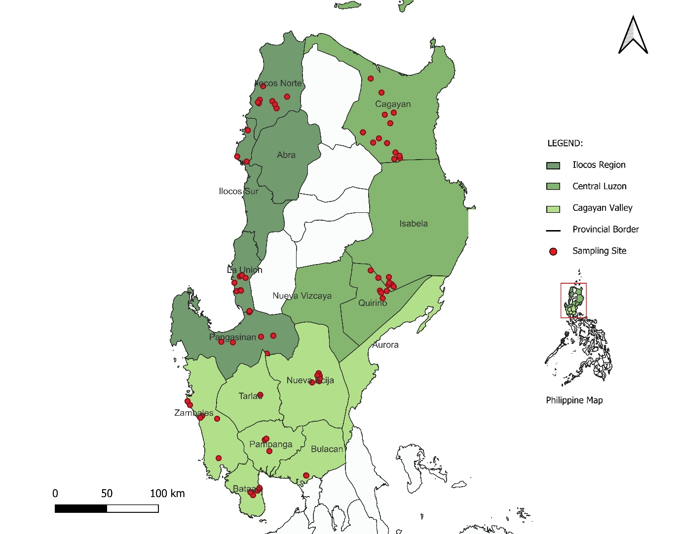
Fig. 1. Map of sampling location.
Bacterial enrichment and isolation
For initial processing, each sample was homogenized and diluted tenfold using sterile buffered peptone water (Nissu Pharm Co. Ltd., Tokyo, Japan). Using 100 µl of the resulting soil slurry, the samples were inoculated and evenly spread onto MacConkey agar plates (HiMedia Laboratories Pvt. Ltd., Nashik, India). Cultures were subsequently incubated at 37°C for 18–24 hours. Five colonies exhibiting the typical E. coli phenotype, characterized by a pink hue and precipitate, were selected from each plate and re-plated onto Eosin-Methylene Blue agar (Laboratorios Conda S. A., Madrid, Spain) for phenotypic confirmation. Further biochemical validation of E. coli was performed using the Vitek® 2 system equipped with the ID-GNB card (bioMérieux, Craponne, France).
Antimicrobial susceptibility testing
The Vitek® 2 Compact System (bioMérieux) was used to conduct a minimum inhibitory concentration (MIC)-based antibiotic susceptibility test (AST) for one isolate from each soil sample. The protocol employed the broth microdilution approach conforming to the Clinical Laboratory Standards Institute guidelines (VET01) (CLSI, 2018) for veterinary pathogens. E. coli was grown in Mueller–Hinton broth (Becton Dickinson, Sparks, MD) for 18 hours at 37°C and diluted with fresh media to a turbidity standard of 0.5 McFarland. Standardized inocula were dispensed into microplate wells preloaded with graded antimicrobial concentrations (VITEK® 2 AST-N233 and AST-XN05 cards). After incubation at 37°C for 18 hours, MIC values were established using CLSI breakpoints for animal bacteria (VET01S) (CLSI, 2020). Escherichia coli ATCC 25922 served as the reference strain for quality control. An isolate was then classified as MDR if it was resistant to at least one agent from three or more antimicrobial categories (Dewi et al., 2024).
Detection of resistance determinants
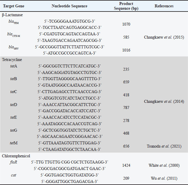
Bacterial DNA was extracted by the boiling method. Briefly, a loopful of bacterial colonies was added to 300 µl TEA buffer and boiled for 10 minutes at 95°C using a heat block. Polymerase chain reaction (PCR)-based screening for resistance determinants was employed to detect the presence of beta-lactamase genes (blaTEM, blaSHV, and blaCTX-M), TET resistance genes (tetA, tetB, tetC, tetD, tetE, tetG, and tetM), and phenicol resistance genes (floR and cat), using the primers listed in Table 1. Each 20 µl PCR reaction contained 1.25 U of Taq DNA polymerase (Promega, Madison, WI), 1× PCR buffer (Promega), 0.2 mM deoxynucleoside triphosphate (Takara Bio Inc.), 2.5 mM MgCl2 (Promega), 1 µM of each primer, and 1 µl of DNA template. For blaTEM, blaSHV, and blaCTX-M amplification, the cycling protocol included an initial denaturation at 94°C for 5 minutes, followed by 35 cycles of denaturation at 94°C for 30 seconds, annealing at 60°C for 30 seconds, extension at 72°C for 1.5 minutes, and a final extension at 72°C for 10 minutes. The TET genes were amplified with initial denaturation at 95°C for 5 minutes, followed by 35 cycles involving different denaturation (98°C for 5 seconds for tetABCDEG and 95°C for 30 seconds for tetM), annealing conditions (55°C for 15 seconds for tetABCDEG, and 56°C for 30 seconds for tetM) depending on the gene subtype, extension at 72°C for 1.5 minutes, and a final extension at 72°C for 10 minutes. The floR and cat resistance genes also followed a separate cycling profile with denaturation at 94°C for 5 minutes, followed by 35 cycles of denaturation at 94°C for 30 seconds, annealing at 48°C for 45 seconds, extension at 72°C for 30 seconds, and a final extension at 72°C for 10 minutes. Using 1.5% (w/v) agarose gel in 1.5% Tris-acetate–EDTA, PCR products were separated by gel electrophoresis, stained with GelRed (Biotium, Hayward, CA), at a 100-volt setting for 30 minutes, and examined under UV illumination.
Table 1. Primers used for the detection of the presence of antibiotic resistance genes.
Multilocus sequence typing (MLST) analysis
All MDR E. coli isolates were characterized using MLST analysis. To characterize the multilocus sequence types (STs) among MDR E. coli isolates, PCR amplification of seven essential housekeeping genes, including adk, fumC, gyrB, icd, mdh, purA, and recA, was performed (Wirth et al., 2006). The total volume and PCR reaction mixtures were the same as above. The PCR protocol consisted of a denaturation step at 95°C for 5 minutes, followed by 30 cycles consisting of denaturation at 95°C for 1 minute, annealing at 54°C (for adk, fumC, icd, and purA), 58°C (for recA), or 60°C (for mdh) for 1 minutes, extension at 72°C for 2 minutes, and final extension at 72°C for 5 minutes. Following purification with ExoSAP® IT (Thermo Fisher Scientific Co., Ltd.), the amplified products underwent bidirectional sequencing via the BigDye® ver. 3.1 Terminator Cycle Sequencing Kit on the SeqStudioTM Genetic Analyzer (Thermo Fisher Scientific Co., Ltd.). Resulting sequence data were analyzed through the MLST database (https://pubmlst.org/escherichia/andenterobase.warwick.ac.uk) to assign STs, singleton assignments, clonal complexes, and ultimately, allele identities.
Statistical analysis
The Statistical Package for Social Sciences software version 22.0 (IBM Corp., Armonk, NY) was used for descriptive data interpretation. The χ2 or Fisher’s exact test was used to assess the proportionality of the comparisons between samples. All analyses were performed at a 95% confidence level, and p-values ≤ 0.05 were considered statistically significant.
Ethical approval
Not needed for this study.
Results
Prevalence of E. coli
The prevalence of E. coli in the three regions was 61.3% (103/168), as shown in Table 2. An in-depth analysis revealed that the prevalence of E. coli was 48.9% in Cagayan Valley, 57.7% in Central Luzon, and 81.4% in soil samples from the Ilocos region. A total of 103 E. coli isolates were identified in three regions in Luzon. All isolates were confirmed using Gram-negative bacterial identification of the VITEK®2 system (bioMérieux, Nurtingen, Germany).
Table 2. Prevalence of E. coli in soil collected from dairy buffalo farm sheds.

Antimicrobial susceptibility of E. coli isolated from the environment
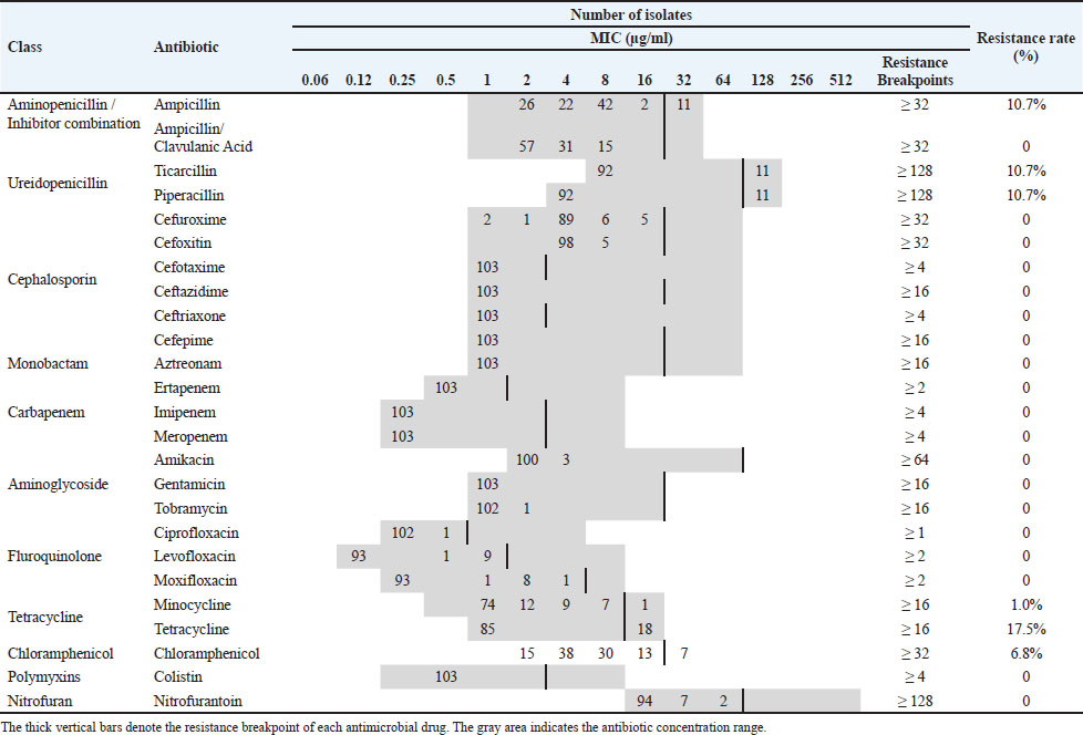
In this study, we analyzed the antimicrobial susceptibility profile of 103 E. coli isolates obtained from dairy buffalo farms. Table 3 shows the MIC distribution of these E. coli isolates to a panel of 25 antibiotics. Of the 25 antibiotics, the isolates showed varying resistance rates to six antibiotic agents. TET had the highest resistance rate (17.5%), followed by ampicillin (AMC) (10.68%), ticarcillin (TIC) (10.68%), piperacillin (PIP) (10.68%), chloramphenicol (CHL) (6.8%), and minocycline (MNO) (1%).
Table 3. Minimum inhibitory concentration distribution for 103 E. coli isolates from the dairy buffalo farm environment.
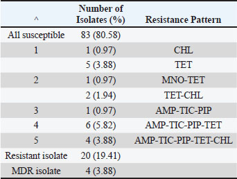
Twenty (19.41%) isolates were resistant, exhibiting different resistance patterns (Table 4). The most frequent pattern of resistance was AMP-TIC-PIP-TET. Of these 20 antimicrobial-resistant isolates, four (3.88%) are multidrug-resistant. The MDR E. coli isolates exhibited resistance to three antimicrobial classes: penicillin, TET, and CHL. Overall, 83 (80.58%) environmental E. coli were susceptible to all tested antibiotics.
Table 4. Resistance pattern of isolated E. coli from dairy buffalo farm environment.
Resistance mechanisms acquired by MDR environmental E. coli
The four MDR isolates, which were found to be resistant to three antimicrobial drug families, were subjected to further analysis to determine their acquired resistance and diversity. Results showed that 100% (4/4) of the patients carried blaTEM and tetA genes (Table 5). No other resistance determinants were detected. Moreover, none of the MDR E. coli isolates were extended-spectrum beta-lactamase (ESBL) producers.
Table 5. Resistance gene and genotype profile of MDR E. coli isolates.

Genetic diversity of environmental MDR E. coli
All four MDR E. coli isolates had identified unique STs. These are ST2690, ST5224, ST2165, and ST662 (Table 5).
Discussion
The detection of E. coli in soil samples from dairy farms and manure and mastitic animals reflects the widespread presence of microorganisms within these farm environments. It is one of the most frequently encountered bacteria in dairy farm ecosystems (Sharma et al., 2018). Contamination of the dairy farm environment poses a risk to public health as it is associated with human illness (Karmali et al., 2010; Mainil, 2013). The growing problem of AMR further compounds this existing threat. In addition to direct animal contact, contamination of animal products by antimicrobial-resistant bacteria and their potential transmission to humans start as E. coli enters the food chain (Loo et al., 2020).
Among the antimicrobials tested in this study, TET has the highest resistance rate. This finding is consistent with previous reports documenting prevalence rates of 86.4% in the Philippines (Bakakew et al., 2021), 57.6% in Southern Vietnam (Hang et al., 2019), and 26% in Canada (Massé et al., 2021). TETs are among the antimicrobials that are widely used in animal husbandry for treatment, growth promotion, and disease prevention (Granados-Chinchilla and Rodríguez, 2017; Bakakew et al., 2021). This reflects the rapid dissemination of TET-resistant bacteria (Michalova et al., 2004). Interestingly, resistance to β-lactam antibiotics such as AMC, TIC, and PIP was also observed among environmental E. coli in this study. This antibiotic group is widely used for mastitis caused by bacterial infections in dairy cattle (Fejzic et al., 2014). However, the emergence of environmental E. coli resistant to β-lactams raises concern regarding the effectiveness of these drugs. In the present work, environmental E. coli also showed remarkable resistance to CHL. Similarly, this was observed in samples from dairy fecal and manure pits, swine farm setups, and even poultry (Kidsley et al., 2018; Massé et al., 2021; Lañada et al., 2022; Mudenda et al., 2023). Despite the global ban on CHL due to its serious side effects, enforced in the Philippines through DA Administrative Order No. 60, Series of 1990, and DOH Administrative Order No. 91, Series of 1991, resistance was still demonstrated. This result indicates that resistance stems not from selection pressure but potentially from cross-resistance among CHL-related drugs, notably florfenicol (Torio and Padilla, 2018). Given the CHL resistance trends revealed here, a comprehensive investigation into the epidemiology, transmission, and molecular dynamics of antibiotic-resistant E. coli resistance mechanisms may be warranted.
Moreover, MDR E. coli strains were also detected in soil samples obtained from dairy buffalo farms. This phenomenon has also been reported in other studies on AMR E. coli in dairy farm settings using samples from cattle with mastitis (My et al., 2023), dairy cows’ milk (Widodo et al., 2022), dairy farm manure pit (Massé et al., 2021), and dairy farm slurry (Ibrahim et al., 2016). This proves that MDR E. coli exists in sick animals and their surroundings, which can be attributed to dairy waste contamination (Baker et al., 2022). The prevalence of MDR isolates in this study was lower than that in previous studies. These findings frequently result from reduced dependence on intense antimicrobial therapies, limited agricultural frameworks, and pastoral grazing practices. In contrast to intensive industrial dairy systems, these farms may exert diminished selective pressure, thereby decreasing the spread of resistant bacteria. Overall, multiple infections caused by environmental E. coli strains can be found concurrently in dairy farm animals. Therefore, strict regulatory measures must be imposed to minimize AMR and ensure the continued effectiveness of drugs against E. coli.
In the current study, environmental MDR E. coli in dairy farms harbored blaTEM and tetA, resistance genes that confer nonsusceptibility to beta-lactam and TET antibiotics, respectively. The acquisition of these genes can be attributed to the extensive use of TET and beta-lactam antibiotics in dairy farms (Bourély et al., 2019). The blaTEM and tetA gene combination in MDR E. coli from the dairy farm soil in this study was identified as the predominant resistance determinant profile, consistent with the pattern detected in E. coli from lake samples in the Philippines (Salvador-Membreve and Rivera, 2021). Furthermore, these resistance determinants are carried predominantly by plasmids and are easily disseminated through horizontal gene transfer rather than chromosomal transfer (Mthembu et al., 2019). Studies have shown that plasmid-borne antibiotic resistance gives bacteria a clear advantage over chromosomal resistance. Plasmids spread quickly between species, can exist in multiple copies, and often carry several resistance genes simultaneously. These offers to host bacteria rapid evolution, adaptability to changing environments, and often carry lower immediate fitness costs (Castañeda-Barba et al., 2024). The presence of blaTEM and tetA in environmental E. coli can be beneficial for their survival even without direct antimicrobial pressure. However, there is a risk that these resistance factors will be transferred to vegetable, livestock, or aquatic animal production systems, eventually reaching humans. Hence, implementing rigorous hygiene and sanitation standards is imperative to limit the spread of AMR in the environment.
On the other hand, bacterial resistance to CHL was reported to be due to enzymatic inactivation by acetylation of antimicrobials through CHL acetyltransferases (CATs) and non-enzymatic action by the CHL exporter cmlA gene (Lu et al., 2018). Resistance to CHL can also be mediated by the flo gene, which serves as the molecular basis for florfenicol resistance (Tadesse et al., 2012). Among these flo genes, floR is the specific gene most commonly detected in E. coli (Karczmarczyk et al., 2011). However, neither cat nor floR genes were present in the MDR E. coli samples. Therefore, environmental MDR E. coli may have developed CHL resistance from additional resistance genes or coresistance with other antimicrobials.
Bacterial genotyping is important for tracing the origin and transmission of antibiotic-resistant strains. In the present study, ST662, ST2165, ST2690, and ST5224 were uniquely identified in E. coli with MDR status. Previous reports have indicated associations of these clones not only with human infections but also with feces and environments inhabited by domestic and wildlife species. ST 690 has been associated with clinical E. coli isolates in pediatric patients (Xiong et al., 2023). A similar study in Indonesia identified ST2690 in ESBL-producing E. coli from chickens and chicken farm workers (Wardoyo et al., 2025). This suggests that the presence of ST may spread across farm environments and into surrounding communities in Southeast Asian agricultural settings. In addition, the ST2165 type was reported in clinical mastitis cases and raw milk, as well as in mcr-1-positive, colistin-resistant E. coli from nursing home environments, and even among poultry and retail products (Tartor et al., 2021). Furthermore, it also harbored multiple plasmid replicon types, indicating a potential role for this ST in the spread of environmental and food-associated AMR. Although not specific to Asia, this illustrates how ST2165 can be associated with MDR profiles that are potentially relevant to livestock and production systems globally. Unlike the other STs, ST5224 and ST662 have not been widely reported in the existing scientific literature and have only been observed in Enterobase metadata from livestock (turkey, cattle, and pig), vegetables, and humans. This could represent novel or atypical lineages emerging in specific environments or geographic niches that are not widely sampled. Although the genotypes currently detected in this study are not globally disseminated, they still pose an imminent threat to public health as zoonotic clones may be transmitted. Therefore, the genotyping analysis employed here uncovers new information on the genetic diversity and potential for cross-species transmission of MDR E. coli in dairy buffalo farm settings in the Philippines, highlighting a One Health perspective that links animal, human, and environmental health in the spread of AMR.
Conclusion
This study concludes that antimicrobial-resistant E. coli is present in dairy buffalo farm environments. This evidence points to the possibility that antimicrobial-resistant bacteria from animals can contaminate and spread to their adjacent environments. In addition to these findings, the presence of MDR E. coli isolates was also confirmed within dairy buffalo farm environments, posing a significant emerging risk to the health and productivity of dairy livestock. Therefore, effective prevention and control of AMR hinge on the strict implementation of antibiotic dispensing regulations and vigilant antimicrobial stewardship to avoid the exploitation of critically important drugs. Robust biosecurity and farm hygiene measures must also be applied to minimize the spread of AMR bacteria.
Acknowledgment
The authors thank DA-BIOTECH for providing financial assistance to this research. The authors would also like to thank the Philippine Carabao Center and Livestock Biotechnology Center for their technical support. Moreover, due credit is greatly indebted to the Biosafety and Environment Section‘s technical staff for their assistance.
Funding
This study was funded by the Department of Agriculture-Biotechnology Program Office through the Livestock Biotechnology Center under Number DABIOTECH-R1803.
Authors’ contribution
LPB, SDG, and CNM designed the study, interpreted the data, and drafted the manuscript. LPB and GASPT were involved in data collection and manuscript preparation. MBSS and CNM participated in preparing and critically checking this manuscript.
Conflicts of interest
The authors have no conflicts of interest to declare.
Data availability
All data generated or analyzed during this study are included in this published article.
References
Almansour, A.M., Alhadlaq, M.A., Alzahrani, K.O., Mukhtar, L.E., Alharbi, A.L. and Alajel, S.M. 2023. The silent threat: antimicrobial-resistant pathogens in food-producing animals and their impact on public health. Microorganisms 22(11), 2127; doi:10.3390/microorganisms11092127
Aquino, D.L., Palacpac, E.P., Molina, A.M., Lacanilao, C.C., Garcia, N.P., Del Barrio, A.N. and Fujihara, T. 2024. Enhancing growth and milk production of dairy buffaloes through home-grown forages and complete nutrient diet. Online. J. Anim. Feed. Res. 14(2), 95–106; doi:10.51227/ojafr.2024.12
Arbab, S., Ullah, H., Wang, W. and Zhang, J. 2022. Antimicrobial drug resistance against Escherichia coli and its harmful effect on animal health. Vet. Med. Sci. 8(4), 1780–1786; doi:10.1002/vms3.825
Bakakew, C.D., Tabuac, J.V. and Torio, H.E. 2021. Phenotypic antimicrobial resistance patterns in Escherichia coli isolated from slaughtered healthy pigs and cattle in Nueva Vizcaya, Philippines. Philipp. J. Vet. Med. 58(1), 30–39.
Baker, M., Williams, A.D., Hooton, S.P.T., Helliwell, R., King, E., Dodsworth, T., María Baena-nogueras, R., Warry, A., Ortori, C.A., Todman, H., Gray-Hammerton, C.J., Pritchard, A.C.W., Iles, E., Cook, R., Emes, R.D., Jones, M.A., Kypraios, T., West, H., Barrett, D.A., Ramsden, S.J., Gomes, R.L., Hudson, C., Millard, A.D., Raman, S., Morris, C., Dodd, C.E.R., Kreft, J.U., Hobman, J.L. and Stekel, D.J. 2022. Antimicrobial resistance in dairy slurry tanks: a critical point for measurement and control. Environ. Int. 169, 107516; doi:10.1016/j.envint.2022.107516
Bedi, J.S., Vijay, D. and Dhaka, P. 2025. Zoonoses associated with buffaloes and their control measures. In Buffalo (Bubalus bubalis) meat. Eds., Chatli, M.K., Kumar, H. and Singh, I. Singapore: Springer; doi:10.1007/978-981-96-8552-3_12
Belotindos, L., Villanueva, M., Miguel, J., Bwalya, P., Harada, T., Kawahara, R., Nakajima, C., Mingala, C. and Suzuki, Y. 2021. Prevalence and characterization of quinolone-resistance determinants in Escherichia coli isolated from food-producing animals and animal-derived food in the Philippines. Antibiotics. 10(4), 413; doi:10.3390/antibiotics10040413
Bourély, C., Cazeau, G., Jarrige, N., Jouy, E., Haenni, M., Lupo, A., Madec, J.Y., Leblond, A. and Gay, E. 2019. Co-resistance to amoxicillin and tetracycline as an indicator of multidrug resistance in Escherichia coli isolates from animals. Front. Microbiol. 10, 2288; doi:10.3389/fmicb.2019.02288
Castañeda-Barba, S., Top, E.M. and Stalder, T. 2024. Plasmids, a molecular cornerstone of antimicrobial resistance in the One Health era. Nat. Rev. Microbiol. 22(1), 18–32; doi:10.1038/s41579-023-00926-x
Changkaew, K., Intarapuk, A., Utrarachkij, F., Nakajima, C., Suthienkul, O. and Suzuki, Y. 2015. Antimicrobial resistance, extended-spectrum β-lactamase productivity, and class 1 integrons in Escherichia coli from healthy swine. J. Food. Prot. 78(8), 1442–1450; doi:10.4315/0362-028X.JFP-14-445
Changkaew, K., Utrarachkij, F., Siripanichgon, K., Nakajima, C., Suthienkul, O. and Suzuki, Y. 2014. Characterization of antibiotic resistance in Escherichia coli isolated from shrimps and their environment. Food. Prot. 77(8), 1394–1401; doi:10.4315/0362-028X.JFP-13-510
Chowdhury, M.S.R., Hossain, H., Rahman, M.N., Rahman, A., Ghosh, P.K., Uddin, M.B., Nazmul Hoque, M., Hossain, M.M. and Rahman, M.M. 2025. Emergence of highly virulent multidrug and extensively drug-resistant Escherichia coli and Klebsiella pneumoniae in buffalo subclinical mastitis cases. Sci. Rep. 15(1), 20241; doi:10.1038/s41598-025-02374-4
Clinical and Laboratory Standards Institute (CLSI). 2018. Performance Standards for Antimicrobial Disk and Dilution Susceptibility Tests for Bacteria Isolated From Animals. 5th ed. CLSI supplement VET01. Wayne, PA: Clinical and Laboratory Standards Institute.
Dadgostar, P. 2019. Antimicrobial resistance: implications and costs. Infect. Drug. Resist. 12, 3903–3910; doi:10.2147/IDR.S234610
Department of Agriculture (DA) and Department of Health (DOH). 1990. Declaring a Ban on the Use of Chloramphenicol in Food-Producing Animals: Department of Agriculture (DA) Administrative Order No. 60, Series of 1990; Department of Health (DOH) Administrative Order No. 91, Series of 1990; Manila, Philippines.
Dewi, R.R., Nuryawan, A., Jajere, S.M., Sihombing, J.M. and Tambunan, I.J. 2024. Antimicrobial resistance profiles of Escherichia coli derived from an integrated agroforestry-livestock system in Deli Serdang Regency, Indonesia. Vet. World 17(3), 690–699; doi:10.14202/vetworld.2024.690-699
Drugea, R.I., Siteavu, M.I., Pitoiu, E., Delcaru, C., Sârbu, E.M., Postolache, C. and Bărăităreanu, S. 2025. Prevalence and antibiotic resistance of Escherichia coli isolated from raw cow’s milk. Microorganisms 13(1), 209; doi:10.3390/microorganisms13010209
Fejzic, N., Begagic, M., Šerić-Haračić, S. and Smajlovic, M. 2014. Beta-lactam antibiotics residues in cow’s milk: comparison of efficacy of three screening tests used in Bosnia and Herzegovina. Bosn. J. Basic Med. Sci. 14(3), 155–159; doi:10.17305/bjbms.2014.3.109
Gautam, A., Bastola, S., Lamsal, K., Kaphle, K., Shrestha, P., Shah, S. and Subedi, D. 2024. Prevalence and risk factors of multidrug resistant (MDR) Escherichia coli isolated from milk of small scale dairy buffaloes in Rupandehi, Nepal. Zoonotic. Dis. 4(3), 174–186; doi:10.3390/zoonoticdis4030016
Geletu, U.S., Usmael, M.A. and Ibrahim, A.M. 2022. Isolation, identification, and susceptibility profile of E. coli, Salmonella, and S. aureus in dairy farm and their public health implication in central Ethiopia. Vet. Med. Int. 2022, 1887977; doi:10.1155/2022/1887977
Granados-Chinchilla, F. and Rodríguez, C. 2017. Tetracyclines in food and feedingstuffs: from regulation to analytical methods, bacterial resistance, and environmental and health implications. J. Anal. Methods Chem. 2017, 1315497; doi:10.1155/2017/1315497
Gundran, R.S., Cardenio, P.A., Salvador, R.T., Sison, F.B., Benigno, C.C., Kreausukon, K., Pichpol, D. and Punyapornwithaya, V. 2020. Prevalence, antibiogram, and resistance profile of extended-spectrum β-lactamase-producing escherichia coli isolates from pig farms in Luzon, Philippines. Microb. Drug. Resist. 26(2), 160–168; doi:10.1089/mdr.2019.0019
Hang, B.P.T., Wredle, E., Börjesson, S., Sjaunja, K.S., Dicksved, J. and Duse, A. 2019. High level of multidrug-resistant Escherichia coli in young dairy calves in southern Vietnam. Trop. Anim. Health Prod. 51(6), 1405–1411; doi:10.1007/s11250-019-01820-6
Holmes, A.H., Moore, L.S.P., Sundsfjord, A., Steinbakk, M., Regmi, S., Karkey, A., Guerin, P.J. and Piddock, L.J.V. 2016. Understanding the mechanisms and drivers of antimicrobial resistance. Lancet 387(10014), 176–187; doi:10.1016/S0140-6736(15)00473-0
Ibrahim, D.R., Dodd, C.E.R., Stekel, D.J., Ramsden, S.J. and Hobman, J.L. 2016. Multidrug-resistant, extended-spectrum β-lactamase (ESBL)-producing Escherichia coli isolated from a dairy farm. FEMS. Microbiol. Ecol. 92(4), 13; doi:10.1093/femsec/fiw013
Karczmarczyk, M., Abbott, Y., Walsh, C., Leonard, N. and Fanning, S. 2011. Characterization of multidrug-resistant Escherichia coli isolates from animals presenting at a university veterinary hospital. Appl. Environ. Microbiol. 77(20), 7104–7112; doi:10.1128/AEM.00599-11
Karmali, M.A., Gannon, V. and Sargeant, J.M. 2010. Verocytotoxin-producing Escherichia coli (VTEC). Vet. Microbiol. 140(3–4), 360–370; doi:10.1016/j.vetmic.2009.04.011
Kidsley, A.K., Abraham, S., Bell, J.M., O’Dea, M., Laird, T.J., Jordan, D., Mitchell, P., McDevitt, C.A. and Trott, D.J. 2018. Antimicrobial susceptibility of Escherichia coli and Salmonella spp. Isolates from healthy pigs in Australia: results of a pilot national survey. Front. Microbiol. 9, 1207; doi:10.3389/fmicb.2018.01207
Lañada, E., Portugaliza, H., Blanza, A., Espejo, M.C. and Pagente, M.D. 2022. Conventional biotyping revealed Mycoplasma and antimicrobial-resistant Escherichia coli and Salmonella isolates during disease outbreak in broiler farms and hatcheries in Leyte, Philippines. Ann. Trop. Res. 44(1), 74–85.
Loo, E., Lai, K. and Mansor, R. 2020. Antimicrobial usage and resistance in dairy cattle production. IntechOpen. doi.org/10.5772/intechopen.81365
Lu, J., Zhang, J., Xu, L., Liu, Y., Li, P., Zhu, T., Cheng, C., Lu, S., Xu, T., Yi, H., Li, K., Zhou, W., Li, P., Ni, L. and Bao, Q. 2018. Spread of the florfenicol resistance floR gene among clinical Klebsiella pneumoniae isolates in China. Antimicrob. Resist. Infect. Control. 7, 127; doi:10.1186/s13756-018-0415-0
Magiorakos, A.P., Srinivasan, A., Carey, R.B., Carmeli, Y., Falagas, M.E., Giske, C.G., Harbarth, S., Hindler, J.F., Kahlmeter, G., Olsson-Liljequist, B., Paterson, D.L., Rice, L.B., Stelling, J., Struelens, M.J., Vatopoulos, A., Weber, J.T. and Monnet, D.L. 2012. Multidrug-resistant, extensively drug-resistant and pan-drug-resistant bacteria: an international expert proposal for interim standard definitions for acquired resistance. Clin. Microbiol. Infect. 18(3), 268–281; doi:10.1111/j.1469-0691.2011.03570.x
Mahmoudi, P., Rashidi, A. and Idrees Hama Faraj, S. 2025. Network meta-analysis of the prevalence and antimicrobial resistance of Escherichia coli isolated from bovine milk and dairy products: a global perspective. J. Dairy Sci. 108(9), 9330–9344; doi: 10.3168/jds.2024-26197
Mainil, J. 2013. Escherichia coli virulence factors. Vet. Immunol. Immunopathol. 152(1–2), 2–12; doi:10.1016/j.vetimm.2012.09.032
Massé, J., Lardé, H., Fairbrother, J.M., Roy, J.P., Francoz, D., Dufour, S. and Archambault, M. 2021. Prevalence of antimicrobial resistance and characteristics of Escherichia coli isolates from fecal and manure pit samples on dairy farms in the Province of Québec, Canada. Front. Vet. Sci. 8, 654125; doi:10.3389/fvets.2021.654125
Michalova, E., Pnovotna, P. and Schlegelova, J. 2004. Tetracyclines in veterinary medicine and bacterial resistance to them. Vet. Med. 49(3), 79–100; doi:10.17221/5681-VETMED
Morales-Ubaldo, A.L., Rivero-Perez, N., Valladares-Carranza, B., Velázquez-Ordoñez, V., Delgadillo-Ruiz, L. and Zaragoza-Bastida, A. 2023. Bovine mastitis, a worldwide impact disease: prevalence, antimicrobial resistance, and viable alternative approaches. Vet. Anim. Sci. 21, 100306; doi:10.1016/j.vas.2023.100306
Mthembu, T.P., Zishiri, O.T. and El Zowalaty, M.E. 2019. Molecular detection of multidrug-resistant Salmonella isolated from livestock production systems in South Africa. Infect. Drug. Resist. 12, 3537–3548; doi:10.2147/IDR.S211618
Mudenda, S., Malama, S., Munyeme, M., Matafwali, S.K., Kapila, P., Katemangwe, P., Mainda, G., Mukubesa, A.N., Hadunka, M.A. and Muma, J.B. 2023. Antimicrobial resistance profiles of Escherichia coli isolated from laying hens in Zambia: implications and significance on One Health. JAC. Antimicrob. Resist. 22(5), 60; doi:10.1093/jacamr/dlad060
My, T.T., Thien, L.V., Manh, V.D., My, B.T.P., Lan, D.T.M., Binh, D.X. and Duc, V.M. 2023. Antimicrobial resistance and molecular characterization of Escherichia coli isolated from bovine mastitis samples in Nghe An province, Vietnam. Vet. World. 16(4), 743–751; doi:10.14202/vetworld.2023.743-751
Obaidat, M.M. and Stringer, A.P. 2019. Prevalence, molecular characterization, and antimicrobial resistance profiles of Listeria monocytogenes, Salmonella enterica, and Escherichia coli O157: h7 on dairy cattle farms in Jordan. J. Dairy Sci. 102(10), 8710–8720; doi:10.3168/jds.2019-16461
Ombarak, R.A., Hinenoya, A., Elbagory, A.R.M. and Yamasaki, S. 2018. Prevalence and molecular characterization of antimicrobial resistance in Escherichia coli isolated from raw milk and raw milk cheese in Egypt. J. Food Prot. 81(2), 226–232; doi:10.4315/0362-028X.JFP-17-277
Salvador-Membreve, D.M. and Rivera, W.L. 2021. Predominance of blaTEM and tetA genes in antibiotic-resistant Escherichia coli isolates from Laguna Lake, Philippines. J. Water. Sanit. Hyg. Dev. 11(5), 814–823; doi:10.2166/washdev.2021.067
Seker, E. and Yardimci, H. 2008. First isolation of Escherichia coli O157: h7 from faecal and milk specimens from Anatolian water buffaloes (Bubalus bubalis) in Turkey. J. S. Afr. Vet. Assoc. 79(4), 167–170; doi:10.4102/jsava.v79i4.267
Sharma, C., Rokana, N., Chandra, M., Singh, B.P., Gulhane, R.D., Gill, J.P.S., Ray, P., Puniya, A.K. and Panwar, H. 2018. Antimicrobial resistance: its surveillance, impact, and alternative management strategies in dairy animals. Front. Vet. Sci. 4, 237; doi:10.3389/fvets.2017.00237
Shoaib, M., He, Z., Geng, X., Tang, M., Hao, R., Wang, S., Shang, R., Wang, X., Zhang, H. and Pu, W. 2023. The emergence of multi-drug resistant and virulence gene carrying Escherichia coli strains in the dairy environment: a rising threat to the environment, animal, and public health. Front. Microbiol. 14, 1197579; doi:10.3389/fmicb.2023.1197579
Skočková, A., Bogdanovičová, K., Koláčková, I. and Karpíšková, R. 2015. Antimicrobial-resistant and extended-spectrum β-lactamase-producing Escherichia coli in raw cow’s milk. J. Food Prot. 78(1), 72–77; doi:10.4315/0362-028X.JFP-14-250
Tadesse, D.A., Zhao, S., Tong, E., Ayers, S., Singh, A., Bartholomew, M.J. and Mcdermott, P.F. 2012. Antimicrobial drug resistance in Escherichia coli from humans and food animals, United States, 1950-2002. Emerg. Infect. Dis. 18(5), 741–739; doi:10.3201/eid1805.111153
Tartor, Y.H., Abd El-aziz, N.K., Gharieb, R.M.A., El Damaty, H.M., Enany, S., Soliman, E.A., Abdellatif, S.S., Attia, A.S.A., Bahnass, M.M., El-Shazly, Y.A., Elbediwi, M. and Ramadan, H. 2021. Whole-genome sequencing of gram-negative bacteria isolated from bovine mastitis and raw milk: the first emergence of colistin mcr-10 and Fosfomycin fosA5 Resistance Genes in Klebsiella pneumoniae in the Middle East. Front. Microbiol. 12(8), 770813; doi:10.3389/fmicb.2021.770813
Torio, H.E. and Padilla, M.A. 2018. Multiple resistance to medically important antimicrobials of commensal Escherichia coli isolated from dressed broiler chickens in Calabarzon, Philippines. Philipp. J. Vet. Med. 55(2), 95–106.
Tsunoda, R., Usui, M., Tagaki, C., Fukuda, A., Boonla, C., Anomasiri, W., Sukpanyatham, N., Akapelwa, M.L., Nakajima, C., Tamura, Y. and Suzuki, Y. 2021. Genetic characterization of coliform bacterial isolates from environmental water in Thailand. J. Infect. Chemother. 27(5), 722–728; doi:10.1016/j.jiac.2020.12.023
Veloo, Y., Rajendiran, S., Zakaria, Z., Ismail, R., Rahman, S.A., Mansor, R. and Thahir, S.S.A. 2025. Prevalence and antimicrobial resistance patterns of Escherichia coli in the environment, cow dung, and milk of selangor dairy farms. Antibiotics (Basel). 14(2), 137; doi:10.3390/antibiotics14020137
Wardoyo, E.H., Sugawara, Y., Nakano, S., Zuo, H., Elahi, S., Arai, C., Kondo, K., Kuntaman, K. and Sugai, M. 2025. Genomic characterization of extended-spectrum β-lactamase-producing Escherichia coli spread among chickens and healthy residents in Lombok, Indonesia. Appl. Environ. Microbiol. 91(5), 236424; doi:10.1128/aem.02364-24
White, D.G., Hudson, C., Maurer, J.J., Ayers, S., Zhao, S., Lee, M.D., Bolton, L., Foley, T. and Sherwood, J. 2000. Characterization of chloramphenicol and florfenicol resistance in Escherichia coli associated with bovine diarrhea. Clin. Microbiol. 38(12), 4593–4598; doi:10.1128/JCM.38.12.4593-4598.2000
Widodo, A., Lamid, M., Effendi, M.H., Khairullah, A.R., Riwu, K.H.P., Yustinasari, L.R., Kurniawan, S.C., Ansori, A.N.M., Silaen, O.S.M. and Dameanti, F.N.A.E.P. 2022. Antibiotic sensitivity profile of multidrug-resistant (MDR) Escherichia coli isolated from dairy cow’s milk in Probolinggo, Indonesia. Biodiversitas 23(10), 4971–4976; doi: 10.13057/biodiv/d231002
Widodo, A., Lamid, M., Effendi, M.H., Tyasningsih, W., Wurlina, W., Al-Arif, M.A., Raharjo, D., Soeharsono, S., Khairullah, A.R., Riwu, K.H.P., Yustinasari, L.R., Kurniawan, S.C., Silaen, O.S.M., Benjamin, M.I. and Afnani, D.A. 2023. Potential hazards of multidrug-resistant Escherichia coli collected from wastewater on dairy farms in East Java, Indonesia. Biodiversitas 24(3), 1900–1907; doi:10.13057/biodiv/d240367
Wirth, T., Falush, D., Lan, R., Colles, F., Mensa, P., Wieler, L.H., Karch, H., Reeves, P.R., Maiden, M.C.J., Ochman, H. and Achtman, M. 2006. Sex and Virulence in Escherichia coli: an Evolutionary Perspective. Mol. Microbiol. 60(5), 1136–1151; doi.org/10.1111%2Fj.1365-2958.2006.05172.x
World Health Organization. 2017. Global priority list of antibiotic-resistant bacteria to guide research. Geneva, Switzerland: WHO.
Wu, R.B., Alexander, T.W., Li, J.Q., Munns, K., Sharma, R. and Mcallister, T.A. 2011. Prevalence and diversity of class 1 integrons and resistance genes in antimicrobial-resistant Escherichia coli originating from beef cattle administered subtherapeutic antimicrobials. J. Appl. Microbiol. 111(2), 511–523; doi:10.1111/j.1365-2672.2011.05066.x
Xiong, Z., Zhang, C., Sarbandi, K., Liang, Z., Mai, J., Liang, B., Cai, H., Chen, X., Gao, F., Lan, F., Liu, X., Liu, S. and Zhou, Z. 2023. Clinical and molecular epidemiology of carbapenem-resistant Enterobacteriaceae in pediatric inpatients in South China. Microbiol. Spectr. 11(6), 283923; doi:10.1128/spectrum.02839-23
Zhang, Z., Wei, M., Jia, B. and Yuan, Y. 2024. Recent advances in antimicrobial resistance: insights from Escherichia coli as a Model Organism. Microorganisms 13(1), 51; doi:10.3390/microorganisms13010051








