


| Case Report | ||
Open Vet. J.. 2025; 15(12): 6805-6808 Open Veterinary Journal, (2025), Vol. 15(12): 6805-6808 Case Report Cervical ectopia cordis in a neonatal camel (Camelus dromedarius): A rare case report from North Kordofan, SudanJadkrem Jahlla Mohamed Ahmed1, Ahmed Mustafa Ahmed Hussein2and Abdelmalik Ibrahim Khalafalla3,4*1Jahlla Vet Clinic and Pharmacy, Um Rawaba, Kordofan, Sudan 2Al-Timawi Farm, Hail, Saudi Arabia 3Department of Microbiology, Faculty of Veterinary Medicine, University of Khartoum, Khartoum North, Sudan 4Abu Dhabi Agriculture and Food Safety Authority, Abu Dhabi, United Arab Emirates *Corresponding Author: Abdelmalik Ibrahim Khalafalla. Abu Dhabi Agriculture and Food Safety Authority, Submitted: 12/08/2025 Revised: 27/10/2025 Accepted: 01/11/2025 Published: 31/12/2025 © 2025 Open Veterinary Journal
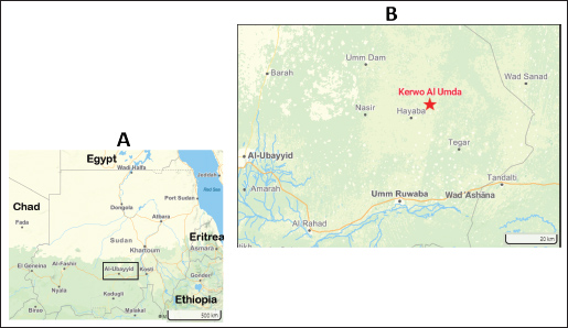
AbstractBackground: Ectopia cordis (EC) is a rare congenital condition in which the heart is partially or completely displaced outside the thoracic cavity. This abnormality is most commonly recognized in humans but has also been observed in dogs, cats, cattle, pigs, and goats. However, it has not been documented in camelids. Case Description: This case report describes the case of a neonatal camel calf born in rural Wad Ashana, North Kordofan, Sudan, who presented with EC. The heart was located in its pericardium outside the thoracic cavity, at the most ventral aspect of the cervical region. The heart exhibited a normal morphology and a normal heartbeat. Despite the absence of any additional malformations, the calf died 2 days after delivery. Conclusion: This case is the first documented instance of EC in camels from Sudan and offers important information for veterinary literature on congenital anomalies. Keywords: Ectopia cordis, Dromedary camels, Sudan. IntroductionCardiac congenital defects related to inheritance and teratogenesis have been reported in both veterinary species and humans worldwide (Cerqueira et al., 2024). Ectopia cordis (EC) is a rare congenital condition characterized by abnormal heart positioning, resulting in partial or complete displacement of the heart outside the thoracic cavity due to a cleft (Hiraga et al., 1993). EC is broadly classified into four main types based on the location of the ectopically positioned heart: cervical, thoracic, thoracoabdominal, and abdominal. It is a complex condition with an unclear etiology. It occurs when the sternum and ventral body wall do not fuse properly during embryonic development, preventing the heart from being properly enclosed within the chest cavity (Shone and Funderburk, 1974; Sadłecki et al., 2011). This failure is often linked to disruptions in the normal migration of somatic muscle cells (SMCs). Although most cases occur sporadically, some are associated with specific chromosomal abnormalities and gene mutations, highlighting the genetic factors that contribute to this rare and serious congenital malformation (Engum, 2008). Although EC is most commonly recognized in humans, it has also been documented in various domestic animals, including dogs, cats, cattle, pigs, and goats, but not in camelids. Therefore, this study describes the EC occurrence in a neonatal dromedary camel. Case DetailsOn July 26, 2025, a male dromedary camel calf (Camelus dromedarius) was born naturally in Kerwo Al Umda, a rural village, north of Wad Ashana city, North Kordofan, Sudan (13.4698471° N, 31.4560806° E), approximately 300 km southwest of Khartoum (Fig. 1). A camel herd was raised for meat and milk in a free-range production system. The owner reported no similar cases within his herd or in nearby herds, claiming that he had never seen such a case in camels.
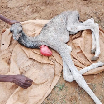
Fig. 1. Locator Map (A) illustrating the national context of Sudan, accompanied by a detailed inset (B) that provides an enlarged view of the North Kordofan region, highlighting the village of Kerwo Al Umda where the case was reported. Map created using Mapcreator (https://mapcreator.io/). Upon examination, the camel calf exhibited complete cardiac displacement outside the thoracic cavity caused by a cleft. On the left side of the animal, the heart was visible and protruded at the most ventral aspect of the cervical region, covered with pericardium and without skin (Fig. 2). Physical examination of the external appearance revealed that the protruding heart appeared normal, and the calf maintained a regular heartbeat, as confirmed by visual observation and video documentation (Video 1-Supplementary Material). The calf was alert, active, and interacted with its mother, but was unable to stand for long and suckle. No other congenital abnormalities or malformations were observed during the physical examination. Supportive care was provided by milking the mother and bottle-feeding the colostrum; however, the calf passed away approximately 2 days after birth. A necropsy was not conducted due to the unavailability of trained personnel, the long distance to the nearest veterinary hospital, and security issues arising from the ongoing civil war.
Fig. 2. Cervical ectopia cordis in a neonatal camel showing a visible protrusion at the neck base on the left side of the animal, North Kordofan, Sudan. Ethical approvalInformed consent was obtained from the owner of the animals to allow for the examination, photography, and use of images and videos for research and training purposes. DiscussionThis is the first reported instance of EC in dromedary camels. Heart conditions that affect camels include necrotic myocarditis, hypertrophic cardiomyopathy, vegetative valvular endocarditis, pericarditis, and congenital abnormalities such as septal anomalies, patent ductus arteriosus, transposition of the aorta and pulmonary artery, persistent aortic trunk, and persistent right aortic arch and sarcocystosis (Tharwat et al., 2012). EC is a rare congenital anomaly in veterinary medicine. The cardiac anomaly identified in this camel calf was classified as complete thoracic EC (TEC), similar to previously reported cases in cattle and sheep. This contrasts with the predominance of thoracoabdominal EC in cats, piglets, and dogs (Cerqueira et al., 2024). EC occurs in humans, appears seldom in cattle, dogs, rabbits, pigs, cats, and goats, and is very uncommon in sheep (Freeman and McGovern, 1984; Hiraga et al., 1993; Cerqueira et al., 2024). The condition can be categorized as cervical, thoracocervical, thoracic, thoracoabdominal, or abdominal based on the location of the ectopic heart. Depending on whether the heart is covered by a pericardial sac, it can also be categorized as partial or total (Cerqueira et al., 2024). Furthermore, there are two categories of EC: internal and external. In internal cases, the heart is displaced entirely into either the cervical or abdominal region, whereas in external cases, it protrudes outside the body, as observed in this instance. Animals with external EC typically do not survive for long, but those with internal EC can survive for extended periods. For example, there have been cases of cows with internal EC, including one that had a normal delivery and another that lived to the age of 14 years (Bowen and Adrian, 1962; Windberger et al., 1992). Additionally, Onda et al. (2011) reported a calf with cervical EC that not only grew normally but also became pregnant and successfully gave birth. In this report, the affected calf survived for 2 days after birth due to the supportive care provided by the owner, which included milking the mother and administering colostrum via bottle-feeding. The cause of death may have been cardiorespiratory complications and bacterial infections. EC generally has a poor prognosis; most cases result in death due to infection, hypoxemia, or heart failure (Bernstein, 2011). ConclusionThis case is the first documented instance of the EC in camels from Sudan and offers important information for veterinary literature on congenital anomalies. This study contributes to the understanding of this rare condition in dromedary camels, highlighting the need for increased awareness among veterinary practitioners and camel farmers in rural regions. Research is needed to explore the environmental conditions, parental history, and potential prenatal or perinatal factors that may have contributed to this anomaly in camels. AcknowledgmentsWe would like to express our gratitude to the owner of the affected camel, his family, and the rural community of Kerwo Al Umda village for reaching out to the first author and for their interest and cooperation. Conflict of interestThe authors declare that they have no competing interests. FundingNone. Authors’ contributionsJadkrem Jahlla Mohamed Ahmed visited the study area and investigated the case. Ahmed Mustafa, Ahmed Hussein, and Abdelmalik Ibrahim Khalafalla wrote the main text of the manuscript. All authors have reviewed and approved the final version of the manuscript. Data availabilityAll data were provided in the manuscript. ReferencesBernstein, D. 2011. Kliegman: nelson textbook of pediatrics. Elsevier Publishers, London, p. 1599. Bowen, J.M. and Adrian, R.W. 1962. Ectopia cordis in cattle. J. Am. Vet. Med. Assoc. 141, 1162–1167. Cerqueira, L.A., Mâcedo, I.L., Sousa, D.E.R., Amorim, H.A.L., Borges, J.R.J., Ximenes, F.H.B., Câmara, A.C.L. and Castro, M.B. 2024. Complete Thoracic Ectopia Cordis in Two Lambs. Animals. Open. Access. J. From. MDPI. 14(15), 2213; doi:10.3390/ani14152213 Engum, S.A. 2008. Embryology, sternal clefts, ectopia cordis, and Cantrell‘s pentalogy. Seminars. Pediatric. Surg. 17(3), 154–160; doi:10.1053/j.sempedsurg.2008.03.004 Freeman, L.E. and McGovern, P.T. 1984. Ectopia cordis thoracoabdominalis in a piglet. Vet. Rec. 115(17), 431–433; doi:10.1136/vr.115.17.431 Hiraga, T., Abe, M., Iwasa, K., Takehana, K. and Tanigaki, A. 1993. Cervical-pectoral ectopia cordis in two Holstein calves. Vet. Path. 30, 529–534; doi:10.1177/030098589303000606 Onda, K., Sugiyama, M., Niho, K., Sato, R., Arai, S., Kaneko, K., Ito, S., Muto, M., Suganuma, T., Wakao, Y. and Wada, Y. 2011. Long-term survival of a cow with cervical ectopia cordis. Can. Vet. J.=La Revue Veterinaire Canadienne 52(6), 667–669. Sadłecki, P., Krekora, M., Krasomski, G., Walentowicz-Sadłecka, M., Grabiec, M., Moll, J. and Respondek-Liberska, M. 2011. Prenatally evolving ectopia cordis with successful surgical treatment. Fetal Diagnosis Therapy 30(1), 70–72; doi:10.1159/000326300 Shone, J.D. and Funderburk. 1974. Ectopia cordis in a dog. J. Am. Vet. Med. Assoc. 165(1), 69–70. Tharwat, M., Al-Sobayil, F., Ali, A. and Buczinski, S. 2012. Echocardiography of the normal camel (Camelus dromedaries) heart: technique and cardiac dimensions. BMC Vet. Res. 8, 130; doi:10.1186/1746-6148-8-130 Windberger, U., Forstenpointner, G., Grabenwöger, F., Kopp, E., Künzel, W., Mayr, B., Pernthaner, A., Simon, P. and Losert, U. 1992. Cardiac function, morphology and chromosomal aberrations in a calf with ectopia cordis cervicalis. Zentralblatt Fur. Veterinarmedizin. Reihe A 39(10), 759–768; doi:10.1111/j.1439-0442.1992.tb00241.x Supplementary materialLive video showing a camel calf with ectopia cordis, exhibiting a morphologically normal heart that maintained a regular heartbeat. | ||
| How to Cite this Article |
| Pubmed Style Ahmed JJM, Hussein AMA, Khalafalla AI. Cervical ectopia cordis in a neonatal camel (Camelus dromedarius): A rare case report from North Kordofan, Sudan. Open Vet. J.. 2025; 15(12): 6805-6808. doi:10.5455/OVJ.2025.v15.i12.59 Web Style Ahmed JJM, Hussein AMA, Khalafalla AI. Cervical ectopia cordis in a neonatal camel (Camelus dromedarius): A rare case report from North Kordofan, Sudan. https://www.openveterinaryjournal.com/?mno=276702 [Access: January 25, 2026]. doi:10.5455/OVJ.2025.v15.i12.59 AMA (American Medical Association) Style Ahmed JJM, Hussein AMA, Khalafalla AI. Cervical ectopia cordis in a neonatal camel (Camelus dromedarius): A rare case report from North Kordofan, Sudan. Open Vet. J.. 2025; 15(12): 6805-6808. doi:10.5455/OVJ.2025.v15.i12.59 Vancouver/ICMJE Style Ahmed JJM, Hussein AMA, Khalafalla AI. Cervical ectopia cordis in a neonatal camel (Camelus dromedarius): A rare case report from North Kordofan, Sudan. Open Vet. J.. (2025), [cited January 25, 2026]; 15(12): 6805-6808. doi:10.5455/OVJ.2025.v15.i12.59 Harvard Style Ahmed, J. J. M., Hussein, . A. M. A. & Khalafalla, . A. I. (2025) Cervical ectopia cordis in a neonatal camel (Camelus dromedarius): A rare case report from North Kordofan, Sudan. Open Vet. J., 15 (12), 6805-6808. doi:10.5455/OVJ.2025.v15.i12.59 Turabian Style Ahmed, Jadkrem Jahlla Mohamed, Ahmed Mustafa Ahmed Hussein, and Abdelmalik Ibrahim Khalafalla. 2025. Cervical ectopia cordis in a neonatal camel (Camelus dromedarius): A rare case report from North Kordofan, Sudan. Open Veterinary Journal, 15 (12), 6805-6808. doi:10.5455/OVJ.2025.v15.i12.59 Chicago Style Ahmed, Jadkrem Jahlla Mohamed, Ahmed Mustafa Ahmed Hussein, and Abdelmalik Ibrahim Khalafalla. "Cervical ectopia cordis in a neonatal camel (Camelus dromedarius): A rare case report from North Kordofan, Sudan." Open Veterinary Journal 15 (2025), 6805-6808. doi:10.5455/OVJ.2025.v15.i12.59 MLA (The Modern Language Association) Style Ahmed, Jadkrem Jahlla Mohamed, Ahmed Mustafa Ahmed Hussein, and Abdelmalik Ibrahim Khalafalla. "Cervical ectopia cordis in a neonatal camel (Camelus dromedarius): A rare case report from North Kordofan, Sudan." Open Veterinary Journal 15.12 (2025), 6805-6808. Print. doi:10.5455/OVJ.2025.v15.i12.59 APA (American Psychological Association) Style Ahmed, J. J. M., Hussein, . A. M. A. & Khalafalla, . A. I. (2025) Cervical ectopia cordis in a neonatal camel (Camelus dromedarius): A rare case report from North Kordofan, Sudan. Open Veterinary Journal, 15 (12), 6805-6808. doi:10.5455/OVJ.2025.v15.i12.59 |

