


| Research Article | ||
Open Vet. J.. 2025; 15(4): 1798-1802 Open Veterinary Journal, (2025), Vol. 15(4): 1798-1802 Research Article First report on equine papillomavirus type 1 in Arabian horses in Saudi Arabia: Clinical, histopathological, and molecular characterizationMohammed Ali Al-Hammadi*Department of Microbiology, College of Veterinary Medicine, King Faisal University, Al-Ahsa, Saudi Arabia *Corresponding Author: Department of Microbiology, College of Veterinary Medicine, King Faisal University, Al-Ahsa, Saudi Arabia. Email: malhammadi [at] kfu.edu.sa Submitted: 12/02/2025 Accepted: 06/04/2025 Published: 30/04/2025 © 2025 Open Veterinary Journal
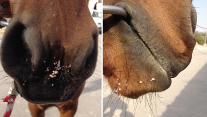
AbstractBackground: Equine papillomavirus type 1 (EcPV-1) causes cutaneous papillomatosis in horses, which is characterized by benign, proliferative epithelial lesions. Despite its global presence, the prevalence and characterization of EcPV-1 in Arabian horses in Saudi Arabia have not been previously reported. Aim: This study is the first to identify and comprehensively analyze EcPV-1 in four Arabian horses presenting with multiple raised, verrucous lesions around the muzzle and lips. Methods: This study involved clinical examination, skin biopsy collection, histopathological examination, and molecular analysis. Results: Clinical examination revealed non-painful, nonpruritic growths ranging in diameter from 2 to 8 mm. Histopathological analysis of skin biopsies showed thickening and hyperplasia of all epidermal layers, accompanied by acanthosis, hyperkeratosis, and the presence of koilocyte within the stratum spinosum and granulosum. Molecular analysis using polymerase chain reaction confirmed the presence of EcPV-1 DNA in all affected horses, with 384 bp amplicons corresponding to the E4 and L2 genes of the viral genome. Gel electrophoresis revealed clear bands in samples from affected horses, whereas viral DNA was not detected in normal control samples. Conclusion: This study provides the first molecular and histopathological evidence of EcPV-1 infection in Arabian horses in Saudi Arabia, highlighting the need for further epidemiological studies to understand the distribution and impact of EcPV-1 in the region. These findings also underscore the importance of integrating molecular diagnostics with clinical and histopathological evaluation for the accurate diagnosis and management of equine papillomatosis. Keywords: Equine papillomavirus (EcPV-1), Arabian horses, Cutaneous papillomatosis, Viral DNA detection, veterinary dermatology. IntroductionPapillomaviruses (PVs) are a diverse group of non-enveloped, double-stranded DNA viruses that induce benign epithelial proliferative tumors in various species, including humans and animals (Bernard et al., 2010; Doorbar et al., 2012). Equine Papillomaviruses (EcPVs) are the causative agents of Equine Papillomatoses, which have been observed in domesticated horses for centuries, with the first molecular identification of EcPV reported in 1986 (O’Banion et al., 1986). EcPVs are associated with several clinical conditions in horses, including classical viral papillomatosis, genital papillomatosis, and aural plaques (Lange et al., 2013; Torres and Koch, 2013). The strict tissue tropism exhibited by PVs indicates that different types of EcPVs are associated with distinct clinical manifestations in horses. To date, seven types of EcPV have been identified and categorized into three molecular genera based on their genetic sequences (Lange et al., 2011). Equine papillomavirus type 1 (EcPV-1), the first type to be identified, has been predominantly associated with classical viral papillomatosis”a condition characterized by benign, self-limiting skin tumors primarily affecting horses under 3 years of age (O’Banion et al., 1986; Ghim et al., 2004). This condition is often transient, with lesions typically regressing spontaneously within a few months, leaving affected horses with lifelong immunity (Sundberg et al., 1985). Other types, such as EcPV-2 and EcPV-7, have been implicated in genital papillomatosis, whereas EcPV-3, EcPV-4, EcPV-5, and EcPV-6 are commonly found in aural plaques (Scase et al., 2010; Lange et al., 2011; Fairley et al., 2014). Despite the clinical significance of equine papillomas, they are rarely submitted for histopathological examination due to their characteristic appearance and typically benign, self-resolving nature (Runnells and Benbroo, 1942). These lesions account for only about 5% of all equine neoplasms submitted to diagnostic laboratories (Ghim et al., 2004). Clinically, cutaneous papillomas present as solitary or multiple elevated, small, circumscribed, horny masses ranging from 2 to 20 mm in diameter (Ghim et al., 2004). Histologically, these papillomas exhibit marked epithelial proliferation on a thin fibrovascular stalk in a papillary pattern. Intranuclear inclusions and abundant keratohyalin-like cytoplasmic granules are characteristic features of the stratum granulosum layer (Ghim et al., 2004). Cutaneous papillomas are heavily pigmented, whereas those affecting mucous membranes, especially in the lower genital tract, are unpigmented and have a thin stratum corneum. Although Equine Papillomatoses have been well-documented historically, there remains a paucity of data on the prevalence and molecular epidemiology of EcPV-1 in regions such as Saudi Arabia. This case report documents the first known case of EcPV-1 infection in Saudi Arabia, highlighting the need for further molecular epidemiological studies to better understand the distribution and impact of EcPV-1 in the region. Materials and MethodsAnimals and sample collectionIn April and May 2017, four Arabian horses (two stallions and two mares), aged between 2 and 4 years and weighing between 320 and 410 kg, were presented to the Veterinary Teaching Hospital at the College of Veterinary Medicine, King Faisal University. The horses exhibited multiple raised, verrucous epidermis-like proliferations around the muzzle and lips. These skin lesions, ranging in size from 2 to 8 mm, were neither pruritic nor painful. All horses were in good physical condition, with normal heart and respiratory rates and blood profiles. According to the owners, the lesions had developed over a period of 2 months, and the horses were not receiving any treatment at the time of presentation. To facilitate diagnosis, two surgical biopsies were performed from the periorbital mass of each horse using a disposable 6-mm punch (Kolplast). One biopsy from each horse was fixed in 10% neutral buffered formalin, processed, and embedded in paraffin for histopathological analysis. The second biopsy was immediately placed in a 1.5 ml sterile, DNA/RNA-free microtube (Axygen), frozen in liquid nitrogen, and stored at –80°C for subsequent molecular biological examination. HistopathologySkin biopsies were fixed in 10% neutral buffered formalin immediately after collection. After 24 hours, the specimens were trimmed, dehydrated in a graded series of absolute ethyl alcohol, cleared in xylene, and embedded in paraffin blocks. Sections of 5 μm thickness were cut from the paraffin blocks, deparaffinized, and stained with Hematoxylin and Eosin (H&E) for microscopic examination. Genomic DNA extractionTotal DNA was extracted from up to 25 mg of the cutaneous skin lesions using a DNeasy Blood and Tissue Kit (QIAGEN, USA) according to the manufacturer’s protocol. Briefly, the specimens were lysed using ATL buffer and proteinase K, followed by the addition of absolute ethanol. The mixture was then transferred to a spin column for purification. Purified DNA was eluted in 150 μl of AE buffer and stored at –20°C until further analysis. Control samples from two healthy horses with no evidence of skin lesions were also collected and subjected to the same procedures to verify the presence of EcPV-1 DNA and confirm the association of the virus with the lesions. Detection of EcPV-1 DNA by polymerase chain reaction (PCR)To detect EcPV-1 DNA, specific primers targeting a 384-bp fragment of the E4 and L2 genes of the EcPV-1 genome were used. The forward primer EcPV1-F (5′-TGC GTT CGC CCC AAT AGT CAT CTT-3′) corresponds to nucleotide positions 3,441 to 3,464, and the reverse primer EcPV1-R (5′-ACC GCC CGC CTC ACC CTT GTC-3′) corresponds to positions 3,804 to 3,824 (Postey et al., 2007). These primers were designed using OligoAnalyzer 3.1 (Integrated DNA Technologies, USA) and synthesized by Metabion International AG, Germany. The extracted DNA samples were then screened for EcPV-1 DNA using a HotStartTaq® Plus Master Mix Kit (QIAGEN, USA). A 2 μl aliquot of each purified DNA sample was amplified in a 20 μl reaction mixture containing 2× HotStartTaq Plus Master Mix, 1.5 mM MgCl2, 200 μM of each dNTP, 1 unit of HotStartTaq Plus DNA polymerase, and 10 μM of each forward and reverse primer. The thermocycling conditions were set as follows: initial denaturation at 95°C for 5 minutes, followed by 35 cycles of 94°C for 30 seconds, 60°C for 30 seconds, and 72°C for 30 seconds, with a final extension step at 72°C for 10 minutes. The PCR products were then subjected to electrophoresis on a 1.5% agarose gel, stained with ethidium bromide, and visualized using a gel documentation system (BIORAD). ResultsEcPV-1 affected horsesThe horses suspected of EcPV-1 infection presented with multiple raised, verrucous lesions on the epidermis, particularly concentrated around the muzzle and lips (Fig. 1). These lesions were characterized by their wart-like appearance, with sizes varying from 2 to 8 mm. The proliferative nature of the lesions was evident, and the affected areas had a roughened texture. Despite the presence of these lesions, the horses did not exhibit signs of discomfort, as the lesions were neither pruritic nor painful. Additionally, all horses maintained normal physiological parameters, including heart rate, respiratory rate, and body condition, suggesting that the infection did not significantly impact their overall health. The distribution and appearance of the lesions are consistent with classical equine papillomatosis, which is typically associated with EcPV-1 infection.
Fig. 1. Typical presentation of wart-like lesions on the muzzle and lips of a 2-year-old Arabian horse.
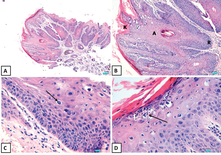
Fig. 2. H&E-stained sections of equine skin mass (bar=50 μm). (A) The exophytic mass is located on the skin surface. (B) The mass consists of proliferating keratinocytes, featuring elongation of the rete ridges (R), acanthosis (A), and hyperkeratosis (K). (C) Koilocytes are multifocally present within the epidermis (arrow). (D) Keratohyaline granules within the stratum granulosum are abundant and exhibit variability in size and shape (arrow). Histopathological analysisHistopathological examination of the collected specimens revealed characteristic exophytic papillomatous masses demonstrating significant thickening and hyperplasia across all layers of the epidermis (Fig. 2A). This was evidenced by the presence of elongated and expanded rete ridges, pronounced acanthosis, and orthokeratotic hyperkeratosis (Fig. 2B). The mitotic activity was minimal, with only 1–2 mitotic figures observed per high-power field. A notable feature was the presence of numerous koilocytes within the stratum spinosum and stratum granulosum. These cells were distinguished by their large size, slightly eccentric nuclei, and the presence of a clear perinuclear halo (Fig. 2C). Additionally, keratohyaline granules within the stratum granulosum were markedly enlarged and exhibited variability in shape and size, further confirming the diagnosis of papillomatosis (Fig. 2D).
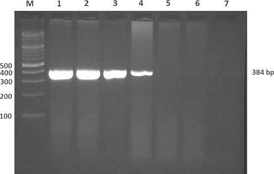
Fig. 3. Ethidium-bromide–stained agarose gel electrophoresis of PCR-amplified EcPV-1 DNA. Lane M: Molecular weight markers (bp); Lanes 1–4: Four suspected EcPV-1 cutaneous samples showing positive amplification; Lanes 5–6: Two normal cutaneous equine samples with no amplification; Lane 7: Non-template control (negative control) showing no amplification. Molecular analysis of EcPV-1 DNAAll four samples with suspected EcPV-1 infection were tested positive for viral DNA using PCR analysis. The presence of a 384-bp sharp band was observed in gel electrophoresis, confirming the presence of EcPV-1 DNA in these samples. In contrast, no bands were detected in the two normal cutaneous samples used as controls, further supporting the specificity of the PCR results (Fig. 3). DiscussionThe identification of EcPV-1 in Arabian horses from Saudi Arabia represents the first documented case of this virus in the region and contributes to the global understanding of papillomavirus infections in equines. PVs are known to cause benign epithelial proliferations in various species, and the clinical presentation observed in this study aligns with classical EcPV-1-induced papillomatosis as previously described in the literature (O’Banion et al., 1986; Ghim et al., 2004; Torres and Koch, 2013). The affected horses in this study exhibited multiple verrucous proliferations on their muzzle and lips, a characteristic finding consistent with EcPV-1 infection. These lesions, neither pruritic nor painful, are typical of cutaneous papillomatosis, which is often seen in younger horses, particularly those under 3 years of age (Sundberg et al., 1985; Postey et al., 2007). The development of these lesions over a 2-month period, as reported by the owners, further supports the diagnosis of papillomatosis, given the rapid onset and progression typical of this condition. Histopathological examination of the lesions revealed significant epithelial hyperplasia, hyperkeratosis, and the presence of koilocyte, all hallmark features of papillomavirus-induced lesions (Sundberg et al., 1985; Ghim et al., 2004). The presence of koilocyte, which are characterized by large cells with clear halos surrounding slightly eccentric nuclei, is particularly indicative of papillomavirus infection and corroborates the molecular findings. The kerato-hyaline granules observed within the stratum granulosum, varying in size and shape, further support the diagnosis and are consistent with previous histopathological descriptions of EcPV-1-induced lesions (Runnells and Benbroo, 1942; Fairley and Haines, 1992). Molecular detection of EcPV-1 DNA through PCR amplification confirmed the presence of the virus in four patients with suspected EcPV-1 infection. The detection of a 384-bp fragment corresponding to the E4 and L2 genes of EcPV-1 aligns with the established molecular diagnostic techniques for this virus (Postey et al., 2007). The absence of viral DNA in control samples from healthy skin further strengthens the association between observed lesions and EcPV-1 infection. The PCR results, coupled with the clinical and histopathological findings, provide a comprehensive confirmation of EcPV-1 as the etiological agent in these cases. The identification of EcPV-1 in Saudi Arabia highlights the need for further epidemiological studies to assess the prevalence and impact of this virus in the region. Although equine papillomatosis is generally self-limiting and does not typically lead to severe complications, understanding its distribution and behavior in different geographical contexts is crucial. This case series underscores the importance of molecular diagnostics for accurately identifying viral pathogens, particularly in regions where such infections may be underreported or misdiagnosed. Moreover, our findings contribute to a broader understanding of papillomavirus-induced diseases in horses. The detailed histopathological and molecular analyses presented here provide valuable insights into the pathogenesis of EcPV-1 and its clinical manifestations. Future studies should focus on the genetic characterization of EcPV-1 isolates from different regions to explore potential variations in viral strains and their associated clinical outcomes. ConclusionThis study documented the first occurrence of EcPV-1 in Arabian horses in Saudi Arabia, expanding the known geographic range of this virus. The combination of clinical, histopathological, and molecular findings provides a robust diagnosis of EcPV-1-induced cutaneous papillomatosis. These results emphasize the importance of vigilance in the diagnosis and reporting of papillomavirus infections in equines, particularly in regions where such cases have not been previously documented. Further research is warranted to explore the epidemiology, genetic diversity, and potential implications of EcPV-1 infection in Saudi Arabia and beyond. Conflicts of interestThe authors declare that they have no conflicts of interest. Ethical considerationAll work was conducted according to the guidelines of the College of Veterinary Medicine, King Faisal University, Al-Ahsa, Saudi Arabia. Author contributionsAll work was conducted by Dr. Mohammed Ali Al-Hammadi. Data availabilityAll data related to this study are included in this manuscript. ReferencesBernard, H.U., Burk, R.D., Chen, Z., van Doorslaer, K., zur Hausen, H. and de Villiers, E.M. 2010. Classification of papillomaviruses (PVs) based on 189 PV types and proposal of taxonomic amendments. Virology 401(1), 70–79. Doorbar, J., Quint, W., Banks, L., Bravo, I.G., Stoler, M., Broker, T.R. and Stanley, M.A. 2012. The biology and life-cycle of human papillomaviruses. Vaccine 30(Suppl 5), F55–F70. Fairley, R.A. and Haines, D.M. 1992. The electron microscopic and immunohistochemical demonstration of a papillomavirus in equine aural plaques. Vet. Pathol. 29(1), 79–81. Fairley, R., Morley, C., Williams, S., Senior, D. and Neill, M. 2014. Aural plaques in two imported horses in New Zealand. New Zealand Vet. J. 62(4), 232–233. Ghim, S.J., Rector, A., Delius, H., Sundberg, J.P., Jenson, A.B. and Van Ranst, M. 2004. Equine papillomavirus type 1: complete nucleotide sequence and characterization of recombinant virus-like particles composed of the EcPV-1 L1 major capsid protein. Biochem. Biophys. Res. Commun. 324(3), 1108–1115. Lange, C.E., Tobler, K., Ackermann, M. and Favrot, C. 2011. Identification of two novel equine papillomavirus sequences suggests three genera in one cluster. Vet. Microbiol. 149(1-2), 85–90. Lange, C.E., Tobler, K., Lehner, A., Grest, P., Welle, M.M., Schwarzwald, C.C. and Favrot, C. 2013. EcPV2 DNA in equine papillomas and in situ and invasive squamous cell carcinomas supports papillomavirus etiology. Vet. Pathol. 50(4), 686–692. O’Banion, M.K., Reichmann, M.E. and Sundberg, J.P. 1986. Cloning and characterization of an equine cutaneous papillomavirus. Virology 152(1), 100–109. Postey, R.C., Appleyard, G.D. and Kidney, B.A. 2007. Evaluation of equine papillomas, aural plaques, and sarcoids for the presence of Equine papillomavirus DNA and Papillomavirus antigen. Can. J. Vet. Res. 71(1), 28–33. Scase, T., Brandt, S., Kainzbauer, C., Sykora, S., Bijmholt, S., Hughes, K., Sharpe, S. and Foote, A. 2010. Equus caballus papillomavirus-2 (EcPV-2): an infectious cause for equine genital cancer? Equine Vet. J. 42(8), 738–745. Sundberg, J.P., Junge, R.E. and el Shazly, M.O. 1985. Oral papillomatosis in New Zealand white rabbits. Am. J. Vet. Res. 46(3), 664–668. Torres, S.M. and Koch, S.N. 2013. Papillomavirus-associated diseases. Vet. Clin. North Am. Equine Pract. 29(3), 643–655. Runnells, R.A. and Benbroo, E.A. 1942. Epithelial tumors of horses. 1942, 176–179. | ||
| How to Cite this Article |
| Pubmed Style Mohammed Ali Al-Hammadi. First report on equine papillomavirus type 1 in Arabian horses in Saudi Arabia: Clinical, histopathological, and molecular characterization. Open Vet. J.. 2025; 15(4): 1798-1802. doi:10.5455/OVJ.2025.v15.i4.32 Web Style Mohammed Ali Al-Hammadi. First report on equine papillomavirus type 1 in Arabian horses in Saudi Arabia: Clinical, histopathological, and molecular characterization. https://www.openveterinaryjournal.com/?mno=247162 [Access: January 25, 2026]. doi:10.5455/OVJ.2025.v15.i4.32 AMA (American Medical Association) Style Mohammed Ali Al-Hammadi. First report on equine papillomavirus type 1 in Arabian horses in Saudi Arabia: Clinical, histopathological, and molecular characterization. Open Vet. J.. 2025; 15(4): 1798-1802. doi:10.5455/OVJ.2025.v15.i4.32 Vancouver/ICMJE Style Mohammed Ali Al-Hammadi. First report on equine papillomavirus type 1 in Arabian horses in Saudi Arabia: Clinical, histopathological, and molecular characterization. Open Vet. J.. (2025), [cited January 25, 2026]; 15(4): 1798-1802. doi:10.5455/OVJ.2025.v15.i4.32 Harvard Style Mohammed Ali Al-Hammadi (2025) First report on equine papillomavirus type 1 in Arabian horses in Saudi Arabia: Clinical, histopathological, and molecular characterization. Open Vet. J., 15 (4), 1798-1802. doi:10.5455/OVJ.2025.v15.i4.32 Turabian Style Mohammed Ali Al-Hammadi. 2025. First report on equine papillomavirus type 1 in Arabian horses in Saudi Arabia: Clinical, histopathological, and molecular characterization. Open Veterinary Journal, 15 (4), 1798-1802. doi:10.5455/OVJ.2025.v15.i4.32 Chicago Style Mohammed Ali Al-Hammadi. "First report on equine papillomavirus type 1 in Arabian horses in Saudi Arabia: Clinical, histopathological, and molecular characterization." Open Veterinary Journal 15 (2025), 1798-1802. doi:10.5455/OVJ.2025.v15.i4.32 MLA (The Modern Language Association) Style Mohammed Ali Al-Hammadi. "First report on equine papillomavirus type 1 in Arabian horses in Saudi Arabia: Clinical, histopathological, and molecular characterization." Open Veterinary Journal 15.4 (2025), 1798-1802. Print. doi:10.5455/OVJ.2025.v15.i4.32 APA (American Psychological Association) Style Mohammed Ali Al-Hammadi (2025) First report on equine papillomavirus type 1 in Arabian horses in Saudi Arabia: Clinical, histopathological, and molecular characterization. Open Veterinary Journal, 15 (4), 1798-1802. doi:10.5455/OVJ.2025.v15.i4.32 |

