


| Case Report | ||
Open Vet J. 2020; 10(3): 272-275 doi: 10.4314/ovj.v10i3.5 Open Veterinary Journal, (2020), Vol. 10(3): 272–275 Case Report http://dx.doi.org/10.4314/ovj.v10i3.5 Splenic T-cell lymphoma in a North American River Otter (Lontra canadensis)Crystal L. Matt1*, Christoph Mans2, Grayson Doss2, Marie Pinkerton3 and Betsy Elsmo41Department of Medical Sciences, School of Veterinary Medicine, University of Wisconsin-Madison, Madison, WI 53706, USA 2Department of Surgical Sciences, School of Veterinary Medicine, University of Wisconsin-Madison, Madison, WI 53706, USA 3Department of Pathobiological Sciences, School of Veterinary Medicine, University of Wisconsin-Madison, Madison, WI 53706, USA 4Wisconsin Veterinary Diagnostic Laboratory, Madison, WI 53706, USA *Corresponding Author: Crystal L. Matt. Department of Medical Sciences, School of Veterinary Medicine, University of Wisconsin-Madison, Madison, WI 53706, USA. Email: clmatt [at] okstate.edu Submitted: 10/12/2019 Accepted: 09/07/2020 Published: 15/08/2020 © 2020 Open Veterinary Journal
AbstractBackground: Splenic lymphoma is commonly reported in domestic ferrets (Mustela putorious furo), but very rarely reported in wild Mustelidae species, including otters. One report described B-cell splenic lymphoma in an Asian small-clawed otter (Aonyx cinerea) that metastasized and was the primary reasoning for humane euthanasia (Stedman and Mills, 2014). Case Description: The current report describes a case of splenic T-cell lymphoma in a captive North American river otter (Lontra canadensis). The otter died several weeks after splenectomy and no evidence of metastasis was found on gross necropsy or histopathological evaluation. Conclusion: The splenectomy performed on this individual was presumptively curative for its splenic lymphoma. Extensive myocardial fibrosis was found, and suspected to have caused severe cardiac arrhythmia leading to acute death. Keywords: Fibrosis, Mustelid, Neoplasia, Splenectomy. IntroductionAlthough there is a great wealth of information regarding neoplasia in domestic ferrets (Mustela putorious furo), reports describing neoplasia in wild Mustelidae species are lacking. Lymphoma is widely recognized as the most common malignant neoplasm in ferrets and has been suggested to have an infectious cause. Although it can originate nearly anywhere, it is most commonly found in the spleen, liver, and lymph nodes in this domestic species (Hess, 2005). Lymphomas are lymphoproliferative diseases that form solid masses, which are typically found in lymph nodes or lymphoid organs. They are closely related to leukemias, which are typically within the blood and bone marrow, as both arise from either B-cell or T-cell lymphocytes (King, 2006). B-cell neoplasms are far more common than T-cell, as B-cells are more predisposed to gene rearrangement. Clonality of B-cell tumors can be demonstrated using flow cytometry. T-cell neoplasms are more likely to present outside of a lymph node. The cytokine-releasing property of T-cells, when exaggerated by neoplastic development, results in the recruitment of a large number of other inflammatory cells. This can make the identification of T-cells difficult, as morphologic findings may only show inflammation. Therefore, flow cytometry may be inadequate to identify clonality for T-cell neoplasia, and PCR for antigen receptor rearrangements is more effective (King, 2006). Reports of neoplasia in any otter species are uncommon, particularly for North American river otters. Previously reported neoplasms in North American river otters include pheochromocytoma (Schlanser et al., 2012), pleural squamous cell carcinoma (de Velde et al., 2019), and T-cell gastrointestinal lymphoma (Hanson, 2007). T-cell gastrointestinal lymphoma has also been reported in a Eurasian otter (Lutra lutra) (Bartlett et al., 2010). T-cell lymphoma in a sea otter (Enhydra lutris) was found as a primary cerebral lesion (Tanaka et al., 2013). This sea otter presented with convulsions, arrhythmia, anorexia, and labored breathing before being found dead one week later. Few reports have noted splenic lymphoma in otters, although one report has described splenic lymphoma of B-cell origin in a 7-year-old, Asian small-clawed otter (Aonyx cinerea) (Stedman and Mills, 2014). This otter underwent a splenectomy, but was eventually euthanized due to severe metastatic disease. The current report describes a case of splenic T-cell lymphoma in a captive North American river otter (Lontra canadensis). Case ReportA 13-year-old, intact female, captive North American river otter was evaluated for a two-day history of lethargy and inappetence. The otter was housed singly at a zoological facility and had no significant medical history. A physical examination, complete blood count, and serum biochemistry, performed under anesthesia 5 months prior to the onset of clinical signs, did not reveal any clinically relevant abnormalities. Upon presentation for lethargy and inappetence, diagnostic testing and physical examination were performed under general anesthesia with ketamine, dexmedetomidine, and midazolam premedication, along with isoflurane and sevoflurane (at different intervals) maintenance. Physical examination revealed an enlarged, palpable spleen, but no other significant findings. A complete blood count, serum biochemistry, total T4, and urinalysis were performed, with the only abnormal finding being an elevated GGT (231 U/L; RI: 8–38 U/L, Tocidlowski et al., 2000). A contrast-enhanced, whole-body CT scan revealed several abnormalities, including splenomegaly with nodules and extracapsular hemorrhage, perihepatic nodules, a nodule in the area of the left thyroid gland, nephroliths, T12–L1 intervertebral disk disease, and dystrophic mineralization of the meninges. All other structures, including the heart, were apparently normal. An abdominal ultrasound was performed to evaluate and perform fine needle aspirates of the splenic and perihepatic nodules for cytological evaluation. The perihepatic nodules were revealed to be sites of necrosis that were not associated with the liver. The nodular changes in the spleen were highly suspect for lymphoma on cytology, with less likely benign lymphoid hyperplasia as a differential. The ultrasound also revealed biliary sludge suggestive of cholangitis. The animal recovered from anesthesia without complications. Surgical removal of the spleen was recommended. Surgical exploratory laparotomy with splenectomy (Fossum, 2013) was performed one month later. The otter was premedicated with hydromorphone (0.1 mg/kg IM), ketamine (3.5 mg/kg IM), midazolam (0.2 mg/kg IM), and alfaxalone (6 mg/kg IM). Anesthesia was induced with alfaxalone (1 mg/kg IV) and maintained with sevoflurane gas. Pre-surgical blood work revealed anemia with a PCV of 27% (one month prior: 34%; RI: 32.2%–60.8%, Tocidlowski et al., 2000). Cefazolin (30 mg/kg IV) was given 30 minutes prior to starting surgery. During abdominal exploratory surgery, a mild amount of free blood was found in the abdominal cavity and a grossly enlarged, heterogeneous, nodular spleen was noted (Fig. 1). The spleen was surgically removed, along with the majority of the perihepatic nodular changes, and both were submitted in 10% neutral buffered formalin for histopathological evaluation. Hemostasis was achieved with bipolar electrocautery throughout the procedure. The body wall was closed in a simple continuous pattern. The skin was closed using an intradermal pattern. Surgical tissue glue was placed over the incision to increase strength during healing and provide protection. An injection of cefovecin sodium (8 mg/kg SC) and an injection of meloxicam (0.15 mg/kg SC) were administered prior to recovery. The otter recovered from anesthesia and surgery without complications. It was discharged with oral meloxicam (0.2 mg/kg PO q 24 hours for 5 days) and instructions for the facility to move the otter to a smaller enclosure to avoid jumping, climbing, and swimming for several days, and prevent all pool access for two weeks.
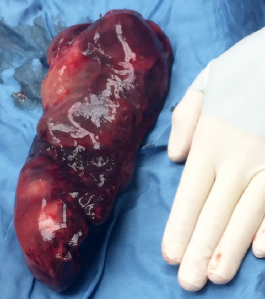
Fig. 1. Gross intraoperative image of neoplastic spleen removed five weeks prior to patient presenting deceased. Spleen was confirmed to be uniformly CD3 positive on immunohistochemistry, indicative of T-cell lymphoma. On histopathologic examination, the spleen was found to have extensive areas of capsular and subcapsular necrosis. The red pulp was diffusely congested, with poorly defined loose sheets of individualized round cells ~2–2.5× the diameter of a red blood cell. These cells had scant amphophilic cytoplasm and a round nucleus with finely stippled chromatin and occasionally a prominent nucleolus. There was mild anisocytosis and anisokaryosis, and up to 2–3 mitotic figures per 400× field. Immunohistochemistry showed the intermediate lymphocyte population to be uniformly CD3 positive, consistent with a T-cell lymphoma. The perihepatic mass was revealed to be hepatic tissue that was diffusely necrotic with mineralization. The final diagnoses were intermediate splenic T-cell lymphoma with severe multifocal necrosis and moderate extramedullary hematopoiesis. Five weeks after surgery, the otter was found deceased in its enclosure without any clinical signs prior to death. The histologic postmortem lesions included extensive myocardial fibrosis (Fig. 2), multifocal renal interstitial fibrosis, hypertrophy and mineralization of the tunica media of meningeal vessels, and incidental sarcocysts scattered throughout myofibers. The cause of death was presumed to be secondary to significant myocardial fibrosis. No evidence of lymphoma was found in the remaining evaluated tissues, including the heart, kidney, liver, mesentery, brain, thyroid, lymph node, small intestine, eye, pawpad, skeletal muscle, tongue, trachea, lung, esophagus, stomach, pancreas, adrenal glands, urinary bladder, ovaries, and uterus.
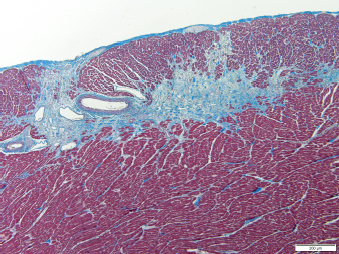
Fig. 2. Histopathologic image of the left ventricular wall in a North American river otter showing extensive areas of myofiber loss with replacement by fibrosis. Artery present displays segmental thickening of the tunica media by amorphous hyaline material or fibrosis. Masson’s trichome stain. Scale bar=200 micrometers. DiscussionThe current report describes a case of splenic T-cell lymphoma in a geriatric, captive North American river otter. Splenic lymphoma has previously been reported in an Asian small-clawed otter, but it was confirmed to be B-cell in origin based on immunohistochemistry (Stedman and Mills, 2014). The B-cell lymphoma had already metastasized to adjacent lymph nodes at the time of splenectomy. Sixteen months later, the Asian small-clawed otter presented with lethargy and inappetence, and was euthanized due to renal failure and evidence of metastatic thoracic and abdominal masses. Gross necropsy revealed enlargement of the mediastinal/tracheobronchial, hepatic, gastric, pancreaticoduodenal, renal, sublumbar, inguinal, axillary, prescapular, and cervical lymph nodes, along with a mass was in the cranial pole of the right kidney, and several masses in several liver lobes. These were all confirmed to be metastatic lymphoma, with a final diagnosis of metastatic marginal zone lymphoma (Stedman and Mills, 2014). Unlike the Asian small-clawed otter, the North American river otter in the case presented here did not have any signs of metastasis either at splenectomy or necropsy. Both were presented for lethargy and inappetence, which are nonspecific clinical signs and therefore unlikely to be helpful in diagnosing lymphoma. At no point did the river otter have peripheral lymphadenopathy, which fits with its diagnosis of hepatosplenic T-cell lymphoma. This is in contrast to the marginal zone lymphoma, which frequently presents with peripheral lymphadenopathy (King, 2006). Clinical signs of lymphoma in ferrets, as in many other species, are often variable and nonspecific. Like in the case presented here, most ferrets present with anorexia and lethargy, often with a long-term history of weight loss (Fox and Marini, 2014). The most common sites of extranodal lymphoma in ferrets are the spleen, kidney, and liver (Fox and Marini, 2014). Ferrets are commonly found to have splenomegaly, but it is typically attributable to extra-medullary hematopoiesis (EMH). However, two-thirds of ferrets with lymphoma are found to have splenic enlargement, although many of these are also the result of EMH as well (Fox and Marini, 2014). Treatment for splenic lymphoma in dogs is typically splenectomy with or without adjunctive chemotherapy (van Stee et al., 2015). In one study of 41 cases of splenic lymphoma treated with splenectomy in dogs, adjunctive chemotherapy was shown to not have any impact on survival times, suggesting that splenectomy alone could be sufficient treatment for lymphoma confined to the spleen (van Stee et al., 2015). Hemoabdomen was a strongly negative prognostic indicator; however, all hemoabdomen cases in this study were associated with B-cell marginal zone lymphoma (van Stee et al., 2015). Because T-cell splenic lymphoma is much less common than B-cell lymphoma, this study had few T-cell cases (n=3; 28 cases in total). As splenic tumors are generally associated with a risk of bleeding, the mild amount of free blood found during splenectomy for this otter was likely secondary to the tumor itself bleeding, despite it being T-cell in origin. This is also more likely when considering that, upon its necropsy five weeks post-splenectomy, the otter had no evidence of hemoabdomen. In the case reported here, the river otter died less than two months after splenectomy. However, no evidence was found that its death was associated with the previously diagnosed splenic T-cell lymphoma. The cause of death was suspected to be secondary to potentially malignant arrhythmias related to the myocardial fibrosis, which has been shown to be a mechanism behind sudden cardiac death in racehorses (Kiryu et al., 1999). There was no evidence of metastasis upon postmortem histopathology. Therefore, removal of the spleen appears to have been curative for the T-cell lymphoma in this animal, suggesting that splenectomy alone is a potentially valid option to consider in future cases of splenic neoplasia in otters. Conflict of InterestThe authors declare that there is no conflict of interest. ReferencesBartlett, S.L., Imai, D.M., Trupkiewicz, J.G., Garner, M.M., Ogasawara, S., Stokol, T., Kiupel, M., Abou-Madi, N., Kollias, G.V. 2010. Intestinal lymphoma of granular lymphocytes in a fisher (Martes pennanti) and a Eurasian otter (Lutra lutra). J. Zoo Wildl. Med. 41, 309–315. de Velde, N.V., Demetrick, D.J., Duignan, P.J. 2019.Primary pleural squamous cell carcinoma in a free-ranging River Otter (Lontra canadensis). J. Wildl. Dis. 55(3), 728–732. Fossum, T.W. 2013. Small animal surgery textbook. Elsevier Health Sciences, St. Louis, Missouri, USA. Fox, J.G., Marini, R.P., 2014. Biology and Diseases of the Ferret. Hoboken, NJ: John Wiley & Sons. Hanson, S. 2007. Gastrointestinal lymphoma in a Captive North American River Otter, Lontra canadensis IAAAM Archive . Hess, L. 2005. Ferret lymphoma: the old and the new, In Seminars in avian and exotic pet medicine, pp: 199–204. King, T. 2006. Elsevier’s integrated pathology. Elsevier Health Sciences, Mosby Inc, USA. Kiryu, K., Machida, N., Kashida, Y., Yoshihara, T., Amada, A., Yamamoto, T. 1999. Pathologic and electrocardiographic findings in sudden cardiac death in racehorses. J. Vet. Med. Sci. 61, 921–928. Schlanser, J.R., Patterson, J.S., Kiupel, M., Hencken, C., Sikarskie, J.G., Harrison, T.M. 2012. Disseminated pheochromocytoma in a North American river otter (Lontra canadensis). J. Zoo Wildl. Med. 43, 407–411. Stedman, N.L., Mills, Z.V. 2014. Splenic Marginal Zone Lymphoma in an Asian Small-Clawed Otter (Aonyx cinerea). J. Zoo Wildl. Med. 45, 719–722. Tanaka, N., Izawa, T., Kashiwagi-Yamamoto, E., Kuwamura, M., Ozaki, M., Nakao, T., Yamate, J. 2013. Primary cerebral T-cell lymphoma in a sea otter (Enhydra lutris). J. Vet. Med. Sci. 75, 1667–1669. Tocidlowski, M.E., Spelman, L.H., Sumner, P.W. and Stoskopf, M.K. 2000. Hematology and serum biochemistry parameters of North American river otters (Lontra canadensis). J. Zoo Wildl. Med. 31, 484–490. van Stee, L.L., Boston, S.E., Singh, A., Romanelli, G., Rubio-Guzman, A., Scase, T.J. 2015. Outcome and prognostic factors for canine splenic lymphoma treated by splenectomy (1995–2011). Vet. Surg. 44, 976–982. | ||
| How to Cite this Article |
| Pubmed Style Matt C, Mans C, Doss G, Pinkerton M, Elsmo B. Splenic T-cell lymphoma in a North American river otter (Lontra canadensis). Open Vet J. 2020; 10(3): 272-275. doi:10.4314/ovj.v10i3.5 Web Style Matt C, Mans C, Doss G, Pinkerton M, Elsmo B. Splenic T-cell lymphoma in a North American river otter (Lontra canadensis). https://www.openveterinaryjournal.com/?mno=77361 [Access: July 15, 2025]. doi:10.4314/ovj.v10i3.5 AMA (American Medical Association) Style Matt C, Mans C, Doss G, Pinkerton M, Elsmo B. Splenic T-cell lymphoma in a North American river otter (Lontra canadensis). Open Vet J. 2020; 10(3): 272-275. doi:10.4314/ovj.v10i3.5 Vancouver/ICMJE Style Matt C, Mans C, Doss G, Pinkerton M, Elsmo B. Splenic T-cell lymphoma in a North American river otter (Lontra canadensis). Open Vet J. (2020), [cited July 15, 2025]; 10(3): 272-275. doi:10.4314/ovj.v10i3.5 Harvard Style Matt, C., Mans, . C., Doss, . G., Pinkerton, . M. & Elsmo, . B. (2020) Splenic T-cell lymphoma in a North American river otter (Lontra canadensis). Open Vet J, 10 (3), 272-275. doi:10.4314/ovj.v10i3.5 Turabian Style Matt, Crystal, Christoph Mans, Grayson Doss, Marie Pinkerton, and Betsy Elsmo. 2020. Splenic T-cell lymphoma in a North American river otter (Lontra canadensis). Open Veterinary Journal, 10 (3), 272-275. doi:10.4314/ovj.v10i3.5 Chicago Style Matt, Crystal, Christoph Mans, Grayson Doss, Marie Pinkerton, and Betsy Elsmo. "Splenic T-cell lymphoma in a North American river otter (Lontra canadensis)." Open Veterinary Journal 10 (2020), 272-275. doi:10.4314/ovj.v10i3.5 MLA (The Modern Language Association) Style Matt, Crystal, Christoph Mans, Grayson Doss, Marie Pinkerton, and Betsy Elsmo. "Splenic T-cell lymphoma in a North American river otter (Lontra canadensis)." Open Veterinary Journal 10.3 (2020), 272-275. Print. doi:10.4314/ovj.v10i3.5 APA (American Psychological Association) Style Matt, C., Mans, . C., Doss, . G., Pinkerton, . M. & Elsmo, . B. (2020) Splenic T-cell lymphoma in a North American river otter (Lontra canadensis). Open Veterinary Journal, 10 (3), 272-275. doi:10.4314/ovj.v10i3.5 |

