


| Original Article | ||
Open Vet J. 2021; 11(1): 52-60 doi: 10.4314/ovj.v11i1.9 Open Veterinary Journal, (2021), Vol. 11(1): 52–60 Original Research http://dx.doi.org/10.4314/ovj.v11i1.9 Electroacupuncture anesthesia for laparotomy in goatsKhalifa Ashour1, Naglaa Abd Elkader Awad2, Sherein S. Abdelgayed3, Amal Zakaria Ahmed Leil4 and Eldessouky Sheta2*1Equine Department of Police General Security, Ministry of Interior, Tripoli, Libya 2Department of Surgery, Anaesthesiology and Radiology, Faculty of Veterinary Medicine, Cairo University, Giza, Egypt 3Department of Pathology, Faculty of Veterinary Medicine, Cairo University,Giza, Egypt 4Department of Field Investigation, Animal Reproduction Research Institute, Agriculture Research Center (ARC), Giza, Egypt *Corresponding Author: Prof. Eldessouky Sheta. Department of Surgery, Anaesthesiology and Radiology, Faculty of Veterinary Medicine, Cairo University, Cairo, 12211 Giza, Egypt. Email: shetaeme [at] hotmail.com Submitted: 02/11/2020 Accepted: 24/12/2020 Published: 24/01/2021 © 2021 Open Veterinary Journal
AbstractBackground: The objective of the study to evaluate the effect of electroacupuncture during laparotomy in goats. Aim: To study the abdominal anesthesia in goats by electroacupuncture using the physiological variables, vital parameters, hematological, biochemical, cortisol hormone, pain threshold, and wound healing for laparotomy in goats. Methods: Fifteen healthy adult bucks were used to receive electroacupuncture in 10 newly selected acupoints. The data (M ± SD) were assessed at intervals (0 minutes) before induction (control group), (5 minutes), (10 minutes), (15 minutes), and (20 minutes) during induction, (30 minutes), (45 minutes), and (60 minutes) throughout surgery and (24 hours) after surgery, cortisol levels in serum at (0), (24 hours), and (72 hours) throughout laparotomy. Results: The goats of the study showed improvement in the rates of eyelid closure, head, and neck relaxation, rumen motility, and tympany which were graded into mild (+), moderate (++), and severe (+++) degrees. The respiratory rates, body temperatures, and capillary fill times were not significantly different. The total mean of hematocrit was (19.9 ± 2.68), the total mean of hemoglobin was (9.9 ± 0.94), the total mean of red blood cells was (7.9 ± 0.8), the total mean of platelets was (244,861.3 ± 138,444.8) and the total mean of SPO2 was (70.5 ± 4.6). ALT and AST showed no significance. The significant mean cortisol level was (2.6 ± 2.01) and the significant mean pain threshold level was (0.02 ± 0.03). The results proved that electroacupuncture had a lot of significant parameters. The wound healing was improved by early epithelization and immature granulation tissue (at 7 days). Thick keratinized epithelization and collagen deposition in the dermal tissue with enhanced angiogenesis (at 14 days). Mild restoration of skin and the dermal tissue was well-organized (at 21 days). Besides, well-formed scar tissue covering a highly cellular organized dermal tissue (at 28 days). Conclusions: Electroacupuncture had been considered a powerful anesthetic for abdominal surgery in goats. Moreover, wound healing proved excellent and better healing. Keywords: Acupuncture, Anesthesia, Electroacupuncture, Goat, Laparotomy. IntroductionElectroacupuncture (EA) involves the electrical stimulation of acupoints via inserted needles. The ancient practice of acupuncture started in China approximately 3,000 years ago. This procedure was first introduced in human practice in 1960 and was successfully used to relieve pain during Caesarean section, gastrectomy, enterectomy, and castration in domestic animals along with maintaining a stable physiologic index (Wang and Jin, 1989; Parmen, 2014). Electroacupuncture is one of the alternative therapies in the production of analgesia for surgical interferences (Groppetti et al., 2011). Abdominal surgery has been performed in equine under electroacupuncture analgesia (Rogers, 1995; Sheta et al., 2015). Pain is blocked by acupoints stimulation with the release of endogenous opioids and neurotransmitters (Janssens et al., 1988), which also affects the blood pressure receptors and be used to increase or decrease blood pressure (Asayama et al., 2012). Electrical stimuli were induced through the acupuncture analgesia points in the peripheral sensory nerves to the spinal cord. The stimuli reached the midbrain through the ascending spinothalamic tracts. In the midbrain, the ascending signals caused release of endorphins, serotonin, and other neurotransmitters, which activated a descending inhibition mechanism and prevented the pain signals from the surgical area from reaching the cerebral cortex (Rogers, 1995; Tian et al., 1997). The purpose of the study reported here was to explore the physiological variables, vital parameters, hematologic, biochemical, cortisol hormone, pain threshold, and histopathological healing of electroacupuncture in goat's laparotomy. Materials and MethodsThe study was carried out on 15 healthy uncastrated bucks (mean ± SD age, 1.72 ± 0.08 years; mean body weight 37.27 ± 0.89 kg). The goats were placed under identical conditions and fed on concentrates, green fodder, and allowed to graze for 4 hours a day. Physiological variables (head and neck relaxation, teeth grinding, tongue protrusion, salivation, close eyelids, rumen motility, and tympany), vital parameters (temperature, heart rate, respiratory rate, and capillary refill time) These parameters are used to evaluate the effect of abdominal anesthesia under electroacupuncture. Hematologic data [hematocrit, red blood cell (RBCs), white blood cells (WBCs), staff cells, segmented cells, lymphocytes, monocytes, eosinophils, hemoglobin, platelets, SPO2], and biochemical data (ALT and AST) were analyzed and recorded at intervals (0 minute) before induction (control group), (5 minutes), (10 minutes), (15 minutes), (20 minutes) during induction, (30 minutes), (45 minutes) (60 minutes), during surgery and (24) hours post-surgery. Cortisol levels in serum at (0), (24 hours), and 72 hours) were calculated (Cadoy and Acorda, 2018). SPO2 was measured by Description Infunix technology Pure scope IP-3050 (Pugh et al., 2020). Experimental surgery designLaparotomy was carried out on 15 bucks, instead of does, to avoid the physiological changes of does on the results, electroacupuncture performed in 10 newly selected acupoints by the authors. For surgery, the goats were starved for 24 hours and water withheld for 6 hours before induction of anesthesia. The start of the experiment was the moment when EA started (0 minutes). During EA stimulation, the first 20 minutes were regarded as the induction period of EA analgesia, followed by 60 minutes as the maintenance period of EA analgesia. This maintenance time was based on our experiences of commonly performed surgeries. Electro acupuncture stimulator KWD 808I- multipurpose Health Device, delivers a bipolar waveform (+) and (−) at each electrode through needles in the correct points. It induces dense dispersed wave output at frequencies 20–30 Hz for surgery (Sheta et al., 2015). Acupuncture sites were clipped, antiseptically prepared with 10% povidone–iodine solution, and the sterile acupuncture needles inserted into the newly selected acupoints, besides two-parallel needles, away from each other inserted subcutaneously and muscle layers at the surgery area (Fig. 1) The 10 newly selected acupoints for laparotomy in goats were Qi-jia (Withers) (+ve) concerted with St 36 (Tsu-San-Li) (−ve); Tianping (+ve) concerted along Da-Kua (Greater Trochanter) (−ve); Gov 20 (Baihui) (+ve) concerted with Sp 6 (Sanyinjao) (−ve); BL 30 (Baihuanshu) (+ve) concerted along Liv 14 (Chimen) (−ve) and BL 30 (Baihuanshu) (+ve) connected to TW 8 (Triple Warmer or Triple Heater) (−ve). In addition to two-parallel needles, away from each other inserted subcutaneously and muscle layers at the surgery area Table 1. Pain threshold recordingIt was measured at the center of the left flank. The site was clipped, washed with soap and water, and rinsed with 75% ethanol. Two electrodes were penetrated the skin 2 cm apart. Digital Multipara meter-monitor, Shenzhen (VL9205A), apparatus was used to deliver the pulsed direct current to the electrodes, which forced potassium ions into the subcutaneous tissues. Voltage was continuously increased. The pain threshold voltage was recorded at the moment when obvious contractions of the local skin and muscles, turning of the head toward the abdomen, hunching of the back, and evasive body movements were observed. The electrical current was then turned off. Measurement of pain threshold was repeated 5 times. Percentage change in the pain threshold was calculated as follows: Percentage change=[(Vn − V0)/V0] × 100% where Vn is the mean voltage for the pain threshold during the experiment and V0 is the mean voltage for the pain threshold before the experiment (time 0). After surgery, a prophylactic antibiotic (250,000 units of amoxicillin penicillin and 250 mg Dihydrostreptomycin per 10 kg body weight), was administered intramuscularly to each goat once a day for five consecutive days. Thereafter, the bucks were put under the same conditions before surgery. Histopathological studyThe wound skin tissue samples were taken by a scalpel on days 7, 14, 21, and 28 for histological observation. The skin tissues were fixed with 10% formalin. After fixation, samples were embedded in paraffin, cut into 3 mm frozen sections with a cryostat microtome then stained with Hematoxylin and Eosin (Bancroft et al., 2012). Collagen fiber, inflammatory cell, blood vessel, fibroblast, and granulation tissue of the goatskin were examined by a light microscope (Olympus BX50, Japan).
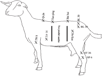
Fig. 1. Showing map of the newly selected acupoints. Table 1. The selected acupoints and location for abdominal surgery in goats.
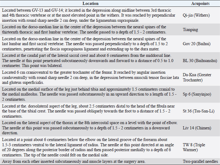
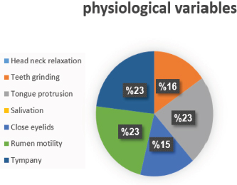
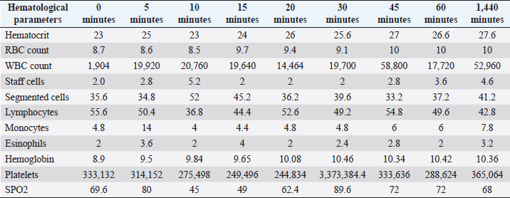
Statistical analysesData were analyzed using PASW Statistics, Version 18.0 software (SPSS Inc., Chicago, IL). Results were analyzed by student-T-test. Significance was set at p ≤ 0.05. Ethical approvalThe Ethical Committee of the Faculty of Veterinary Medicine, Cairo University, Cairo, Egypt (No. of experimental protocol CU06202019053) approved the study of “Electroacupuncture anesthesia for laparotomy in goats” to be performed at the Department of Surgery, Anesthesiology and Radiology. ResultsPhysiological variables findingsTo draw the physiological variables, assumed as (+) equal to 10, and (++) equal to 20. The eyelid closure rates were significantly different. The rate of head and neck relaxation was not significantly different (15%). The rate of tympany was graded into mild (+), moderate (++), and severe (+++), were significantly low (23%). The head and neck relaxation were observed mild at 5 minutes, then severe at 15 to 20 minutes, teeth granting appeared mild at 5 minutes and reach to sever at 15 minutes. then disappeared. Tongue protrusion (23%) appeared moderate at 5 minutes and reached sever at 20 minutes, then moderate at 30–60 minutes, and then disappeared completely. Salivation was appeared moderate at 10 minutes, then sever at 15 minutes, and mild at 20 minutes, absent to 45 minutes, after that disappeared. Closed eyelids (15%) appeared at 15 minutes, then the sever at 20–30 minutes, moderate at 45 minutes Rumen motility (23%) was auscultated at 5 minutes, the moderate from in 10–60 minutes, after that disappeared or Absent. Tympany (23%) was appeared mild at 5–10 minutes, then moderate at 20–60 minutes (Fig. 2). Vital parameters findingsThe vital parameters showed that the body temp was high significantly (p ≤ 0.059). The heart rate was significantly increase (p ≤ 0.246). The respiratory rate exhibited significance increase (p ≤ 0.043). The CRT demonstrated significance increase (p ≤ 0.691) (Fig. 3). Hematological findingsThe hematocrit level was significantly high (21.5 ± 4.7). The hemoglobin concentration was significantly high (9.9 ± 0.9). The RBCs (8.4 ± 1.5), platelets (239164.1 ± 136567.1), staff cells (13.3 ± 2.96), and SPO2 (60.5 ± 4.14) levels were significantly increased. The eosinophils (4.1 ± 0.89), WBCs, lymphocytes, monocytes, and segmented cells showed no significance Table 2.
Fig. 2. The physiological variables of the electroacupuncture.
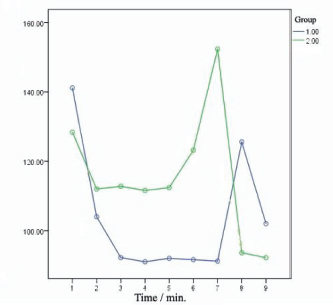
Fig. 3. Vital parameters of the electroacupuncture. Table 2. Hematological changes.
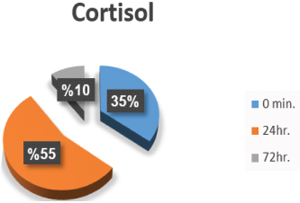
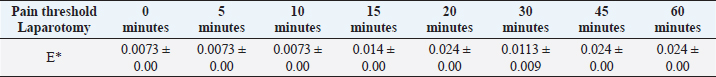
Fig. 4. Biochemical parameters of the electroacupuncture Group 1 (ALT), Group 2 (AST). Biochemical findingsThe ALT (41.2 ± 18.6) and AST (123.3 ± 46.5) showed no significance (Fig. 4). Hormonal findingsThe cortisol was significantly increased (7.36 ± 1.00) (Fig. 5). Pain threshold findingsThe pain threshold showed significance from (0.007 ± 0.00) to (0.02 ± 0.00) (Table 3). Histopathological findings of wound healingAt 7 days post wounding; the inflammatory reaction was moderate and characterized by polymorphonuclear cells and macrophages infiltration with the presence of small scab covering the wound surface. Early evidence of epithelization was detected and represented by the proliferation of epidermal epithelium under the scab. There was marked thickening and hyperplastic proliferation of epidermal epithelium at the free wound edge (Fig. 6). The dermal tissue underlying the wound area showed few polymorphonuclear cells infiltration and more abundant mononuclear cells. Fibroplasia and angiogenesis were detected in the dermis and more extensive in deeper areas of the dermis which characterized by fibroblast and angioblast proliferation forming immature young granulation tissue (Fig. 7). At 14 days post wounding, the wound area was covered by a complete layer of epidermal epithelium indicating a good epithelization rate of the wound. Proliferation of cutaneous appendages was detected. The newly formed epithelium was thick and showed partial keratinization and epithelial differentiated stratum containing polyhedral keratinocyte with the appearance of keratohyalin granules in their cytoplasm (Fig. 8). The dermal tissue showed deposition of parallel well organized and interconnected collagen bundles that arranged parallel to epidermis associated with enhanced angiogenesis represented by well-developed blood capillaries (Fig. 9). At 21 days post wounding, remodeling of the formed epithelium with relatively small scar formation and mild restoration of skin appendages. Keratinized and differentiated stratum comprising the covering epithelium and the newly formed skin appendages from the basal epidermal epithelium were detected (Fig. 10). The dermal tissue revealed well-formed organized tissue consisted of parallel compact and interconnected bundles of collagen with scant angiogenesis (Fig. 11). At 28 days post wounding, well-formed scar tissue covering highly cellular organized tissue was detected (Fig. 12). The dermal tissue revealed well-arranged compact collagen bundles containing scarce blood vessels (Fig. 13).
Fig. 5. Percentage of cortisol levels the electroacupuncture. Table 3. Pain threshold values of the electroacupuncture of anaesthesia for laparotomy (M ± SD).
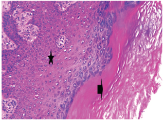
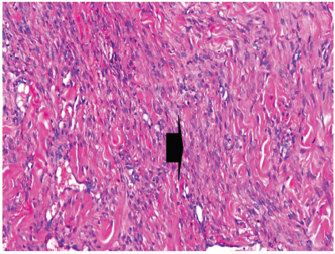
Fig. 6. Skin of a goat from electroacupuncture group at 7 days post wounding showing early evidence of epithelization, hyperplastic proliferation of epidermal epithelium (arrow head) at the free wound edge, and moderate inflammatory cells infiltrations in the dermis (arrow), (HE ×100).
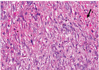
Fig. 7. Skin of a goat from electroacupuncture group at 7 days post wounding showing fibrin exudation at wound surface with dermal edema, hemorrhages (arrow) with moderate mononuclear cells infiltration associated with formation of immature granulation tissue consisted of fibroblast and angioblast forming small blood channels (arrow head), (HE ×200).
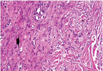
Fig. 8. Skin of a goat from electroacupuncture group at 14 days post wounding showing enhanced reepithelization evidenced by differentiated polyhedral keratinocytes in the stratum spinosum and keratohyalin granules in the stratum granulosum (arrow) and keratinization (arrow head), (HE ×100).
Fig. 9. Skin of a goat from electroacupuncture group at 14 days post wounding showing well organized and interconnected collagen bundles that arranged parallel to each other and to epidermis (arrow head) associated with enhanced angiogenesis represented by well-developed blood capillaries (arrow), (HE ×200).
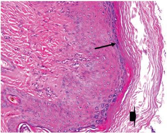
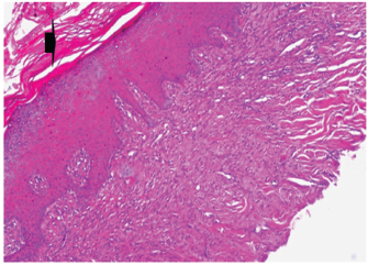
Fig. 10. Skin of a goat from electroacupuncture group at 21 days post wounding showing keratinized (arrow head) and differentiated stratum comprising the covering epithelium (*) and the newly formed skin appendages from the basal epidermal epithelium (HE ×200).
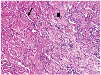
Fig. 11. Skin of a goat from electroacupuncture group at 21 days post wounding showing well-formed organized tissue consisted of parallel compact and interconnected bundles of collagen with scant angiogenesis (arrow head) (HE ×200). DiscussionThe goats of the present study showed statistically significance interactions concerned with rates of eyelid closure [F(4.69, 28.15)=38.99, p < 0.0001], head and neck relaxation [F(5.60, 33.60)=47.87, p < 0.0001], rumen motility [F(2, 12)=24.52, p < 0.0001], salivation [F(2, 12)=49.46, p < 0.0001], teeth grinding [F(2.15, 12.92)=15.36, p < 0.0001], and tympany [F(2.95, 17.67)=23.59, p < 0.0001]. However, there was non-significant interactions between the groups and the overtime rates of tongue protrusion [F(2, 12)=1.00, p=0.39]. Laparotomy blood test showed statistically significant interactions between the goats and the overtime changes of respiratory rates [F(4.49, 26.93)=16.27, p < 0.0001], ALT levels [F(5.19, 30.99)=4.28, p-value=0.004], AST levels [F(3.17, 19.03)=15.36, p < 0.0001], hematocrit [F(5.62, 33.74)=18.03, p < 0.0001], hemoglobin concentrations [F(7.41, 44.47)=2.27, p=0.043], RBCs counts [F(8.04, 48.21)=5.67, p < 0.0001], WBCs counts [F(4.83, 28.96)=2.95, p=0.030], lymphocytes [F(5.5, 32.97)=3.23, p=0.015], monocytes [F(4.11, 24.68)=6.41, p=0.001], segmented cells [F(4.02, 24.14)=3.32, p=0.027], SPO2 [F(5.04, 30.25)=4.498, p=0.003], and cortisol levels [F(2.66, 15.98)=7.1, p=0.004]. However, there were non-significant interactions between the goats and the overtime changes of body temperatures [F(3.25, 15.90)=2.88, p=0.059], heart rates [F(2.80, 16.77)=1.52, p=0.246], capillary refill times [F(2.65, 15.87)=0.461, p=0.691], eosinophils [F(6.61, 39.64)=14.74, p=0.711], platelets counts [F(3.38, 20.27)=1.95, p=0.149], and staff cells [F(66.26, 13.37)=1.165, p=0.347]. There was a statistically significant interaction between the goats and the overtime changes of pain threshold [F (2.32, 13.9)=37.9, p < 0.0001].
Fig. 12. Skin of a goat from electroacupuncture group at 28 days post wounding showing well-formed scar tissue (arrow head) covering highly cellular organized tissue (HE ×100).
Fig. 13. Skin of a goat from electroacupuncture group at 28 days post wounding showing well arranged compact collagen bundles (arrow head) containing scarce blood vessels (HE ×200). The physiological variables in laparotomy have statistically significant interactions between the goats (Qu, et al. 1996; Han, 1998). Electroacupuncture showed a significant rise in the rectal temperature, it may be due to initial apprehension of the animal for the needles and the current that believed to have been mediated at the hypothalamic level, and increased basal metabolic rate of animals. The antifebrile effect is believed to have been mediated at the hypothermic level (Rogers and Bossy, 1981). Nevertheless, electroacupuncture maintains sufficient excitation of the central nervous system (Han, 1998; Latimer et al., 2004). However, laparotomy showed a gradual reduction in temperature that may have been attributable to a decrease in skeletal muscle tone, a decrease in metabolic rate, or muscle relaxation along with depression of thermoregulatory centers (Durga Prasanna Babu, 1988). Heart rate in laparotomy is superior, this parasympathetic stimulation affects the cardiovascular system (Lee et al., 1974) which led to a transient fall in the blood pressure and consequent rise of the heart rate. But the electroacupuncture maintains heart rate normal till the end of surgery as mentioned by Sali (2010) and Shah et al. (2016). The respiratory rate of laparotomy is superior, this rise might be attributed to initial apprehension of the animal to the needles and current, and also due to increased heart rate (Wright and McGrath, 1981). In the present study, it was found that the significant increase in hemodynamic led to rosy red CRT. The goats exhibited significant increase in hematocrit, and gradual increase of red blood cell, which stated that acupuncture increases red blood cell, and platelets were significantly increase group Secondary or reactive thrombocytosis assisted in blood clotting (Habibu et al., 2017). The WBC count and staff cells showed is non-significant. Electroacupuncture also can suppress the production of adrenocortical hormones in response to stress Electroacupuncture-induced transient changes in the present study may have been associated with constriction of the splenic sinuses are similar to that reported in normal goats (Egbe-Nwiyi et al., 2000). The significant increases of blood indices confirmed the data published in the National Institutes of Health Conference which stated that acupuncture increases red blood cells and treats thrombocytopenia, and increases immunity, also corresponding with World Health Organization recommendations that endorse acupuncture for treating leukopenia (Pais et al., 2014). The study found increased levels of hematocrits, granulocytes, and monocytes before and after treatment, but this difference is not significant and also, the increases of hematological variables are within the normal values by the study of (Aiello et al., 2016). Eosinophils in laparotomy was not significant and maintain it until the end of the surgery, this helps the movement to inflamed areas, trapping substances, killing cells, anti-parasitic, and bactericidal activity, participating in immediate allergic reactions, and altering inflammatory responses (Murphy and Weaver, 2016). In laparotomy showed no significance. The hemoglobin concentration was significantly high based on the results of hematologic evaluations, there were no changes in hemoglobin concentration for treated goats (Shah et al., 2016). SPO2 levels in laparotomy were significantly high. The increased permeability induced by oxygen in the alveolar-capillary wall as a development that increased permeability of the endothelial cell lining is needed before circulating antibodies, the oxyhemoglobin dissociation curve describes the relationship between the partial pressure of oxygen and the percent of hemoglobin saturated with oxygen and varies with chemical and physical (Burk and Orr, 2020). ALT and AST are the levels of liver enzymes. It plays a crucial role in metabolism, the process that turns food into energy (Watkins et al., 2006). When elevated ALT levels are found in the blood, the possible underlying causes can be further narrowed down by measuring other enzymes. The ALT and AST levels in laparotomy were not significantly different (Shah et al., 2016). The significant increases in serum cortisol levels after the end of the therapy are attributed to its effect on the central and peripheral nervous systems to activate the body's endogenous or natural pain relief mechanisms. It stimulates the release of several neurotransmitters, chemical secretions that cause a favorable reaction in another nerve, a muscle, or a gland. It also causes a release of β-endorphins and adrenocorticotropic hormone from the pituitary gland that induces the release of cortisol from the adrenal gland; this is in agreement with (Cadoy and Acorda, 2018). In the study, cortisol level was significantly high. Also, the cortisol levels are attributed to its effect on the central and peripheral nervous systems to activate the body endogenous or natural pain relief mechanisms. It stimulates the release of several neurotransmitters, chemical secretions that cause a favorable reaction in another nerve, a muscle, or a gland. It also causes a release of β-endorphins and adrenocorticotropic hormone from the pituitary gland that induces the release of cortisol from the adrenal gland (Padilha et al., 2017). Moreover, Cortisol regulates a wide range of vital processes throughout the body, including metabolism and the immune response. It also has a very important role in helping the body respond to stress (Ramamoorthy and Cidlowski, 2016). Several investigators measured the pain threshold in goats with an algesimetry method based on a leg-lifting response to a subcutaneous electric stimulus. This method is not an involuntary reflex but represented cognitive behavior. Additionally, this technique could not be used for restrained animals. Potassium iontophoresis is a convenient and reliable pain stimulus that can be provided rapidly and repeatedly with minimal loss inconsistency of a subject’s reported pain level (Humphries et al., 1994), this quantitative method is used to measure the pain thresholds. Electroacupuncture induced is sufficiently strong to completely block sharp, acute pain attributable to major surgery in some individuals (Han, 2015). The present work showed high significance in pain threshold which was induced by ACTCH that stimulated production of cortisol which acted as anti-inflammatory and painkilling. The evidence of electroacupuncture analgesia using 10 newly chosen acupoints can produce sufficient analgesia for right flank laparotomy in goats. Electroacupuncture showed improvement in the rates of eyelid closure, head and neck relaxation, rumen motility, and tympany. The respiratory rates, body temperatures, and capillary fill times increase significance. Hematocrit, Hemoglobin, RBCs, Platelets, and SPO2 were significant. ALT and AST showed no significance. The cortisol and pain threshold showed a significant difference. The healing of wounds in electroacupuncture is improved. At 7 days, the electroacupuncture group was characterized by early epithelization and immature granulation tissue. At 14 days, covered by thick epithelization and keratinization. The dermal tissue showed the deposition of collagen and enhanced angiogenesis. At 21 days, mild restoration of skin, and the dermal tissue was a well-organized tissue, at 28 days, showed well-formed scar tissue covering highly cellular organized dermal tissue, as previously mentioned by (Wang et al., 2009) who stated that acupuncture enhances wound healing accelerators such as fibroblast growth factors and platelet-derived growth factors in experimental models. ConclusionElectroacupuncture anesthesia using the 10 newly selected acupoints and two-parallel needles, at the surgery area, can produce sufficient analgesia to the flank laparotomy in goats. Electroacupuncture works more potent and achieves sufficient abdominal anesthesia suitable for goat abdomen surgery superior. Also, the wound healing in laparotomy goats proved excellent and better healing with electroacupuncture regime. AcknowledgmentsThis study is a part of the PhD thesis of Khalifa Ashour. Associate Prof. Dr. Alshimaa Ismal helped with preparation of statistical analyses from the Faculty of Veterinary Medicine, Cairo University, Egypt. Authors would like to thank Anas Ibrahim Otify for her help in preparation of figures and tables. Conflict of interestThe authors declare that there is no conflict of interest. ReferencesAiello, S.E., Moses, M.A. and Allen, D.G. 2016. The Merck veterinary manual Merck, pp: 3174–3185. Asayama, K., Ohkubo, T., Metoki, H., Obara, T., Inoue, R., Kikuya, M., Thijs, L., Staessen, J.A. and Imai, Y. 2012. Hypertension Objective Treatment Based on Measurement by Electrical Devices of Blood Pressure (HOMED-BP). Cardiovascular outcomes in the first trial of antihypertensive therapy guided by self-measured home blood pressure. Hypertens. Res. 35(11), 1102–1110. Bancroft, D., Stevens, A. and Turner, R. 2012. Theory and practice of histological technique, 4th ed, Edinburgh, London, Melbourne: Churchill Livingstone. Burk, K.M. and Orr, J.A. 2020. A procedure for determining subject-specific pulse oxygen saturation response. Med. Biol. Eng. Comput. 58(4):753–761. Cadoy, J.M. and Acorda, J.A. 2018. Physiological responses, cortisol level and analgesia in goats subjected to exploratory laparotomy under acupuncture and regional analgesia. J. Vet. Med. 55(1), 35–44. Durga Prasanna Babu, V. 1988. Studies on acupuncture anaesthesia in sheep with particular reference to abdominal cavity. M.V.Sc thesis submitted to Andhra Pradesh Agricultural University, Hyderabad. Egbe-Nwiyi, T.N., Nwaosu, S.C. and Salami, H.A. 2000. Haematological values of apparently healthy sheep and goats as influenced by age and sex in arid zone of Nigeria. Afr. J. Biomed. Res. 3(2), 109–115. Groppetti, D., Pecile, A.M., Sacerdote, P., Bronzo, V. and Ravasio, G. 2011. Effectiveness of electroacupuncture analgesia compared with opioid administration in a dog model: a pilot study. Br. J. Anaesth. 107(4), 612–618. Habibu, B., Abdullahi, A., Yaqub, L.S., Makun, H.J. and Kawu, M.U. 2017. Variations in platelet count and total protein in relation to differences in sex, age, breed and reproductive status of goats during the cold-dry season (Harmattan). J. Dairy Vet. Anim. Res. 5(3), 00141. Han, J.S. 1998. The neurochemical basis of pain relief by acupuncture, Vol. 2. Hubei, China: Hubei Science and Technology Press. Han, J.S. 2015. Acupuncture. In Treatment of chronic pain by integrative approaches. New York: Springer, pp: 123–136. Humphries, S.A., Long, N.R. and Johnson, M.H. 1994. Iontophoretically applied potassium ions as an experimental pain stimulus for investigating pain mechanisms. Percept Psychophys. 56(6), 637–648. Janssens, L.A., Rogers, P.A. and Schoen, A.M. 1988. Acupuncture analgesia: a review. Vet. Rec. 122(15), 355–358. Latimer, J., Maher, C. and Refshauge, K. 2004. The attitudes and beliefs of physiotherapy students to chronic back pain. Clin. J. Pain 20(1), 45–50. Lee, D.C., Lee, M.O. and Clifford, D.H. 1974. Cardiovascular effects of acupuncture in anesthetized dogs. Am. J. Chin. Med. 2(03), 271–282. Murphy, K. and Weaver, C. 2016. Janeway's immunobiology. New York: Garland science. Padilha, F.G.F., Dimache, L.A.G., Almeida, F.Q.D. and Ferreira, A.M.R. 2017. Blood biochemical parameters of Brazilian sport horses under training in tropical climate. Rev. Bras. de Zootec. 46(8), 678–682. Pais, I., Correia, N., Pimentel, I., Teles, M.J., Neves, E., Vasconcelos, J. and Efferth, T. 2014. Effects of acupuncture on leucopenia, neutropenia, NK, and B cells in cancer patients: a randomized pilot study. Evid. Based Complement Alternat. Med. 2014, 217397. doi: 10.1155/2014/217397. Parmen, V. 2014. Electroacupuncture analgesia in a rabbit ovariohysterectomy. J. Acupunct. Meridian. Stud. 7(1), 15–24. Pugh, D.G., Baird, N.N., Edmondson, M. and Passler, T. 2020. Sheep, goat, and cervid medicine-E-Book. Amsterdam, The Netherlands: Elsevier Health Sciences. Qu, G.L., Zhuang, X.L., Xu, G.H., Yang, L.F., Wang, Z.T., Chen, S.L. and Xie, T. 1996. Clinical observation on combined anesthetics acupuncture anesthesia in 50 patients undergoing renal transplantation. Chin. J. Pain Med. 2, 72–77. Ramamoorthy, S. and Cidlowski, J.A. 2016. Corticosteroids: mechanisms of action in health and disease. Rheum. Dis. Clin. North Am. 42(1), 15–31. Rogers, P.A.M. 1995. Acupuncture analgesia for surgery in animals. In Acupuncture in animals. Ed., Bryden D.I. Sydney, Australia: University of Sydney, pp: 341–352. Rogers, P.A.M. and Bossy, J. 1981. Activation of the defense system of the body in animals and man by acupuncture and moxibustion. Acupunt. Res. Q. 5, 47–54. Sali, K.M. 2010. Determination of anatomical location to the parts of female genital system of Awassi ewes by using laparoscopy. J. Vet. Anat. 3(2), 47–54. Shah, Z., Hu, M.L., Qiu, Z.Y., Zhou, F.Y., Zeng, J., Wan, J. and Ding, M.X. 2016. Physiologic and biochemical effects of electroacupuncture combined with intramuscular administration of dexmedetomidine to provide analgesia in goats. Am. J. Vet. Res. 77(3), 252–259. Sheta, E., Ragab, S., Farghali, H. and EL-Sherif, A. 2015. Successful practice of electroacupuncture analgesia in equine surgery. J. Acupunct. Meridian Stud. 8(1), 30–39. Tian, J.H., Xu, W., Zhang, W., Fang, Y., Grisel, J.E., Mogil, J.S., Grandy, D.K. and Han, J.S. 1997. Involvement of endogenous orphanin FQ in electroacupuncture-induced analgesia. Neuroreport 8, 497–500. Wang, D.W. and Jin, Y.H. 1989. Present status of cesarean section under acupuncture anesthesia in China. Fukushima J. Med. Sci. 35(2), 45–52. Wang, X.Y., Li, X.L., Hong, S.Q., Xi-Yang, Y.B. and Wang, T.H. 2009. Electroacupuncture induced spinal plasticity is linked to multiple gene expressions in dorsal root deafferented rats. J. Mol. Neurosci. 37(2), 97–110. Watkins, P.B., Kaplowitz, N., Slattery, J.T., Colonese, C.R., Colucci, S.V., Stewart, P.W. and Harris, S.C. 2006. Aminotransferase elevations in healthy adults receiving 4 grams of acetaminophen daily: a randomized controlled trial. J. Acupunct. Meridian Stud. 296(1), 87–93. Wright, M. and McGrath, C.J. 1981. Physiologic and analgesic effects of acupuncture in the dog. J. Am. Vet. Med. Assoc. 178(5), 502–507. | ||

| How to Cite this Article |
| Pubmed Style Ashour K, Awad NAE, Abdelgayed SS, Leil AZA, Sheta E. ELECTROACUPUNCTURE ANAESTHESIA FOR LAPAROTOMY IN GOATS. Open Vet J. 2021; 11(1): 52-60. doi:10.4314/ovj.v11i1.9 Web Style Ashour K, Awad NAE, Abdelgayed SS, Leil AZA, Sheta E. ELECTROACUPUNCTURE ANAESTHESIA FOR LAPAROTOMY IN GOATS. https://www.openveterinaryjournal.com/?mno=25036 [Access: July 15, 2025]. doi:10.4314/ovj.v11i1.9 AMA (American Medical Association) Style Ashour K, Awad NAE, Abdelgayed SS, Leil AZA, Sheta E. ELECTROACUPUNCTURE ANAESTHESIA FOR LAPAROTOMY IN GOATS. Open Vet J. 2021; 11(1): 52-60. doi:10.4314/ovj.v11i1.9 Vancouver/ICMJE Style Ashour K, Awad NAE, Abdelgayed SS, Leil AZA, Sheta E. ELECTROACUPUNCTURE ANAESTHESIA FOR LAPAROTOMY IN GOATS. Open Vet J. (2021), [cited July 15, 2025]; 11(1): 52-60. doi:10.4314/ovj.v11i1.9 Harvard Style Ashour, K., Awad, . N. A. E., Abdelgayed, . S. S., Leil, . A. Z. A. & Sheta, . E. (2021) ELECTROACUPUNCTURE ANAESTHESIA FOR LAPAROTOMY IN GOATS. Open Vet J, 11 (1), 52-60. doi:10.4314/ovj.v11i1.9 Turabian Style Ashour, Khalifa, Naglaa Abd Elkader Awad, Sherein Saeid Abdelgayed, Amal Zakaria Ahmed Leil, and Eldessouky Sheta. 2021. ELECTROACUPUNCTURE ANAESTHESIA FOR LAPAROTOMY IN GOATS. Open Veterinary Journal, 11 (1), 52-60. doi:10.4314/ovj.v11i1.9 Chicago Style Ashour, Khalifa, Naglaa Abd Elkader Awad, Sherein Saeid Abdelgayed, Amal Zakaria Ahmed Leil, and Eldessouky Sheta. "ELECTROACUPUNCTURE ANAESTHESIA FOR LAPAROTOMY IN GOATS." Open Veterinary Journal 11 (2021), 52-60. doi:10.4314/ovj.v11i1.9 MLA (The Modern Language Association) Style Ashour, Khalifa, Naglaa Abd Elkader Awad, Sherein Saeid Abdelgayed, Amal Zakaria Ahmed Leil, and Eldessouky Sheta. "ELECTROACUPUNCTURE ANAESTHESIA FOR LAPAROTOMY IN GOATS." Open Veterinary Journal 11.1 (2021), 52-60. Print. doi:10.4314/ovj.v11i1.9 APA (American Psychological Association) Style Ashour, K., Awad, . N. A. E., Abdelgayed, . S. S., Leil, . A. Z. A. & Sheta, . E. (2021) ELECTROACUPUNCTURE ANAESTHESIA FOR LAPAROTOMY IN GOATS. Open Veterinary Journal, 11 (1), 52-60. doi:10.4314/ovj.v11i1.9 |

