





| Research Article | ||
Open Vet. J.. 2026; 16(8): 5850-5862 !
Open Veterinary Journal, (2026), Vol. 16(8): 5850–5862 Research Article Size-dependent reproductive toxicity of zinc oxide nanoparticles in adult male rats: Alterations in sperm quality, steroidogenesis, and testicular microvasculatureMayan A. Saleem1, Zeravan A. Mohammed1* and Shayma Z. Ameen21Department of Veterinary Clinical Sciences, College of Veterinary Medicine, University of Duhok, Duhok, Kurdistan, Iraq 2Department of Physiology and Anatomy, College of Veterinary Medicine, University of Duhok, Duhok, Iraq *Corresponding Author: Zeravan A. Mohammed. Department of Veterinary Clinical Sciences, College of Veterinary Medicine, University of Duhok, Duhok, Iraq. Email: Zeravan.mohammed [at] uod.ac Submitted: 23/06/2026 Revised: 27/07/2026 Accepted: 03/08/2026 Published: 20/08/2026 © 2025 Open Veterinary Journal
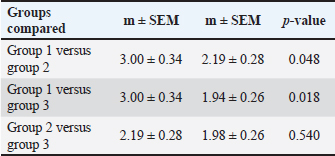
AbstractBackground: Zinc oxide nanoparticles (ZnO Np) are widely used in industrial and biomedical applications; however, their potential reproductive toxicity remains a concern. Aim: This study investigated the size-dependent effects of ZnO Np (20 and 40 nm) on male reproductive function in adult rats, focusing on sperm characteristics, hormonal profile, testicular histology, and immunohistochemical markers. Methods: Forty-five adult male albino rats were randomly divided into three groups (n=15/group): group 1 (control), 20-nm ZnO Np-treated, and 40-nm ZnO Np-treated groups. ZnO Np was orally administered at 100 mg/kg body weight/day for 21 consecutive days. Body and testicular weights, sperm motility and morphology, and serum testosterone levels (Cobas 6000) were assessed. Testicular tissues were examined by H&E staining and immunohistochemistry for Ki-67, Bcl-2, and von Willebrand factor (vWF). Data were analyzed using one-way analysis of variance followed by Tukey’s post hoc test (p < 0.05). Results: ZnO NP exposure significantly impaired reproductive parameters, resulting in reduced sperm motility, altered sperm morphology, and decreased serum testosterone levels compared with controls (p < 0.05). Histopathological examination revealed seminiferous tubule degeneration, spermatogenic cell depletion, germinal epithelium disruption, and vascular alterations in the treated groups. Quantitative immunohistochemical analysis (QIHC) demonstrated a significant reduction in Ki-67 expression, indicating decreased germ cell proliferative activity, with the lowest expression observed in the 40-nm ZnO NP group (9.81% ± 0.73%) compared with the control group (35.84% ± 1.27%) (p < 0.001). In contrast, Bcl-2 immunoreactivity was significantly increased following ZnO NP exposure, reaching the highest level in the 40 nm ZnO NP group (31.47% ± 1.12%) compared with controls (8.72% ± 0.61%) (p < 0.001), suggesting activation of cellular survival responses under nanoparticle-induced stress. Furthermore, vWF expression was significantly altered, with the highest immunoreactivity observed in the 20 nm ZnO NP group (47.6% ± 1.9%) compared with controls (20.4% ± 1.5%) (p < 0.001), indicating changes in testicular microvascular responses. Conclusion: ZnO nanoparticles caused size-dependent reproductive toxicity in male rats, affecting sperm quality, testosterone levels, testicular structure, proliferation, apoptosis regulation, and vascular integrity. Keywords: Rats, Reproductive toxicity, Sperm motility, Testosterone, Zinc oxide nanoparticles. IntroductionNanotechnology is rapidly expanding in the medical, agricultural, cosmetic, food packaging, and pharmaceutical sectors. Zinc oxide nanoparticles (ZnO Np) are widely used because of their antibacterial activity, UV-blocking properties, chemical stability, and cost-effectiveness (Sharma et al., 2024). They are commonly incorporated into sunscreens, drug delivery systems, dietary supplements, and veterinary products. However, their increasing production and environmental exposure have raised concerns regarding their potential toxicity, particularly their adverse effects on the male reproductive system and fertility (Pinho et al., 2020). Due to their small size (1–100 nm), ZnO Np possess a high surface area and enhanced biological reactivity, enabling them to cross biological barriers and accumulate in organs, including the liver, kidneys, brain, and reproductive tissues (Wang et al., 2008; Ma et al., 2013). Their accumulation induces excessive reactive oxygen species (ROS) generation, leading to oxidative stress, mitochondrial dysfunction, inflammation, and apoptosis (Sharma et al., 2012; Goma et al., 2021). The male reproductive system is particularly susceptible to oxidative damage due to the rapid proliferation of spermatogenic cells and the high content of polyunsaturated fatty acids in sperm membranes (Pinho et al., 2020). Excessive ROS impairs sperm motility, morphology, viability, membrane integrity, and testosterone production (Agarwal et al., 2014). Previous studies have shown that ZnO NP exposure reduces sperm quality and testosterone levels and induces histopathological alterations in the testes (Ma et al., 2013; Goma et al., 2021; Hong et al., 2022; Sun et al., 2025). Although smaller NPs generally exhibit greater cellular uptake and biological activity than larger particles (Goma et al., 2021), comparative studies evaluating the reproductive effects of different ZnO NP sizes using integrated hormonal, spermatological, histopathological, and immunohistochemical analyses remain limited. Ki-67 is a cell proliferation marker (Scholzen and Gerdes, 2000), whereas Bcl-2 regulates apoptosis (Cory and Adams, 2002). von Willebrand factor (vWF) is an endothelial marker used to evaluate testicular microvascular integrity (Wagner and Frenette, 2008). Therefore, this study aimed to compare the size-dependent effects of 20- and 40-nm ZnO nanoparticles on male reproductive function in adult rats. Materials and MethodsMaterialsZnO Np, with dimensions of 20–40 nm and a virtually spherical form akin to white powder, were obtained from the Danish Bonyan Company and commercially purchased by the University of Duhok. The nanoparticle powder was measured in a controlled setting based on the weight of the rats and then placed in a 5 ml Eppendorf tube (Germany) dissolved in distilled water, then mechanically agitated for 10–15 minutes to improve dispersion and minimize clumping. The experiments were conducted in January 2025. Regarding the adsorption of the nanoparticles to the walls of the dosing tubes, ZnO Np were freshly suspended immediately before administration and delivered by oral gavage; therefore, adsorption to the gavage equipment was not assessed. AnimalsUpon arrival, the rats were acclimatized to the animal facility for 7 days under standard laboratory conditions (temperature, 21°C–25°C; relative humidity, 40%–70%; and a 10:14 hours light/dark cycle) with free access to standard laboratory chow and water before the initiation of the experiment. This experimental study was conducted using 45 clinically healthy adult male albino rats aged10–12 weeks, with body weights ranging from 268.67 ± 34.8g. The animals were obtained from the animal research facility of the College of Veterinary Medicine, University of Duhok, and housed in individual cages. Environmental conditions were maintained under controlled temperature (21°C–25°C), relative humidity (40%–70%), and a 10:14-hour light/dark cycle. The rats were provided with food and water ad libitum throughout the experimental period. All animals were allowed to acclimatize to laboratory conditions for at least 30 minutes before the initiation of the experiment. Study designThe experimental animals were randomly allocated into three groups (n=15 per group): group 1 (Control), which received distilled water by oral gavage once daily for 21 days; group 2 (20-nm ZnO Np), which received zinc oxide nanoparticles (20 nm) at a dose of 100 mg/kg body weight/day by oral gavage for 21 consecutive days; and group 3 (40-nm ZnO Np), which received zinc oxide nanoparticles (40 nm) at the same dose (100 mg/kg body weight/day) by oral gavage once daily for 21 consecutive days. Dose and nanoparticle size selection rationaleThe dose of 100 mg/kg/day was selected based on previous studies demonstrating reproducible reproductive toxicity without excessive systemic toxicity. ZnO nanoparticles (20 and 40 nm) were chosen to evaluate size-dependent toxicity, as differences in particle size influence cellular uptake and biological reactivity (Hong et al., 2022; Ahmed and El-Sayed, 2025; Ahmed et al., 2025). Body weight and hormone assayThe body weights of all experimental rats were recorded at the beginning of the study, prior to treatment administration, and again at the end of the experimental period using a precision digital laboratory balance (Sartorius). All measurements were documented for subsequent statistical analysis. At the end of the experiment, the rats were anesthetized using CO2 inhalation (Turner et al., 2020). Blood samples (4–5 ml) were collected using capillary tubes from the medial canthus of the eye. The collected blood was centrifuged at 2,000–3,000 rpm for 10 minutes to obtain serum. The separated serum was carefully aspirated and stored at −20°C until use in hormonal analysis. Hormonal assayThe Cobas 6000 analyzer (Roche Diagnostics, Mannheim, Germany) was used to quantify the serum testosterone concentrations. Hormonal assays were performed using ECLIA equipment following the manufacturer’s instructions. The analyzer was calibrated prior to the analysis, and internal quality controls were implemented to ensure the accuracy of the assay. The system software autonomously calculates hormone concentrations, displaying them in nmol/l for testosterone. Sperm analysisEpididymal sperm from each rat was obtained immediately by cutting the cauda epididymis and vas deferens to squeeze the sperm content into a sterile Petri dish containing 1 ml of isotonic saline at 37°C. Sperm viability, count, motility, and morphology were evaluated. Sperm motility was determined as the proportion of progressive and non-progressive spermatozoa, with motile sperm (robust and fastest swimming sperm directed straight lines) and non-progressive motile (the motion of the sperm forward tends to be curved or twisted) and immotile sperm (non-movable sperm at all) in the same field using a light microscope. The motility and deviations of the spermatozoa cells were estimated according to a previous study (Adamkovicova et al., 2016). Thin smears of sperm suspension were obtained from experimental groups of animals afterward, and 1 volume was mixed with 2 volumes of 1% eosin and nigrosin stain. For each smear, approximately 400 sperms/animals were counted (Khafaji, 2023). The sperm morphological study was conducted using a sperm smear on a glass slide, which was air-dried and then fixed with absolute methanol. The slides were then stained with 1% aqueous Eosin-Nigrosin solution lastly mounted with Canada balsam, and covered with coverslips. Under a light microscope, the abnormalities of the sperm in the head and tail and the changes in the midpiece of the sperm were recorded and photographed using a digital microscopic camera at 40 magnification. Furthermore, sperm viability was determined as live sperm showing non-stained sperm, while dead sperm showing stained sperm were evaluated among the 200 sperm. Then, the ratio of sperm motility was calculated and statistically analyzed. Histopathological examinationTestes from each animal were dissected and fixed in 10% neutral-buffered formalin for 48 hours, cleaned, dehydrated, clarified, and then embedded in paraffin wax. Then, 5-µm-thick paraffin sections were stained with hematoxylin and eosin (Suvarna et al., 2018). The embedded tissues were obtained using a microtome (Leica RM 2245, Germany). The sections were then floated onto water (40ºC) onto Superfrost Plus, positively charge slides. The sections were then placed on a hot plate (50ºC) to dry overnight (Suvarna et al., 2018). A light microscope was used to study the stained slide, and images were captured using a Bresser Art. No. 5722100 microscope, equipped with a Swift 18.0 MP USB digital camera (code SC 1803R). ImmunohistochemistryFor immunohistochemical analysis, the paraffin-embedded sections were deparaffinized and rehydrated in a descending alcohol concentration. To perform heat-induced antigen retrieval, the sections were immersed in citrate buffer (pH 6.0) and boiled 3 times for 5 minutes each using a microwave oven at 700 W. Blocking was achieved by incubating tissue in 3% (H2O2) solution in methanol for 10 minutes at room temperature. Sections were washed with phosphate-buffered saline (PBS) for 10 minutes and blocked with 1% bovine serum albumin for 30 minutes at room temperature to block nonspecific primary antibody. Ki-67, monoclonal mouse–anti-human antigen (Clone MIB-1, Code No. M7240, Dako, Glostrup, Denmark), diluted 1:150 in PBS, was used to determine the proliferation index. Bcl-2 monoclonal mouse–anti-human (Clone 124, Code Number IR124, Dako, Glostrup, Denmark), diluted 1:150 in PBS. For VWF, polyclonal Rabbit Anti-Human VWF (Code Number A0082, Dako, Glostrup, Denmark), diluted 1:200 in PBS, was used. After incubation with primary antibody, the sections were washed with PBS (2 times). The Ki-67 sections were treated with rabbit anti-mouse IgG HRP-tagged secondary antibodies diluted 1:150 in PBS. The antibodies were applied to the slides and incubated at room temperature for 30 minutes. Subsequently, 2 rinses with PBS were performed, each lasting 5 minutes (Mohammed, 2016). The antibody binding sites were visualized using 3,3′-diaminobenzidine solution. The tissue sections were counterstained with hematoxylin and eosin for 20 s at RT. Images were captured using a Bresser Art camera. No. 5722100 ImageJ analysis software (Digital Camera18.0) MP USB) for each slide (Mohammed, 2016). For quantification, the average antibody expression was measured in 20 seminiferous tubules for each section. Blinding of the outcome assessmentSperm analysis, histopathological examination, and immunohistochemical evaluation were performed by investigators who were blinded to the treatment allocation to minimize observer bias. Tissue sections and sperm samples were coded before analysis, and group identities were revealed only after all observations and quantitative measurements were completed. Quantitative immunohistochemical analysisImmunohistochemical expression of Ki-67, Bcl-2, and vWF was quantified using ImageJ software (version 1.54, National Institutes of Health, Bethesda, MD, USA). Digital photomicrographs were captured under identical microscope and camera settings at ×400 magnification. Five randomly selected nonoverlapping fields from each tissue section were analyzed for each animal. Statistical analysesStatistical analyses were performed using Genstat 19.0 (VSN International, UK). Shapiro–Wilk and Levene’s tests were used to determine data normality and homogeneity. One-way ANOVA and Tukey’s post hoc test were used to compare groups. The independent samples t-test was used for certain pairwise comparisons. The pairwise chi-square test was used to determine the percentage of sperm motility among the groups. Results are presented as mean ± SEM, with significant differences at p < 0.05. Superscript letters (a–c) indicate significant group differences. Ethical approvalEthical approval for this study was obtained from the College of Veterinary Medicine, University of Duhok (Rerf. No. CVM2025/0201UoD). ResultsThe comparative statistical analysis demonstrated that exposure to ZnO Np significantly influenced sperm morphology in the experimental groups. Regarding normal sperm morphology, group 1 exhibited the greatest mean value (3.00 ± 0.34, p=0.048), whereas groups 2 and 3 demonstrated reduced mean values of (2.19 ± 0.28, p=0.018) and (1.94 ± 0.26, p=0.540), respectively (Table 1). The results revealed a significant difference between group 1 versus groups 2 and 3. However, a statistically insignificant difference was observed between Groups 2 and 3, as shown in Table 1. Table 1. Effect of ZnO Np on normal sperm morphology among groups.
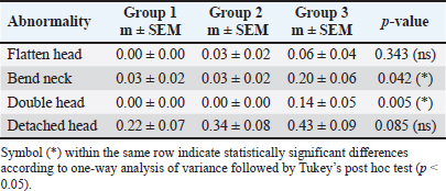
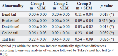
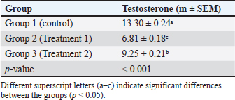
Concerning total abnormalities (Table 2), head abnormalities demonstrated a progressive rise from group 1 (0.41 ± 0.08) to group 3 (0.57 ± 0.10); however, this increase was not significantly different (p=0.248). In contrast, tail anomalies exhibited a statistically significant increase among the groups (p=0.026), with group 3 (0.60 ± 0.12) demonstrating the greatest mean value, followed by group 2 (0.51 ± 0.11), compared with group 1 (0.25 ± 0.07). This suggests that tail structures are more susceptible to damage caused by ZnO Np than head structures (Table 2). A thorough evaluation of specific head abnormalities (Table 3) clarified these variations. Although flattened head (p=0.343) and detached head (p=0.085) did not demonstrate significant differences across the groups, bent neck anomalies were substantially elevated in group 3 (0.20 ± 0.06) compared with those in groups 1 and 2 (p=0.042). The double head abnormality was exclusively observed in group 3 (0.14 ± 0.05), indicating a statistically significant difference between the groups (p=0.005). Increased exposure levels were directly associated with structural anomalies in the head and neck regions of sperm. Certain tail anomalies (Table 4) demonstrated notable disparities. Bent tail (p=0.019), double tail (p=0.015), coiled tail (p=0.039), and tailless sperm (p=0.026) demonstrated statistically significant differences among the groups. Group 2 exhibited the greatest average values for bent tail and double tail anomalies, whereas group 3 showed the most pronounced increase in coiled tail and tailless forms. The broken tail anomaly did not significantly differ (p=0.315). A significant disparity in serum testosterone levels was observed across the experimental groups (p < 0.001; Table 5). Group 1 exhibited the highest testosterone levels (13.30 ± 0.00), whereas group 2 experienced a significant decline in testosterone (6.81 ± 0.00) (Table 5). Group 3 exhibited a mean value of 9.25 ± 0.00, significantly lower than that of group 1 but considerably higher than that of group 2. A thorough evaluation of specific head abnormalities (Table 3) clarified these variations. Although flattened head (p=0.343) and detached head (p=0.085) did not demonstrate significant differences across the groups, bent neck anomalies were substantially elevated in group 3 (0.20 ± 0.06) compared with those in groups 1 and 2 (p=0.042). The double head abnormality was exclusively observed in group 3 (0.14 ± 0.05), indicating a statistically significant difference between the groups (p=0.005). Increased exposure levels were directly associated with structural anomalies in the head and neck regions of sperm. Certain tail anomalies (Table 4) demonstrated notable disparities. Bent tail (p=0.019), double tail (p=0.015), coiled tail (p=0.039), and tailless sperm (p=0.026) demonstrated statistically significant differences among the groups. Group 2 exhibited the greatest average values for bent tail and double tail anomalies, whereas group 3 showed the most pronounced increase in coiled tail and tailless forms. The broken tail anomaly did not significantly differ (p=0.315). Table 2. Effect of ZnO Np on the overall head and tail abnormalities of the sperm among the groups.
Table 3. The effect of ZnO Np on specific head abnormalities of sperm among groups.
Table 4. Effect of ZnO Np on the specific tail abnormalities of the sperms among the groups.
Table 5. The effect of ZnO Np on serum testosterone levels in male rats.
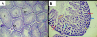
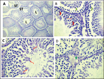
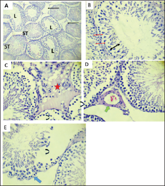
A significant disparity in serum testosterone levels was observed across the experimental groups (p < 0.001; Table 5). Group 1 exhibited the highest testosterone levels (13.30 ± 0.00), whereas group 2 experienced a significant decline in testosterone (6.81 ± 0.00) (Table 5). Group 3 exhibited a mean value of 9.25 ± 0.00, significantly lower than that of group 1 but considerably higher than that of group 2. Effects on the body weightThe results revealed significant differences in baseline body weight between groups (p=0.001, Table 6). Group 2 had a much lower mean weight (215.4 ± 8.08 g) than that of groups 1 (334.2 ± 16.60 g) and 3 (256.4 ± 23.64 g). After treatment, both experimental groups (2 and 3) showed a significant difference in body weight compared to group 1 (p=0.004). Specifically, group 2 had lower body weight (254.5 ± 10.50 g) than group 1 (342.6 ± 15.44 g), and group 3 had lower body weight (223.4 ± 22.85 g), indicating that the weight increase was stopped. Notably, there were no significant differences in the weights of the right (p=0.718) or left testis (p=0.494) between the groups. The average weights of the right testis were 2.02 ± 0.21 g (group 1), 1.98 ± 0.22 g (group 2), and 1.83 ± 0.12 g (group 3). The average weights of the left testis were 1.71 ± 0.14 g, 2.03 ± 0.37 g, and 1.86 ± 0.09 g, respectively. The results showed that the treatment had an impact on sperm motility (p < 0.001, Table 7). Group 2 had a much lower level of progressive motility (10.0%) than group 1 (55.0%) and group 3 (46.0%), indicating that sperm motility was much lower than that in group 1 (Table 7). In contrast, group 2 had the most nonprogressive motility (38.0%), group 3 had the lowest (26.0%), and group 1 had the highest (35.0%). Total motility exhibited a similar trend, with group 2 demonstrating a notable decrease (45.0%) compared to groups 1 (90.0%) and 3 (72.0%) (Table 7). HistopathologyHistological examination of testicular sections from group 1 demonstrated normal arrangement of the seminiferous tubules. They appear uniform in size and shape and are covered by a layer of stratified epithelium called germinal epithelium supported by Sertoli cells. The germinal epithelium was predominantly composed of well-organized layers of spermatogonia cells, primary and secondary spermatocytes, spermatids, and spermatozoa. The spermatogonia were the outermost layer, predominantly comprising dark-stained nuclei, resting on a thin layer of the basement membrane. The next layer, primary spermatocytes, had a large nucleus appeared to contain condensed chromatin. Spermatids and spermatozoa were detected and directed to the inferior part of the seminiferous tubules. The basement membrane appeared smooth (regular) and intact. The interstitial connective tissue was primarily composed of fibroblasts, blood vessels, and Leydig cells (Fig. 1). The testes of rats in the ZnO Np group revealed that some histological degeneration occurred in the testicular tissue compared with that in group 1. Including mild changes in the structure of the seminiferous tubules and interstitial connective tissue. Table 6. Impact of different sizes on body and testicular weight (m ± SEM).
Table 7. Size-dependent effects of ZnO nanoparticles on sperm motility percentages in adult male rats.
Fig. 1. Photomicrograph of rat testes in group 1 (A) illustrating normal histoarchitecture of seminiferous tubules (ST) with their lumens (L) occupied with sperm. Hematoxylin and eosin staining of interstitial connective tissue (black arrows) (10). (B) The germinal epithelium mainly consists of spermatogonia (black thin arrow), primary spermatocytes (yellow thin arrow), spermatids (blue thin arrow), spermatozoa (orange thin arrow), Sertoli cells (red thin arrow), and intact basement membranes (blue thick arrow) (hematoxylin and eosin,40). The germinal layer in some tubules was reduced in thickness, and sloughing and separation of germinal cells from the basement membrane occurred (Fig. 2). The testes of rats in the ZnO Np group revealed marked and more deterioration in the spermatogenic cells, severe depletion in the number of spermatogenic cells, a massive acidophilic hyaline substance, and lipid droplets in the interstitial connective tissue. Congestion of blood vessels. Additionally, a marked disintegration of the basement membrane is observed (Fig. 3). Immunohistochemical localization of Ki-67 in the rat testisImmunohistochemical analysis revealed marked nuclear expression of Ki-67 within the seminiferous tubules of rat testes, with notable differences among the experimental groups. In group 1, the expression of Ki-67 immunoreactivity was strong and widely distributed in the epithelium of the seminiferous tubules. Many germ cells exhibited intense brown nuclear staining, mainly in the spermatogonia and primary spermatocytes. The epithelium in the tubules was thick, compact, and well-ordered, indicating typical mitotic and meiotic processes and normal spermatogenesis (Fig. 4A).
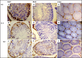
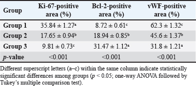
Fig. 2. Photomicrograph of rat testes in the group treated with 20nm ZnO Np. (A) The lumen in some seminiferous tubules was clear, and the thickness of the germinal epithelium was reduced (10X). (B) Note the separation (green head arrow) of basal and luminal cellular compartments in some ST; normal appearance of Sertoli cells (red thin arrow), spermatogenic cells surround by vacuoles (red head arrow), and a small number of sperm (40x). (C) Multiple giant spermatid cells in the lumen of the tubule (red curved arrows) (40X). (D), the interstitial space between the tubules increased and became somewhat edematous (green star) (40X)H&E. In contrast, group 2 showed a noticeably weak Ki-67 staining expression. The population of Ki-67-positive germ cells was markedly reduced compared with that in group 1. The nuclei of both spermatogonia and primary spermatocytes showed weak immunoreactivity (Fig. 4B). Interestingly, group 3 demonstrated low Ki-67 expression levels in its tubules in contrast with group 1 and treated group 2 (Fig. 4C). The expression was confirmed in sparse germ cells. Nuclear staining was observed in only a few scattered germ cells throughout seminiferous tubules. The immunohistochemical expression of apoptosis-related protein Bcl-2 in the germinal epithelium of the seminiferous tubules revealed no immune-reactive cells in groups 1 and 2 (Fig. 4D and E). Immunohistochemical evaluation of testicular sections from group 1 demonstrated weak to moderate Bcl-2 immunoreactivity within the germinal epithelium of the seminiferous tubules. Rats in the treated 2 group exhibited a strong effect of BCL-2 expression (Fig. 4F). Strong expression of the BCL-2 protein was observed only in scattered spermatogonia and early spermatocytes in the basal compartment. The positive reaction appears as dark brown nuclear and light brown cytoplasmic reactions (Fig. 4F). Immunohistochemical analysis of vWF showed noticeable variations in endothelial integrity among the experimental groups (Fig. 4G–I). This study showed that vWF expression was weak and restricted to the vascular endothelium within the interstitial spaces in group 1 (Fig. 4G). However, group 2 exhibited a marked increase in vWF expression in the interstitial regions and around the seminiferous tubules (Fig. 4H). Group 3 (Fig. 4I) had moderately elevated vWF expression compared with group 1, but markedly lower than that detected in group 2. The staining pattern was more localized and less intense, indicating a comparatively attenuated endothelial response. Quantitative immunohistochemical analysis demonstrated significant differences in the expression of Ki-67, Bcl-2, and vWF among the experimental groups (Table 8). Ki-67 immunoreactivity was significantly reduced in ZnO NP-treated rats compared with the control group (p < 0.001), with the lowest expression observed in group 2 (17.65 ± 0.94), followed by group 3 (9.81 ± 0.73), whereas the control group exhibited the highest Ki-67 expression (35.84 ± 1.27). In contrast, Bcl-2 expression was significantly increased following ZnO NP exposure (p < 0.001), reaching its highest level in group 3 (31.47 ± 1.12), followed by group (18.94 ± 0.85), compared with the control group (8.72 ± 0.61). Similarly, vWF immunoreactivity was significantly elevated in the treated groups (p < 0.001), with the greatest expression detected in group 2 (47.6 ± 1.9), followed by group 3 (28.4 ± 1.3), while the control group showed the lowest expression (20.4 ± 1.5). These findings indicate that exposure to 20 nm ZnO Np produced the most pronounced reduction in cellular proliferation and increased antiapoptotic and vascular immunoreactivity compared with the 40 nm ZnO NP-treated group.
Fig. 3. Photomicrograph of a testes rat treated with 40nm ZnO Np. (A): Notable degeneration in the spermatogenic cells in the seminiferous tubules (ST) (10X). (B): Impaired spermatogenic cell integrity (double-headed black arrow), Sertoli cells (red thin arrow), and marked reduction in the number of sperm (40). (C): The interstitial space was increased and became more edematous and contained lipid droplets (red star). There was intense deterioration in the progression of spermatogenesis (curved black arrows) and desquamation of cells into the lumen. (D): congestion of the Interstitial blood vessel (thick green arrow) (40). (E): Fully depleted spermatogenic cells (black curved arrow) in some parts of seminiferous tubules, severe irregularity in the basement membranes, undulate in shape (blue thick arrow) 40x.
Fig. 4. Immunohistochemical analysis of Ki67, Bcl2, and vWF in testicular tissue sections. In the group, there is strong expression of Ki67 (A), negative expression of Bcl2 (D), and moderate expression of vWF (G). In group 2: showed moderate expression of Ki67 (B), weak Bcl2 expression (E), and increased vWF immunoreactivity (H). In group 3, low Ki67 expression (C), strong Bcl2 expression (F), and moderate vWF expression (I), scale bar 50 µm. Table 8. Quantitative immunohistochemical analysis of Ki-67, Bcl-2, and vWF in the testes of control and ZnO nanoparticle-treated rats values are expressed as mean ± SEM (n=15).
DiscussionTo isolate the effect of nanoparticle size, ZnO nanoparticles (20 nm and 40 nm) were administered at the same dose (100 mg/kg/day). This design ensured that differences in reproductive toxicity were attributable to particle size, as smaller nanoparticles exhibit greater surface area, cellular uptake, and biological reactivity than larger particles (Hong et al., 2022; Ahmed and El-Sayed, 2025; Ahmed et al., 2025). This study demonstrates that ZnO Np adversely affect sperm morphology in a size-dependent manner, resulting in significant tail abnormalities and minor head defects, indicating disrupted spermatogenesis and spermiogenesis. These results agree with previous rodent studies demonstrating diminished normal sperm, impaired motility, and damage to seminiferous tubules (Chen et al., 2023; Ahmed and El-Sayed, 2025; Ahmed et al., 2025). Similarly, Goma et al. (2021) reported that ZnO nanoparticle exposure impaired sperm quality, induced oxidative stress, and produced histopathological alterations in the testes of male rats, supporting the findings of the present study. However, they also observed that the severity of reproductive toxicity depended on nanoparticle formulation and exposure conditions, suggesting that differences in particle characteristics, dose, and treatment duration may contribute to variability among studies. Tail anomalies are probably due to oxidative stress and mitochondrial problems. This might be because the sperm flagellum is especially sensitive to ROS-induced lipid peroxidation (Agarwal et al., 2014; Pinho et al., 2020). Structural damage to Sertoli cells and seminiferous epithelium may further hinder spermatogenesis. While several metrics stabilized at heightened exposure levels, the cumulative morphological damage indicates potential reproductive detriment. Future studies should look at sperm motility, DNA integrity, and oxidative markers to determine how these things work and how to keep nanoparticles safe. The effects of ZnO Np on body weight, testicular weight, and sperm motility in adult male rats were examined. Exposure to ZnO Np resulted in considerable reductions in sperm motility, whereas testicular weight remained statistically constant, indicating that nanoparticles predominantly influence functional reproductive characteristics rather than overall testicular morphology. ZnO Np-administered rats demonstrated decreased body weight compared with controls. These results agree with the findings of a previous study, which found that nanoparticle accumulation can provoke oxidative stress, disrupt metabolic functions, and compromise systemic physiology (Mozaffari et al., 2020; Li et al., 2022; Ahmed and El-Sayed, 2025; Ahmed et al., 2025). Despite these systemic effects, testicular weight remained consistent between groups, supporting prior findings that early reproductive toxicity may present functionally rather than structurally (Chen et al., 2023; Ahmed and El-Sayed, 2025; Ahmed et al., 2025). These findings highlight the significance of evaluating functional endpoints, such as sperm motility and spermatogenesis, as more sensitive indications of reproductive dysfunction than organ weight alone. This study noted substantial decreases in sperm motility metrics, but no alterations in testicular weight were observed. Group 1 displayed the highest levels of progressive and total motility, whereas ZnO Np-treated rats experienced significant reductions. These findings agree with previous studies indicating the harmful effects of ZnO Np on sperm quality, including motility, viability, and morphology in male rats (Ahmed and El-Sayed, 2025; Ahmed et al., 2025). Impaired sperm motility is a significant predictor of diminished male fertility because sperm mobility is crucial for successful fertilization. The findings of the current study are also consistent with those of Hong et al. (2022), who demonstrated that oral ZnO NP exposure disrupted spermatogenesis and significantly reduced sperm quality in rats. In contrast to Goma et al. (2021), who reported only mild changes in sperm parameters following lower doses or shorter exposure periods, reproductive toxicity is influenced by nanoparticle size, dose, and exposure duration. The observed reduction in sperm motility may be ascribed to oxidative stress caused by ZnO Np. These particles produce an excess of ROS, resulting in oxidative damage to sperm cells (Li et al., 2022). Sperm membranes are especially susceptible to lipid peroxidation, mitochondrial malfunction, and DNA damage due to their elevated levels of polyunsaturated fatty acids, which hinder motility (Mozaffari et al., 2020; Nowicka-Bauer and Nixon, 2020; Chen et al., 2023; Rehman et al., 2024). Direct ZnO Np deposition in testicular tissue may further impair reproductive function. Nanoparticles can penetrate biological barriers and accumulate in the testes, potentially impacting Sertoli and germ cells and disrupting the blood-testis barrier, compromising the spermatogenic milieu (Li et al., 2022; Ahmed and El-Sayed, 2025; Ahmed et al., 2025). Histopathological investigations validate these observations, indicating degenerative alterations in seminiferous tubules, germinal epithelium vacuolization, and a decrease in germ cell populations following ZnO Np exposure. These findings are consistent with the mechanisms summarized by previous studies (Pinho et al., 2020; Rehman et al., 2024; Kian et al., 2025) that reported that ZnO nanoparticles impair male fertility through oxidative stress, apoptosis, and spermatogenesis disruption. Collectively, these findings suggest that ZnO Np predominantly influences the functional characteristics of male reproduction rather than the overall shape of the testes. Considering the increasing use of ZnO Np in the industrial, pharmaceutical, and biomedical sectors, a better assessment of its reproductive toxicity is imperative. To further understand the dangers linked to ZnO Np exposure, future research should examine prolonged exposure, size-dependent effects, and the underlying molecular pathways. The current investigation revealed a notable decrease in blood testosterone levels in rats treated with ZnO Np, with the lowest levels recorded in group 2 and a substantial decrease in group 3. Testosterone, synthesized by Leydig cells, is crucial for spermatogenesis, the development of male reproductive organs, and fertility. These findings agree with those of Goma et al. (2021) and Hong et al. (2022) who also reported significant reductions in serum testosterone following ZnO NP exposure. However, some studies have reported minimal hormonal changes after lower-dose or shorter-term exposure, suggesting that endocrine toxicity depends on nanoparticle characteristics and experimental conditions. Mozaffari et al. (2020); Mustafa et al. (2021); Rehman et al. (2024) and (Ahmed and El-Sayed, 2025; Ahmed et al., 2025) reported that the reduction in testosterone indicates compromised testicular endocrine function and altered steroidogenesis, presumably due to ZnO Np impacting Leydig cells and essential steroidogenic enzymes. The elevated testosterone levels in group 3 relative to group 2 indicate a size-dependent influence of ZnO Np on endocrine function, supporting earlier findings (Kian et al., 2025), who found that ZnO Np adversely affects male reproductive hormones by inducing oxidative stress and inflammation, inhibiting steroidogenic enzymes, and directly damaging Leydig cells, thereby underscoring its capacity to disturb testicular endocrine function. The current investigation revealed that ZnO Np causes size-dependent histopathological changes in rat testes. The control rats displayed typical seminiferous tubule architecture, characterized by preserved germinal epithelium and structured spermatogenic layers. The ZnO Np-treated groups demonstrated differing levels of degeneration: the low-size group displayed moderate decreases in germinal epithelium thickness and exfoliation of spermatogenic cells, whereas the high-size group exhibited significant loss of spermatogenic cells, basement membrane damage, hyaline degeneration, lipid droplet accumulation, and vascular congestion. These alterations are associated with oxidative stress-induced cellular damage, as ROS can harm DNA, lipids, and proteins, and nanoparticle buildup impairs the blood-testis barrier and Sertoli-germ cell interactions (Pinho et al., 2020; Chen et al., 2023). Interstitial lipid droplets and hyaline substances may signify Leydig cell malfunction and endocrine abnormalities, whereas vascular congestion indicates inflammation or compromised microcirculation (Ma et al., 2013). These data affirm that ZnO Np induces size-dependent reproductive harm through oxidative stress, apoptosis, structural alteration of seminiferous tubules, and perhaps endocrine disruption, underscoring the need for careful management of ZnO Np exposure. The current study demonstrated the immunohistochemical localization of Ki-67 and Bcl-2 in rat testicular tissue, revealing considerable disparities in proliferative and antiapoptotic activity among the experimental groups, indicating unique effects on spermatogenesis. In group 1, the seminiferous epithelium showed strong nuclear Ki-67 expression, indicating a high germ cell proliferative activity. These results agree with normal rat spermatogenesis, where spermatogonia and primary spermatocytes divide to maintain the process (Lokman et al., 2021). Previous research has established robust Ki-67 immunoreactivity in spermatogonia and primary spermatocytes in normal adult rat testes (Lokman et al., 2021; Zakariah et al., 2022). In contrast, group 2 showed markedly reduced Ki-67 staining with only a few immunoreactive nuclei. This weak proliferative signal indicates a major impairment of germ cell proliferation. Other researchers have shown that toxicants or stressors that compromise testicular function reduce Ki-67 expression in seminiferous tubules, reflecting decreased mitotic activity among spermatogonia (Gautam et al., 2024; Maroto et al., 2025). Exposure to environmental contaminants reduces Ki-67 expression and is associated with germinal epithelium degeneration and decreased sperm production (Gautam et al., 2024). Group 3 similarly showed minimal Ki-67 expression with only a few stained germ cells, indicating a lower proliferative index, possibly more pronounced than that in group 2. These findings are in agreement with those of previous studies suggesting that prolonged stress, systemic illness, or agents that disrupt the hypothalamic–pituitary–testicular axis can suppress germ cell proliferation (Mostafa-Hedeab et al., 2023). This suggests that the treatments administered in groups 2 and 3 adversely affected spermatogonial proliferation, resulting in a reduced number of germ cells. Bcl-2 is a key antiapoptotic protein that promotes cell survival by inhibiting mitochondrial-mediated apoptosis (Cory and Adams, 2002). This study showed that Bcl-2 expression was absent in group 2, whereas control rats exhibited weak to moderate expression in the germinal epithelium. Basal spermatogonia and early spermatocytes express Bcl-2 to maintain survival during active proliferation (Sakkas et al., 2003; Zakariah et al., 2022). The loss of Bcl-2 expression in group 2 may indicate an increased susceptibility of germ cells to apoptosis, which is consistent with testicular degeneration. This finding agrees with previous studies showing that reduced Bcl-2 expression is associated with increased germ cell apoptosis and impaired spermatogenesis (Zakariah et al., 2022; Maroto et al., 2025). Interestingly, group 3 showed focal but strong Bcl-2 expression in some spermatogonia and early spermatocytes. This pattern suggests a compensatory upregulation of survival mechanisms in a subset of germ cells exposed to treatment. Similar increases in Bcl-2 expression have been reported in testicular models exposed to toxic stimuli, where surviving germ cells upregulate antiapoptotic pathways to resist apoptosis (Maroto et al., 2025). However, the localized nature of Bcl-2 immunoreactivity indicates that, although some germ cells may adapt to stress, overall spermatogenesis remains compromised. The distinct expression patterns of Ki-67 and Bcl-2 in this study indicate a strong relationship between spermatogenesis regulation and cellular proliferation and programmed cell death. Under stress conditions, reduced Ki-67 expression is associated with decreased spermatogenic efficiency and increased apoptosis (O’Donnell, 2001; De Rooij and Russell, 2013; Zakariah et al., 2022). Furthermore, the elevated Bcl-2 expression observed in group 3 is consistent with findings suggesting that moderate stress can activate cellular survival pathways; however, prolonged or severe stress eventually overcomes these protective mechanisms, leading to germ cell loss (Roufayel, 2016). This study also illustrates that ZnO Np influences endothelial function in rat testes in a size-dependent manner, as demonstrated by vWF immunoreactivity. In controls, vWF was localized in the endothelial cells of interstitial blood vessels, consistent with its role in vascular integrity and testicular perfusion (Maroto et al., 2025). Administration of ZnO Np at lower exposure levels markedly increased vWF expression, indicating enhanced endothelial activation and angiogenic activity. These findings are consistent with those of studies showing that ZnO nanoparticles stimulate endothelial proliferation and angiogenesis through vascular endothelial growth factor-related pathways (Paszek et al., 2012). Conversely, higher ZnO Np exposure reduced vWF expression compared with lower exposure, suggesting endothelial dysfunction and vascular damage. This may be due to oxidative stress induced by excessive accumulation of nanoparticles (Chen et al., 2023). This biphasic response is consistent with previous studies demonstrating that low ZnO Np sizes enhance cellular function, whereas high ZnO Np sizes exert toxic effects (Chuang et al., 2016; Ahmed and El-Sayed, 2025; Ahmed et al., 2025). Study limitations1- One limitation of this study is that the authors did not perform the physicochemical characterization of the zinc oxide nanoparticles independently. ZnO nanoparticles were commercially obtained and used according to the manufacturer’s specifications regarding particle size and purity. Therefore, additional characterization techniques, including Transmission electron microscopy, dynamic light scattering, X-ray diffraction, zeta potential analysis, and band gap determination, were not conducted. Consequently, the findings should be interpreted based on the nanoparticle characteristics reported by the manufacturer. 2- Another limitation of this study is that the ZnO nanoparticle suspensions were dispersed by mechanical agitation rather than sonication. Although vortex mixing provided a homogeneous suspension for administration, sonication may have further reduced nanoparticle agglomeration and improved dispersion. Future studies should incorporate sonication and physicochemical characterization of the NP suspension before administration. ConclusionThis study concludes that ZnO nanoparticles induce size-dependent reproductive toxicity in adult male rats, with smaller nanoparticles producing more pronounced adverse effects on sperm quality, reproductive hormone levels, testicular histology, cell proliferation, apoptosis, and microvascular integrity. Reduced Ki-67 expression, increased Bcl-2 immunoreactivity, and altered vWF expression indicate that ZnO nanoparticles disrupt the balance between cellular proliferation, survival, and vascular homeostasis within the testes. These findings provide further evidence that nanoparticle size is an important determinant of reproductive toxicity. The molecular mechanisms underlying these alterations, including oxidative stress, inflammatory responses, mitochondrial dysfunction, and signaling pathways regulating apoptosis and spermatogenesis, should be investigated in future studies. Long-term exposure studies, detailed dose–response analyses, and evaluations of potential protective agents, such as antioxidants, are warranted to better understand the reproductive risks associated with ZnO nanoparticles and to support the safe development of nanomaterials for biomedical and industrial applications. AcknowledgmentsThe authors express sincere gratitude to the Dean and staff of the College of Veterinary Medicine, University of Duhok, Duhok Medical Research Center staff (DMRS), and the Animal House at the College of Veterinary Medicine, University of Duhok. The authors are grateful to the Vin Laboratory staff for their kind assistance. The authors would also like to thank Duhok City for their kind cooperation and assistance. Conflict of interestThere are no conflicts of interest to declare. FundingThis study was funded by the authors only. Authors’ contributionsZeravan A. Mohammed contributed to the conception, study design, and the study methodology. All authors performed the sampling and laboratory tests. The authors interpreted the statistical analyses and read the histological and immunohistochemical data, microscopic slides, and electron microscopic results. The authors have drafted the main text. Zeravan A. Mohammed has revised the text. The authors have read and approved the final version of the manuscript for publication. Data availabilityThe data supporting the findings of this study are available within the manuscript. ReferencesAdamkovicova, M., Toman, R., Martiniakova, M., Omelka, R., Babosova, R., Krajcovicova, V., Grosskopf, B. and Massanyi, P. 2016. Sperm motility and morphology changes in rats exposed to cadmium and diazinon. Reproductive Biol. Endocrinol. 14(1), 42; doi:10.1186/s12958-016-0177-6 Agarwal, A., Virk, G., Ong, C. and Du Plessis, S.S. 2014. Effect of oxidative stress on male reproduction. World J. Men’s Health 32(1), 1–17; doi:10.5534/wjmh.2014.32.1.1 Ahmed, D.H. and El-Sayed, W.M. 2025. Large diameter zinc oxide nanoparticles reduced testicular CYP17A1 and viability and elevated testicular inflammation in adult male rats. J. Basic. Appl. Zool. 86, 102; doi:10.1186/s41936-025-00527-5 Ahmed, D.H., El-Beih, N.M., El-Hussieny, E.A. and El-Sayed, W.M. 2025. Zinc oxide nanoparticles induced testicular toxicity through inflammation and reducing testosterone and cell viability in adult male rats. Biol. Trace. Element. Res. 203(4), 1934–1948; doi:10.1007/s12011-024-04330 Chen, T., Zhang, L., Yao, L., Luan, J., Zhou, X., Cong, R., Guo, X., Qin, C. and Song, N. 2023. Zinc oxide nanoparticles-induced testis damage at single-cell resolution: depletion of spermatogonia reservoir and disorder of Sertoli cell homeostasis. Environ. Int. 181, 108292; doi: 10.1016/j.envint.2023.108292 Chuang, K.J., Lee, K.Y., Pan, C.H., Lai, C.H., Lin, L.Y., Ho, S.C., Ho, K.F. and Chuang, H.C. 2016. Effects of zinc oxide nanoparticles on human coronary artery endothelial cells. Food Chem. Toxicol. 93, 138–144; doi:10.1016/j.fct.2016.05.008 Cory, S. and Adams, J.M. 2002. The Bcl-2 family: regulators of the cellular life-or-death switch. Nature. Rev. Cancer. 2(9), 647–656; doi:10.1038/nrc883 De Rooij, D.G. and Russell, L.D. 2013. All you wanted to know about spermatogonia but were afraid to ask. J. Androl. 21(6), 776–798; doi:10.1002/j.1939-4640.2000.tb03408.x Gautam, R., Priyadarshini, E., Patel, A.K. and Arora, T. 2024. Assessing the impact and mechanisms of environmental pollutants (heavy metals and pesticides) on the male reproductive system: a comprehensive review. J. Environ. Sci. Health Part. C. 42(2), 126–153; doi:10.1080/26896583.2024.2302738 Goma, A.A., Tohamy, H.G., El-Kazaz, S.E., Soliman, M.M., Shukry, M., Elgazzar, A.M. and Rashed, R.R. 2021. Insight study on the comparison between zinc oxide nanoparticles and its bulk impact on reproductive performance, antioxidant levels, gene expression, and histopathology of testes in male rats. Antioxidants 10(1), 41; doi:10.3390/antiox10010041 Hong, X., Shao, N., Yin, L., Li, C., Tao, G., Sun, Y., Qian, K., Yang, J., Xiao, P., Yu, X. and Zhou, Z. 2022. Exposure to zinc oxide nanoparticles affects testicular structure, reproductive development and spermatogenesis in parental and offspring male rats. Ann. Translational. Med. 10(13), 751; doi:10.21037/atm-22-3047 Khafaji, S. 2023. Antioxidant, anti-inflammatory, and anti-reprotoxic effects of kaempferol and vitamin E on lead acetate-induced testicular toxicity in male rats. Open. Vet. J. 13(12), 1683–1695; doi:10.5455/OVJ.2023.v13.i12.17 Kian, M., Soleimanzadeh, A., Javadi, S. and Najafi, G. 2025. Zinc oxide nanoparticles as a chemical castration agent in male rats. Scientific. Rep. 15, 44702; doi: 10.1038/s41598-025-28667-2 Li, J., Ning, M., Zhang, Y., Liu, Q., Liu, K., Zhang, H., Zhao, Y., Chen, C. and Liu, Y. 2022. The potential for nanomaterial toxicity affecting the male reproductive system. WIREs Nanomed. Nanobiotechnol. 14(6), 1806; doi:10.1002/wnan.1806 Lokman, M., Ashraf, E., Kassab, R.B., Abdel Moneim, A.E. and El-Yamany, N.A. 2021. Aluminum chloride–induced reproductive toxicity in rats: the protective role of zinc oxide nanoparticles. Biol. Trace Element Res. 200(9), 4035–4044; doi:10.1007/s12011-021-03010-8 Ma, H., Williams, P.L. and Diamond, S.A. 2013. Ecotoxicity of manufactured ZnO Np nanoparticles—a review. Environ. Pollut. 172, 76–85; doi:10.1016/j.envpol.2012.08.011 Maroto, M., Torvisco, S.N., García-Merino, C., Fernández-González, R. and Pericuesta, E. 2025. Mechanisms of hormonal, genetic, and temperature regulation of germ cell proliferation, differentiation, and death during spermatogenesis. Biomolecules 15(4), 500; doi:10.3390/biom15040500 Mohammed, Z.A. 2016. Mechanism of fertility failure in high yielding dairy cows. PhD thesis. University of Nottingham, United Kingdom. Available via http://eprints.nottingham.ac.uk/37350/1 Mostafa-Hedeab, G., Behairy, A., Abd-Elhakim, Y.M., Mohamed, A.A.R., Noreldin, A.E., Dahran, N., Gaber, R.A., Alqahtani, L.S., Essawi, W.M. and Eskandrani, A.A. 2023. Green synthesized zinc oxide nanoparticles using Moringa oleifera ethanolic extract lessens acrylamide-induced testicular damage, apoptosis, and steroidogenesis-related gene dysregulation in adult rats. Antioxidants 12(2), 361; doi:10.3390/antiox12020361 Mozaffari, Z., Parivar, K., Roodbari, N.H. and Irani, S. 2020. The impact of zinc oxide nanoparticles on LH, FSH, and testosterone hormones in mature male rats. J. Pharm. Res. Int. 32(4), 30–37; doi:10.9734/jpri/2020/v32i430422 Mustafa, A.A., Ahmed, K.N., Hamoudi, T.A. and Hadi, A.M. 2021. Effect of zinc nanoparticles on reproductive hormones and histological structure of testes in rats. Indian. J. Forensic. Med. Toxicol. 15(3), 810–814; doi: 10.37506/ijfmt.v15i3.15410 Nowicka-Bauer, K. and Nixon, B. 2020. Molecular changes induced by oxidative stress that impair human sperm motility. Antioxidants 9(2), 134; doi:10.3390/antiox9020134 O’Donnell, L. 2001. Estrogen and spermatogenesis. Endocrine. Rev. 22(3), 289–318; doi:10.1210/edrv.22.3.0431 Paszek, E., Czyz, J., Woźnicka, O., Jakubiak, D., Wojnarowicz, J., Łojkowski, W. and Stepień, E. 2012. Zinc oxide nanoparticles impair the integrity of human umbilical vein endothelial cell monolayer in vitro. J. Biomed. Nanotechnol. 8(6), 957–967; doi:10.1166/jbn.2012.1463 Pinho, A.R., Rebelo, S. and Pereira, M.D.L. 2020. The impact of zinc oxide nanoparticles on male (in)fertility. Materials 13(4), 849; doi:10.3390/ma13040849 Rehman, N., Jabeen, F., Asad, M., Nijabat, A., Ali, A., Khan, S.U. and Ahmad, A. 2024. Exposure to zinc oxide nanoparticles induced reproductive toxicities in male Sprague Dawley rats. J. Trace Elements Med. Biol. 83, 127411; doi: 10.1016/j.jtemb.2024.127411 Roufayel, R. 2016. Regulation of stressed-induced cell death by the Bcl-2 family of apoptotic proteins. Mol. Membrane Biol. 33(6–8), 89–99; doi:10.1080/09687688.2017.1400600 Sakkas, D., Seli, E., Bizzaro, D., Tarozzi, N. and Manicardi, G.C. 2003. Abnormal spermatozoa in the ejaculate: abortive apoptosis and faulty nuclear remodelling during spermatogenesis. Reproductive Biomed. Online 7(4), 428–432; doi:10.1016/S1472-6483(10)61886-X Scholzen, T. and Gerdes, J. 2000. The Ki-67 protein: from the known and the unknown. J. Cellular. Physiol. 182(3), 311–322. Sharma, N., Kurmi, B.D., Singh, D., Mehan, S., Khanna, K., Karwasra, R., Kumar, S., Dua, K. and Kakkar, D. 2024. Nanoparticles toxicity: an overview of its mechanism and plausible mitigation strategies. Drug. Chem. Toxicol. 47(5), 457–469; doi:10.1080/1061186X.2024.2316785 Sharma, V., Anderson, D. and Dhawan, A. 2012. Zinc oxide nanoparticles induce oxidative DNA damage and ROS-triggered mitochondria-mediated apoptosis in human liver cells (HepG2). Apoptosis 17(8), 852–870; doi:10.1007/s10495-012-0705-6 Sun, A., Ma, S. and Wang, W.X. 2025. Two-sided cellular and physiological effects of zinc oxide nanoparticles (nZnO): a critical review. Environ. Sci. Nano. 12, 1059–1078; doi:10.1039/D4EN00676C Suvarna, K.S., Layton, C. and Bancroft, J.D. 2018. Bancroft’s theory and practice of histological techniques. 8th ed. Oxford, United Kingdom: Elsevier, p: 704. Turner, P.V., Brabb, T., Pekow, C. and Vasbinder, M.A. 2020. Administration of substances to laboratory animals: routes of administration and factors to consider. J. Am. Assoc. For. Lab. Anim. Sci. 59(5), 506–513. Wagner, D.D. and Frenette, P.S. 2008. The vessel wall and its interactions. Blood 111(11), 5271–5281; doi:10.1182/blood-2008-01-078204 Wang, B., Feng, W., Wang, M., Wang, T., Gu, Y., Zhu, M., Ouyang, H., Shi, J., Zhang, F., Zhao, Y., Chai, Z., Wang, H. and Wang, J. 2008. Acute toxicological impact of nano- and submicro-scaled zinc oxide powder on healthy adult mice. J. Nanoparticle Res. 10(2), 263–276; doi:10.1007/s11051-007-9245-3 Zakariah, M., Molele, R.A., Mahdy, M.A.A., Ibrahim, M.I.A. and Mcgaw, L.J. 2022. Regulation of spermatogenic cell apoptosis by the pro-apoptotic proteins in the testicular tissues of mammalian and avian species. Anim. Reprod. Sci. 247, 1; doi:10.1016/j.anireprosci.2022.107158 | ||



| How to Cite this Article |
| Pubmed Style Saleem MA, Mohammed ZA, Ameen SZ. Size-dependent reproductive toxicity of zinc oxide nanoparticles in adult male rats: Alterations in sperm quality, steroidogenesis, and testicular microvasculature. Open Vet. J.. 2026; 16(8): 5850-5862. doi:10.5455/OVJ.2026.v16.i8.73 Web Style Saleem MA, Mohammed ZA, Ameen SZ. Size-dependent reproductive toxicity of zinc oxide nanoparticles in adult male rats: Alterations in sperm quality, steroidogenesis, and testicular microvasculature. https://www.openveterinaryjournal.com/?mno=325425 [Access: September 04, 2026]. doi:10.5455/OVJ.2026.v16.i8.73 AMA (American Medical Association) Style Saleem MA, Mohammed ZA, Ameen SZ. Size-dependent reproductive toxicity of zinc oxide nanoparticles in adult male rats: Alterations in sperm quality, steroidogenesis, and testicular microvasculature. Open Vet. J.. 2026; 16(8): 5850-5862. doi:10.5455/OVJ.2026.v16.i8.73 Vancouver/ICMJE Style Saleem MA, Mohammed ZA, Ameen SZ. Size-dependent reproductive toxicity of zinc oxide nanoparticles in adult male rats: Alterations in sperm quality, steroidogenesis, and testicular microvasculature. Open Vet. J.. (2026), [cited September 04, 2026]; 16(8): 5850-5862. doi:10.5455/OVJ.2026.v16.i8.73 Harvard Style Saleem, M. A., Mohammed, . Z. A. & Ameen, . S. Z. (2026) Size-dependent reproductive toxicity of zinc oxide nanoparticles in adult male rats: Alterations in sperm quality, steroidogenesis, and testicular microvasculature. Open Vet. J., 16 (8), 5850-5862. doi:10.5455/OVJ.2026.v16.i8.73 Turabian Style Saleem, Mayan A., Zeravan A. Mohammed, and Shayma Z. Ameen. 2026. Size-dependent reproductive toxicity of zinc oxide nanoparticles in adult male rats: Alterations in sperm quality, steroidogenesis, and testicular microvasculature. Open Veterinary Journal, 16 (8), 5850-5862. doi:10.5455/OVJ.2026.v16.i8.73 Chicago Style Saleem, Mayan A., Zeravan A. Mohammed, and Shayma Z. Ameen. "Size-dependent reproductive toxicity of zinc oxide nanoparticles in adult male rats: Alterations in sperm quality, steroidogenesis, and testicular microvasculature." Open Veterinary Journal 16 (2026), 5850-5862. doi:10.5455/OVJ.2026.v16.i8.73 MLA (The Modern Language Association) Style Saleem, Mayan A., Zeravan A. Mohammed, and Shayma Z. Ameen. "Size-dependent reproductive toxicity of zinc oxide nanoparticles in adult male rats: Alterations in sperm quality, steroidogenesis, and testicular microvasculature." Open Veterinary Journal 16.8 (2026), 5850-5862. Print. doi:10.5455/OVJ.2026.v16.i8.73 APA (American Psychological Association) Style Saleem, M. A., Mohammed, . Z. A. & Ameen, . S. Z. (2026) Size-dependent reproductive toxicity of zinc oxide nanoparticles in adult male rats: Alterations in sperm quality, steroidogenesis, and testicular microvasculature. Open Veterinary Journal, 16 (8), 5850-5862. doi:10.5455/OVJ.2026.v16.i8.73 |


