





| Research Article | ||
Open Vet. J.. 2026; 16(8): 5841-5849 !
Open Veterinary Journal, (2026), Vol. 16(8): 5841–5849 Research Article Isolation, identification, and whole-genome genetic characterization of two waterfowl-origin H6N6 subtype avian influenza virusesHonghong Guo1, Jin Jin2, Zexiao Ma1, Dizhou Luo1, Binyi Sun2, Weiping Zhang1, Chenggang Liu2*1Shanwei Animal Disease Control Center; Shanwei, China 2Shanwei Academy of Agricultural Sciences; Shanwei, China *Corresponding Author: Chenggang Liu. Shanwei Academy of Agricultural Sciences; Shanwei, China. Email: lcg85 [at] 126.com Submitted: 31/05/2026 Revised: 15/07/2026 Accepted: 25/07/2026 Published: 20/08/2026 © 2025 Open Veterinary Journal
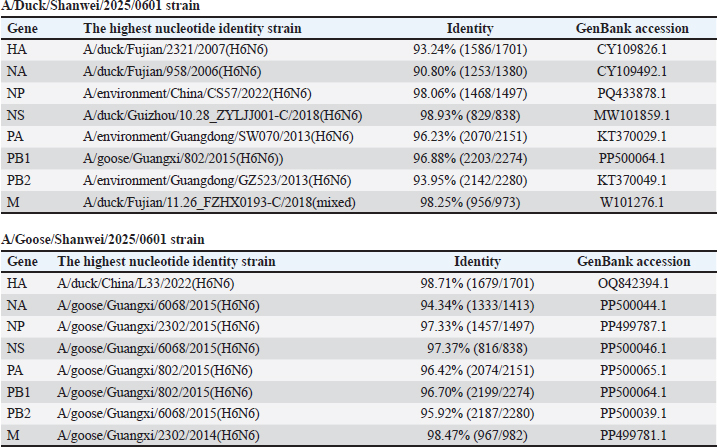
AbstractBackground: H6N6 subtype avian influenza viruses (AIVs) are widely circulating in waterfowl and exhibit considerable genetic diversity. Their frequent reassortment and potential host adaptation highlight the need for molecular surveillance in waterfowl populations. Aim: This study aimed to investigate the genetic characteristics and molecular evolutionary relationships of waterfowl-origin H6N6 subtype AIVs isolated in Shanwei, Guangdong Province, China. Methods: Whole-genome sequencing was performed on one duck-origin and one goose-origin H6N6 subtype AIV isolate. Sequence homology of the eight gene segments was analyzed using BLAST. Phylogenetic trees were constructed based on the HA and NA gene sequences. In addition, key amino acid residues were analyzed to evaluate molecular features associated with receptor binding, mammalian adaptation, drug susceptibility, and pathogenicity. Results: Both isolates were identified as H6N6 subtype AIVs. BLAST analysis showed that the closest reference strains differed among the gene segments of the two isolates. The HA and NA segments of the duck-origin isolate showed the highest similarity to H6N6 viruses isolated from ducks in Fujian Province, whereas several gene segments of the goose-origin isolate were closely related to H6N6 viruses isolated from geese in Guangxi Province. Both isolates retained 226Q and 228G at the HA receptor-binding sites, 627E and 701D in the PB2 protein, and 31S in the M2 protein. The goose-origin isolate possessed 66S in the PB1-F2 protein, whereas the duck-origin isolate had a deletion at positions 62–73 in the NA protein. Conclusion: These findings indicate the circulation of H6N6 subtype AIVs among waterfowl in Shanwei. Overall, the two isolates retained molecular characteristics typical of avian-origin low-pathogenicity influenza viruses. However, their complex genetic origins and several notable molecular markers highlight the need for continuous surveillance and further biological risk assessment. Keywords: Avian influenza virus, Genetic evolution, H6N6 subtype, Waterfowl, Whole-genome sequencing. IntroductionAvian influenza viruses (AIVs) belong to the genus Influenza A virus within the family Orthomyxoviridae. They are segmented, negative-sense RNA viruses that can infect wild waterfowl, domestic poultry, and various mammalian species (AbuBakar et al., 2023; Bruno et al., 2026; Zhao et al., 2026). AIVs are generally classified as low-pathogenicity avian influenza viruses (LPAIVs) or highly pathogenic avian influenza viruses (HPAIVs) based on their pathogenicity in poultry and the molecular features of the hemagglutinin (HA) cleavage site (Spackman, 2020). Among these viruses, H6 subtype AIVs are generally classified as LPAIVs. However, they have a broad host range, complex genotypes, and are continuously detected in wild birds, domestic poultry, and environmental samples, making them an important component of the AIV ecosystem (Cui et al., 2022b). Previous studies have shown that H6 subtype viruses are widely circulating among wild birds and domestic poultry worldwide (Lin et al., 2023). Their relatively high detection rate in poultry populations provides a basis for their continuous circulation and genetic evolution. H6 subtype AIVs exhibit substantial genetic diversity and can combine with multiple NA subtypes to form different subtypes, including H6N1, H6N2, H6N5, H6N6, and H6N8 (Hou et al., 2017; Wu et al., 2018; Yan et al., 2023). Among these viruses, the H6N6 subtype has gradually become one of the more commonly detected H6 subtype AIVs in poultry populations in several regions of China in recent years (Cui et al., 2022b). Some studies have reported that H6N2 was the predominant subtype among duck-origin H6 AIVs in China during the early period, whereas H6N6 viruses have gradually increased since 2009 and have become one of the major circulating H6 subtype AIVs in recent years (Xu et al., 2023). In addition, H6N6 viruses have been detected not only in domestic poultry and wild birds but also in mammals, indicating that their potential risk of cross-species transmission should not be overlooked (Wan et al., 2022). The AIV genome consists of eight gene segments, which can readily undergo genetic reassortment when different viral subtypes co-infect the same host or coexist within the same ecological environment (Ito, 2025). The continuous circulation of H6N6 subtype viruses in poultry populations in China suggests that they may serve as important gene donors or intermediate vectors for other AIVs. Recent whole-genome studies have shown that H6N6 viruses in China have complex genotypic compositions and can reassort with different subtypes, including H6N2, H5N6, H7N9, H5N2, H4N2, and H6N8, resulting in multiple novel reassortment events (Du et al., 2024). Therefore, whole-genome sequencing, phylogenetic analysis, and analysis of key amino acid residues in newly isolated H6N6 viruses are important for understanding their genetic origins, molecular characteristics, and potential public health risks. Shanwei City is located on the southeastern coast of Guangdong Province, where waterfowl farming, tidal flat wetlands, and migratory bird activities coexist. These ecological factors may provide favorable conditions for the maintenance, transmission, and reassortment of AIVs. The Guangdong Haifeng Bird Provincial Nature Reserve, located in Haifeng County, Shanwei, is an important bird reserve along the southeastern coast of China. It consists of diverse wetland ecosystems and provides suitable habitats for waterfowl feeding, resting, breeding, and overwintering. Moreover, the Haifeng Bird Nature Reserve is an important part of the East Asian–Australasian Flyway. In this context, surveillance of waterfowl-origin AIVs in Shanwei is of practical significance for understanding the regional epidemiological dynamics of AIVs and assessing their potential transmission risks. In the present epidemiological investigation, one duck-origin and one goose-origin H6N6 subtype AIV were isolated using SPF embryonated chicken eggs. BLAST similarity analysis of the eight gene segments, phylogenetic analysis of the HA and NA genes, and molecular characterization of key amino acid residues were subsequently performed (Fig. 2). By comparing the genetic relationships of the surface and internal genes of the two isolates, this study aimed to preliminarily clarify the genetic origins, molecular characteristics, and potential risks of waterfowl-origin H6N6 subtype AIVs in Shanwei, thereby providing baseline data for continuous surveillance and integrated prevention and control of AIVs in this region. Materials and MethodsSample collection and virus isolationFrom April to July 2025, active surveillance was conducted in ducks and geese from waterfowl farms in Shanwei. Throat and cloacal swabs collected from the same waterfowl were pooled in 1 mL of sterile phosphate-buffered saline. Viral nucleic acids were extracted using a rapid animal virus DNA/RNA extraction kit (TIANLONG, Xi’an, China). Original samples that tested positive for AIV by RT-PCR using universal primers targeting the AIV M gene (Tao et al., 2009) were thoroughly vortexed and centrifuged at 4,000 rpm for 5 minutes. The supernatants were filtered through 0.22-μm filters, and 0.1 mL of each filtrate was inoculated into the allantoic cavity of 9-day-old specific pathogen-free (SPF) embryonated chicken eggs using a 1-mL syringe. The inoculation site was sealed with wax, and the eggs were incubated at 37°C for 72 hours, followed by overnight chilling at 4°C. Allantoic fluid was aseptically collected and stored at −70°C until further use. One-day-old SPF embryonated chicken eggs were purchased from the SPF Laboratory Animal Center of Xinxing Dahuanong Poultry and Egg Co., Ltd. Hemagglutination activity in the allantoic fluid was detected using a hemagglutination assay, and hemagglutination-positive samples were further identified by RT-PCR for influenza A virus. Following virus isolation and molecular identification, two H6N6 subtype AIV isolates were obtained and designated A/Duck/Shanwei/2025/0601 and A/Goose/Shanwei/2025/0601, respectively. Viral RNA extraction and whole-genome sequencingViral nucleic acids were extracted using a rapid animal virus DNA/RNA extraction kit. Reverse transcription was performed using the HiScript II 1st Strand cDNA Synthesis Kit (+gDNA wiper) (Vazyme, Nanjing, China) with the Uni12 primer (Cui et al., 2022a), according to the manufacturer’s instructions. Using the reverse-transcribed cDNA as the template, the eight gene segments of influenza A virus were amplified by PCR with the MBTuni-12 and MBTuni-13 primers, as described by Zhou et al (Zhou et al., 2009). The PCR products were submitted to Novogene Co., Ltd. (Beijing, China) for NGS sequencing. The raw paired-end FASTQ data were first processed using Trim Galore (v0.6.5) for adapter removal and low-quality base trimming. The default Phred quality threshold for trimming was set to 20, and FastQC was used after trimming for quality control assessment. Data cleaning was performed using the following parameters: --paired--quality 20--stringency 1--length 50--trim-n--fastqc. After quality control, reads with adapter contamination, low-quality bases, excessive N bases, or insufficient length were removed, and the resulting clean reads were used for subsequent influenza virus genome assembly. The quality-controlled clean reads were assembled using the FLU module of IRMA (CDC Iterative Refinement Meta-Assembler). Assembly was performed with default parameters using the following command format: IRMA FLU sample_R1_val_1.fq.gz sample_R2_val_2.fq.gz sample_name. After assembly, consensus sequences of the eight gene segments, including PB2, PB1, PA, HA, NP, NA, M, and NS, were extracted for each sample. The assembly results were further evaluated based on sequencing depth and sequence completeness. Regions with insufficient coverage or poor assembly quality were excluded from subsequent analyses or marked as missing nucleotides. BLAST similarity analysisThe coding regions of the eight gene segments, including HA, NA, PB1, PB2, NS, NP, M, and PA, from the two H6N6 subtype AIV isolates were submitted separately to the NCBI BLAST database for nucleotide sequence homology analysis. The nucleotide BLAST program was used, and publicly available influenza A virus sequences in the GenBank database were selected as reference sequences for comparison. Based on sequence identity, query coverage, E-value, and total score in the BLAST results, the representative strain with the highest similarity was selected for each gene segment. When multiple strains showed comparable similarity for the same gene segment, sequences with higher coverage, better completeness, and more complete strain background information were preferentially selected as representative references. Phylogenetic analysis of the HA and NA genesTo analyze the genetic evolutionary relationships of the HA and NA genes of the two H6N6 subtype AIV isolates obtained in this study, all available H6 subtype HA gene sequences and N6 subtype NA gene sequences were downloaded from the GISAID EpiFlu database. Sequences that were too short, contained substantial deletions, showed excessive redundancy, or lacked complete background information were excluded. The HA and NA gene sequences obtained in this study were combined separately with the downloaded reference sequences and aligned using MEGA 7.0. After alignment, regions with inconsistent lengths at both ends were manually trimmed to ensure that the sequence regions used for phylogenetic analysis were consistent. Phylogenetic trees were constructed using the Neighbor-Joining method with 1,000 bootstrap replicates. The two H6N6 virus isolates obtained in this study were highlighted in red font or marked in red in the phylogenetic trees. The HA gene phylogenetic tree was used to determine the genetic lineage of the HA genes of the two isolates and their relationships with H6 reference strains from China and other countries. The NA gene phylogenetic tree was used to analyze the evolutionary origin of the N6 genes of the two isolates. Analysis of key amino acid residuesThe open reading frames of the eight gene segments of the two H6N6 virus isolates were translated using SnapGene Viewer 5.3.2 to obtain the amino acid sequences of viral proteins, including PB2, PB1, PB1-F2, PA, HA, NP, NA, M1, M2, NS1, and NS2. The amino acid sequences of the isolates were then compared with those of reference strains and previously reported key molecular markers of AIVs. Molecular characteristics associated with receptor-binding properties, mammalian adaptation, virulence-related residues, antiviral drug susceptibility, and NA stalk deletion were analyzed. Ethical approvalNot needed for this study. ResultsWhole-genome BLAST similarity analysis of two H6N6 subtype avian influenza virus isolatesBLAST similarity analysis was performed for the eight gene segments of the two H6N6 subtype AIV isolates, A/Duck/Shanwei/2025/0601 and A/Goose/Shanwei/2025/0601. The results showed that the strains with the highest similarity differed among the gene segments of the two isolates (Table 1), suggesting that different gene segments may have distinct genetic origins. The HA gene of A/Duck/Shanwei/2025/0601 showed the highest similarity to A/duck/Fujian/2321/2007 (H6N6), with a nucleotide identity of 93.24%, whereas its NA gene showed the highest similarity to A/duck/Fujian/958/2006 (H6N6), with a nucleotide identity of 90.80%. The NP, NS, and M genes of this isolate showed relatively high similarities of 98.06%, 98.93%, and 98.25%, respectively. In contrast, the PB2 gene showed 93.95% similarity, while the PA and PB1 genes showed 96.23% and 96.88% similarity, respectively. Overall, the surface genes of the duck-origin isolate, particularly the NA gene, showed relatively low similarity to known reference strains, suggesting a certain genetic distance from the currently available closely related sequences in the database. For the A/Goose/Shanwei/2025/0601 isolate, the HA gene showed the highest similarity to A/duck/China/L33/2022 (H6N6), with a nucleotide identity of 98.71%, whereas the NA gene was most similar to A/goose/Guangxi/6068/2015 (H6N6), with a nucleotide identity of 94.34%. Among its internal genes, the M gene showed the highest similarity, at 98.47%. The NP, NS, PA, PB1, and PB2 genes showed similarities of 97.33%, 97.37%, 96.42%, 96.70%, and 95.92%, respectively. Compared with the duck-origin isolate, the HA gene of the goose-origin isolate was more closely related to recently circulating waterfowl-origin H6N6 viruses in China. However, its NA gene and several internal genes were most closely related to goose-origin H6N6 viruses from Guangxi, suggesting that this isolate may have a close genetic relationship with waterfowl-origin H6N6 viruses circulating in southern China. Table 1. BLAST similarity analysis of various gene fragments.
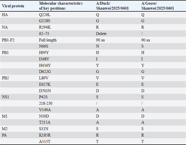
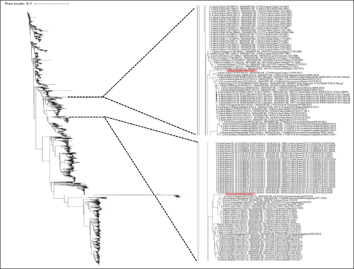
Phylogenetic analysis of the H6 geneThe H6 phylogenetic tree constructed based on the HA gene showed that H6 subtype AIVs could be divided into the North American lineage and the Eurasian lineage (Fig. 1). The two isolates obtained in this study, A/Duck/Shanwei/2025/0601 and A/Goose/Shanwei/2025/0601, both belonged to the Eurasian lineage. However, they did not cluster within the same subclade, indicating that the HA genes of the two isolates may have different genetic backgrounds. The HA gene of A/Duck/Shanwei/2025/0601 was located near a clade mainly composed of duck-origin H6N6 viruses isolated from Fujian Province during 2006–2007, and was closely related to several H6-related viruses from Fujian, Guangdong, and environmental sources. This finding was consistent with the BLAST results, which showed that its HA gene had the highest similarity to A/duck/Fujian/2321/2007. In contrast, the HA gene of A/Goose/Shanwei/2025/0601 clustered within a clade containing recently identified waterfowl-origin H6N6 viruses from China, including A/duck/China/L33/2022, A/duck/China/L31/2022, A/duck/China/L32/2022, and A/duck/China/L83/2022. This result was also consistent with the high similarity between its HA gene and that of A/duck/China/L33/2022 in the BLAST analysis. These findings indicate that, although both H6N6 isolates belonged to the Eurasian H6 lineage, their HA genes were not derived from the same closely related subclade. Molecular characterization of key amino acid residues in the two H6N6 virus isolatesAnalysis of key amino acid residues in the two H6N6 viruses showed that both isolates possessed 226Q and 228G at the HA receptor-binding sites, with no Q226L or G228S substitutions, suggesting that they retained the receptor-binding characteristics of avian-origin influenza viruses (Table 2). Both isolates carried R at position 294 of the NA protein, with no R294K substitution. Notably, A/Duck/Shanwei/2025/0601 had an amino acid deletion at positions 62–73 in the NA stalk region, whereas this deletion was not observed in A/Goose/Shanwei/2025/0601. For polymerase-related proteins, both viruses retained 627E and 701D in PB2, with no typical mammalian-adaptive substitutions such as E627K or D701N. Both isolates also carried V at position 89 of PB2. In the PB1 protein, both viruses possessed 99H, 368I, 436Y, and 622G. The PB1-F2 protein of both isolates was full-length, consisting of 90 amino acids; however, A/Goose/Shanwei/2025/0601 carried the N66S substitution, whereas A/Duck/Shanwei/2025/0601 retained 66N. For the other gene segments, both isolates possessed 42S and 149A in NS1, with no deletion at positions 218–230. The M1 protein carried 30D and 215A in both isolates, and the M2 protein retained 31S, with no S31N substitution. The PA protein contained 185R and 515T in both viruses. Overall, typical molecular markers associated with mammalian adaptation or antiviral resistance, including HA-Q226L/G228S, PB2-E627K/D701N, and M2-S31N, were not detected in either isolate. However, the NA stalk deletion in the duck-origin isolate and the PB1-F2 N66S substitution in the goose-origin isolate indicate differences between the two viruses at several key molecular sites.
Fig. 1. Phylogenetic analysis of AIV H6 genes. The red taxon line represents the H6N6 subtype isolated in the laboratory. DiscussionIn this study, two H6N6 subtype AIV isolates, designated A/Duck/Shanwei/2025/0601 and A/Goose/Shanwei/2025/0601, were isolated from waterfowl samples collected in Shanwei. Their whole-genome sequences, HA/NA phylogenetic relationships, and key amino acid residues were analyzed. BLAST analysis showed that the closest reference strains differed among the eight gene segments of the two isolates, suggesting that their genomes may have distinct genetic origins. The HA and NA genes of A/Duck/Shanwei/2025/0601 showed the highest similarity to early duck-origin H6N6 viruses from Fujian Province, with nucleotide identities of only 93.24% and 90.80%, respectively. In contrast, the HA gene of A/Goose/Shanwei/2025/0601 showed the highest similarity to A/duck/China/L33/2022, with a nucleotide identity of 98.71%, whereas its NA gene and several internal genes were more closely related to goose-origin H6N6 viruses from Guangxi Province. These findings suggest that the duck-origin and goose-origin H6N6 viruses isolated from the same region during the same period were not derived from a single closely related transmission chain, but may represent the coexistence or introduction of H6N6 viruses with different genetic backgrounds in local waterfowl. Phylogenetic analysis of the HA and NA genes further supported this observation. The H6 phylogenetic tree showed that both isolates belonged to the Eurasian lineage but were located in different subclades. Similarly, the N6 phylogenetic tree showed that the NA genes of the two isolates did not cluster within the same closely related clade. Combined with the BLAST results shown in Table 1, the HA and NA genes of the duck-origin isolate were more closely related to early duck-origin H6N6 viruses from Fujian Province, whereas the goose-origin isolate was more closely related to recently circulating waterfowl-origin H6N6 viruses from China or to H6N6 viruses from Guangxi and Guangdong. These results indicate that waterfowl-origin H6N6 viruses in Shanwei may be influenced by multiple viral introductions, regional waterfowl movement, and local environmental circulation. Previous studies have shown that H6N6 viruses have undergone long-term evolution and multiple reassortment events in poultry populations in China, resulting in diverse genotypes, and may act as intermediate carriers during the evolution of viruses such as H5N6 and H7N9 (Du et al., 2024). Therefore, the inconsistent phylogenetic positions of the HA and NA genes of the two isolates observed in this study are consistent with the complex genetic origins and frequent reassortment characteristics of H6N6 subtype viruses. Table 2. Analysis of the important amino acids of the HA, NA, PB1-F2, PB1, PB2, NS1, M1, M2, and PA proteins.
From the perspective of surface gene characteristics, both viruses possessed 226Q and 228G at the receptor-binding sites of the HA protein, with no Q226L or G228S substitutions, suggesting that they retained the molecular features of AIVs that preferentially bind to α-2,3-linked sialic acid receptors. Receptor-binding specificity is an important factor affecting the cross-species transmission potential of AIVs. AIVs generally preferentially bind to α-2,3-linked sialic acid receptors, whereas human influenza viruses predominantly bind to α-2,6-linked sialic acid receptors (Zhao and Pu, 2022). Mutations at key residues in the HA receptor-binding region can alter viral host tropism and tissue tropism (Imai and Kawaoka, 2012). Therefore, the absence of typical human receptor-binding-associated substitutions in the two isolates suggests that they currently do not show clear molecular markers of adaptation to human-type receptors. Analysis of polymerase-related residues showed that both viruses possessed 627E and 701D in the PB2 protein, with no classical mammalian-adaptive substitutions such as PB2-E627K or PB2-D701N (Tammiranta et al., 2023; Chakraborty et al., 2025). PB2-E627K and PB2-D701N are considered important molecular markers for assessing the cross-species transmission risk of AIVs, as they are associated with enhanced replication, host adaptation, and increased pathogenicity in mammalian cells (Min et al., 2013). In the present study, both isolates retained avian-like residues at these positions, suggesting a relatively limited potential for mammalian adaptation. However, cross-species transmission of AIVs is not determined by a single mutation, but rather by multiple factors, including HA receptor-binding specificity, HA–NA functional balance, polymerase activity, and internal gene constellation. Therefore, the presence of PB2-627E and PB2-701D alone does not completely exclude the possibility of mammalian infection.
Fig. 2. Phylogenetic analysis of AIV N6 genes. The red taxon line represents the H6N6 subtype isolated in the laboratory. Analysis of the NA protein showed that A/Duck/Shanwei/2025/0601 had an amino acid deletion at positions 62–73 in the NA stalk region, whereas this deletion was not observed in A/Goose/Shanwei/2025/0601. NA stalk deletion is generally considered to be associated with the adaptation of AIVs from wild waterfowl to domestic poultry and may affect NA enzymatic activity, HA–NA functional balance, virus release efficiency, and host adaptation (Park et al., 2017). Previous studies have shown that NA stalk truncation or changes in glycosylation can influence viral replication and pathogenicity in cell culture and animal models; however, these effects are closely related to the viral genetic background, host species, and HA–NA compatibility (Li et al., 2011; Xu et al., 2025). Previous studies have reported that a chicken-origin H9N2 AIV was lethal to mice due to the synergistic effects of the PB2 E627V substitution and a deletion at amino acid position 217 in HA (Zhao et al., 2025). Therefore, the NA stalk deletion observed in the duck-origin isolate warrants further attention, although its biological significance should be further validated using viral growth kinetics and experimental infection studies in chickens or ducks. For the PB1-F2 protein, both viruses encoded a full-length PB1-F2 protein of 90 amino acids. However, A/Goose/Shanwei/2025/0601 carried the N66S substitution, whereas A/Duck/Shanwei/2025/0601 retained 66N. Previous studies have shown that PB1-F2 can affect viral pathogenicity, cell death, and host innate immune responses. Among these changes, the N66S substitution has been reported to enhance the virulence of some influenza viruses and suppress the early interferon response (Conenello et al., 2011). However, the effect of PB1-F2 N66S is also strain-background dependent; therefore, increased virulence of the goose-origin isolate cannot be inferred solely from this residue. The present findings suggest that the goose-origin isolate possesses a molecular feature in PB1-F2 that warrants continued monitoring. Further studies, including mouse infection experiments, cytokine expression analysis, and viral replication assays, are needed to evaluate its biological significance. Regarding antiviral drug-related residues, both viruses possessed R at position 294 of the NA protein, with no R294K substitution, and S at position 31 of the M2 protein, with no S31N substitution. M2-S31N is an important molecular marker associated with adamantane resistance in influenza A viruses and has been detected in multiple influenza A virus subtypes worldwide (Bright et al., 2006). The absence of M2-S31N in the two isolates indicates that they did not exhibit typical molecular features associated with adamantane resistance. However, given the continuous evolution of AIVs in natural environments and poultry populations, NA- and M2-related antiviral resistance markers should continue to be monitored in future surveillance studies. From a regional ecological perspective, Shanwei is located along the coast of Guangdong Province, and the Haifeng Bird Nature Reserve provides an important habitat for waterfowl and migratory birds along the East Asian–Australasian Flyway. Factors such as migratory bird movement, waterfowl farming, mixed rearing of geese and ducks, live poultry trade, and environmental contamination on farms may facilitate the contact, transmission, and reassortment of AIVs from different sources within local areas. The Guangdong Haifeng Bird Provincial Nature Reserve consists of complex wetland ecosystems, including the Gongping, Dahu, and Dongguan–Lian’anwei areas, and represents an important site for waterfowl feeding, resting, and overwintering. Therefore, continuous surveillance of waterfowl-origin AIVs in Shanwei is important not only for understanding viral circulation in poultry farming systems but also for assessing the risk of viral introduction and reassortment at the migratory bird–waterfowl–domestic poultry interface. ConclusionThe two H6N6 subtype AIVs isolated in this study were waterfowl-origin viruses related to the Eurasian lineage. However, their HA and NA genes belonged to different genetic branches, and the closest reference strains for their eight gene segments were not completely consistent, suggesting the possible co-circulation or multiple introductions of H6N6 viruses with different genetic backgrounds in waterfowl in Shanwei. Overall, both viruses retained the molecular characteristics of avian-origin low-pathogenicity influenza viruses and lacked typical markers associated with mammalian adaptation or antiviral resistance, such as HA-Q226L/G228S, PB2-E627K/D701N, and M2-S31N. However, the NA stalk deletion in the duck-origin isolate and the PB1-F2 N66S substitution in the goose-origin isolate indicate that some molecular features remain worthy of further investigation. Future studies should expand the sample size and include different seasons, host species, and environmental samples for continuous surveillance. In addition, virus isolation, whole-genome sequencing, animal infection experiments, and receptor-binding assays should be integrated to systematically evaluate the genetic evolution, host adaptation, and potential transmission risk of H6N6 subtype AIVs in Shanwei. FundingThis work was supported by Guangdong Basic and Applied Basic Research Foundation (2024A1515012917), Guangdong Provincial Department of Science and Technology Project (2024TQ08N105, KTP20240371), and the Presidential Foundation of the Shanwei Academy of Agricultural Sciences (SWNK2025A01). Authors’ ContributionsConceptualization, C.L.; methodology, H.G., Z.M., and Z.L.; software, J.J. and B.S.; validation, B.S., D.L., and J.J.; formal analysis, H.G., Z.L., and Z.M.; investigation, H.G., Z.M., D.L., and W.Z.; resources, C.L. and W.P.; data curation, C.L. and H.G.; writing—original draft preparation, H.G.; writing—review and editing, C.L.; visualization, B.S.; supervision, C.L.; project administration, C.L., W.Z.; funding acquisition, C.L. All authors have read and agreed to the published version of the manuscript. Conflict of InterestThe authors declare no conflicts of interest. Data AvailabilityThe data from the experiments will be available from the authors if required. ReferencesAbuBakar, U., Amrani, L., Kamarulzaman, F.A., Karsani, S.A., Hassandarvish, P. and Khairat, J.E., 2023. Avian influenza virus Tropism in humans. Viruses 15, 833, 1–27. Bright, R.A., Shay, D.K., Shu, B., Cox, N.J. and Klimov, A.I. 2006. Adamantane resistance among influenza A viruses isolated early during the 2005-2006 influenza season in the United States. JAMA 295, 891–894. Bruno, L., Nappo, M.A., Frontoso, R., Montinaro, S., Di Lecce, R., Guarnieri, C., Ferrari, L. and Corradi, A. 2026. Avian Influenza Viruses: global panzootic, host range expansion and emerging one-health threats. Vet. Sci. 13 67, 1–68. Chakraborty, C., Das, A., Bhattacharya, M. and Islam, M.A. 2025. Emerging H5N1 mutations (PB2-D701N, PB2-E627K, HA-S110N, NA-V116X) in a recent outbreak in U.S. dairy cows: need for continuous monitoring. Ann. Med. Surg. (2012) 87, 5347–5350. Conenello, G.M., Tisoncik, J.R., Rosenzweig, E., Varga, Z.T., Palese, P. and Katze, M.G., 2011. A single N66S mutation in the PB1-F2 protein of influenza A virus increases virulence by inhibiting the early interferon response in vivo. J. Virol. 85, 652–662. Cui, J., Cui, P., Shi, J., Fan, W., Xing, X., Gu, W., Zhang, Y., Zhang, Y., Zeng, X., Jiang, Y., Chen, P., Yang, H., Chen, Y., Liu, J., Liu, L., Tian, G., Lu, Y., Chen, H., Li, C. and Deng, G., 2022a. Continued evolution of H6 Avian influenza viruses isolated from farms in China between 2014 and 2018. Transbound. Emerg. Dis. 69, 2156–2172. Cui, M., Huang, Y., Wang, X., Bian, X., Du, L., Yan, Y., Gu, J., Dong, W., Zhou, J. and Liao, M. 2022b. Genetic characterization and evolution of H6N6 subtype Avian influenza viruses. Front. Microbiol. 13, 963218. Du, Y., Xia, J., Wang, Z., Xu, J., Ji, Y., Jin, Y., Pu, L. and Xu, S. 2024. Evolution of H6N6 viruses in China between 2014 and 2019 involves multiple reassortment events. Emerg. Microbes. Infect. 13, 2341142. Hou, G., Li, J., Peng, C., Wang, S., Chen, J. and Jiang, W. 2017. Complete Genome sequence of a Novel Reassortant H6N8 Avian Influenza Virus isolated from wild waterfowl in Poyang Lake, China. Genome Announc. 5, 2. Imai, M. and Kawaoka, Y. 2012. The role of receptor binding specificity in interspecies transmission of influenza viruses. Curr. Opin. Virol. 2, 160–167. Ito, T. 2025. Pathogenicity and host range of avian influenza viruses: molecular determinants and virological perspectives. J. Vet. Med. Sci. 87, 1259–1265. Li, J., Zu Dohna, H., Cardona, C.J., Miller, J. and Carpenter, T.E. 2011. Emergence and genetic variation of neuraminidase stalk deletions in avian influenza viruses. PloS One 6, e14722. Lin, M., Yao, Q.C., Liu, J., Huo, M., Zhou, Y., Chen, M., Li, Y., Gao, Y. and Ge, Y. 2023. Evolution and reassortment of H6 subtype Avian Influenza Viruses. Viruses 15, 1–14. Min, J.Y., Santos, C., Fitch, A., Twaddle, A., Toyoda, Y., DePasse, J.V., Ghedin, E. and Subbarao, K. 2013. Mammalian adaptation in the PB2 gene of avian H5N1 influenza virus. J. Virol. 87, 10884–10888. Park, S., Il Kim, J., Lee, I., Bae, J.Y., Yoo, K., Nam, M., Kim, J., Sook Park, M., Song, K.J., Song, J.W., Kee, S.H. and Park, M.S. 2017. Adaptive mutations of neuraminidase stalk truncation and deglycosylation confer enhanced pathogenicity of influenza A viruses. Sci. Rep. 7, 10928. Spackman, E., 2020. A Brief Introduction to Avian Influenza Virus. Methods Mol. Biol. (Clifton, N.J.) 2123, 83–92. Tammiranta, N., Isomursu, M., Fusaro, A., Nylund, M., Nokireki, T., Giussani, E., Zecchin, B., Terregino, C. and Gadd, T., 2023. Highly pathogenic avian influenza A (H5N1) virus infections in wild carnivores connected to mass mortalities of pheasants in Finland. Infect. Genet. Evol. J. Mol. Epidemiol. Evol. Genet. Infect. Dis. 111, 105423. Tao, Q., Wang, X., Bao, H., Wu, J., Shi, L., Li, Y., Qiao, C., Yakovlevich, S.A., Mikhaylovna, P.N. and Chen, H. 2009. Detection and differentiation of four poultry diseases using asymmetric reverse transcription polymerase chain reaction in combination with oligonucleotide microarrays. J. Vet. Diagn. Invest. 21, 623–632. Wan, Z., Gong, J., Sang, J., Jiang, W., Zhao, Z., Lian, M., Tang, T., Li, Y., Kan, Q., Xie, Q., Li, T., Shao, H., Gao, W., Qin, A. and Ye, J. 2022. Mouse adaptation of H6 avian influenza viruses and their molecular characteristics. Front. Microbiol. 13, 1049979. Wu, H., Yang, F., Liu, F., Lu, R., Peng, X., Chen, B., Yao, H. and Wu, N. 2018. Isolation and characterization of novel reassortant H6N1 avian influenza viruses from chickens in Eastern China. Virol. J. 15, 164. Xu, N., Chen, Y., Wu, Y., Guo, Y., Wang, C., Qin, T., Chen, S., Peng, D. and Liu, X. 2025. The evolution of hemagglutinin-158 and neuraminidase-88 glycosylation sites modulates antigenicity and pathogenicity of clade 2.3.2.1 H5N1 avian influenza viruses. Vet. Microbiol. 300, 110333. Xu, X., Chen, Q., Tan, M., Liu, J., Li, X., Yang, L., Shu, Y., Wang, D. and Zhu, W. 2023. Epidemiology, evolution, and biological characteristics of H6 avian influenza viruses in China. Emerg. Microbes. Infect. 12, 2151380. Yan, Z., Li, Y., Huang, S. and Wen, F. 2023. Global distribution, receptor binding, and cross-species transmission of H6 influenza viruses: risks and implications for humans. J. Virol. 97, e0137023. Zhao, C. and Pu, J. 2022. Influence of host sialic acid receptors structure on the host specificity of influenza viruses. Viruses 14, 2141, 1-15. Zhao, L., Tian, M., Hu, X., Fan, M., Hou, C. and Ping, J. 2025. PB2 627V and HA 217 sites synergistically affect the lethality of H9N2 in mice. Virol. Sin. 40, 35–49. Zhao, Y., Liu, C., Li, Y., Wang, Y., Hou, B., Gao, H., Liu, Z., Wu, X., Lu, M., Shen, Y., Yang, P., Yuan, H., Hou, Y. and Xiao, Y., 2026. Genetic and biological characterization of H9N2 avian influenza viruses isolated from swine in China. BMC Vet. Res. 22, 384, 1-10 Zhou, B., Donnelly, M.E., Scholes, D.T., St George, K., Hatta, M., Kawaoka, Y. and Wentworth, D.E., 2009. Single-reaction genomic amplification accelerates sequencing and vaccine production for classical and Swine origin human influenza a viruses. J. Virol. 83, 10309–10313. | ||

| How to Cite this Article |
| Pubmed Style Guo H, Jin J, Ma Z, Luo D, Sun B, Zhang W, Liu C. Isolation, identification, and whole-genome genetic characterization of two waterfowl-origin H6N6 subtype avian influenza viruses. Open Vet. J.. 2026; 16(8): 5841-5849. doi:10.5455/OVJ.2026.v16.i8.72 Web Style Guo H, Jin J, Ma Z, Luo D, Sun B, Zhang W, Liu C. Isolation, identification, and whole-genome genetic characterization of two waterfowl-origin H6N6 subtype avian influenza viruses. https://www.openveterinaryjournal.com/?mno=322707 [Access: September 03, 2026]. doi:10.5455/OVJ.2026.v16.i8.72 AMA (American Medical Association) Style Guo H, Jin J, Ma Z, Luo D, Sun B, Zhang W, Liu C. Isolation, identification, and whole-genome genetic characterization of two waterfowl-origin H6N6 subtype avian influenza viruses. Open Vet. J.. 2026; 16(8): 5841-5849. doi:10.5455/OVJ.2026.v16.i8.72 Vancouver/ICMJE Style Guo H, Jin J, Ma Z, Luo D, Sun B, Zhang W, Liu C. Isolation, identification, and whole-genome genetic characterization of two waterfowl-origin H6N6 subtype avian influenza viruses. Open Vet. J.. (2026), [cited September 03, 2026]; 16(8): 5841-5849. doi:10.5455/OVJ.2026.v16.i8.72 Harvard Style Guo, H., Jin, . J., Ma, . Z., Luo, . D., Sun, . B., Zhang, . W. & Liu, . C. (2026) Isolation, identification, and whole-genome genetic characterization of two waterfowl-origin H6N6 subtype avian influenza viruses. Open Vet. J., 16 (8), 5841-5849. doi:10.5455/OVJ.2026.v16.i8.72 Turabian Style Guo, Honghong, Jin Jin, Zexiao Ma, Dizhou Luo, Binyi Sun, Weiping Zhang, and Chenggang Liu. 2026. Isolation, identification, and whole-genome genetic characterization of two waterfowl-origin H6N6 subtype avian influenza viruses. Open Veterinary Journal, 16 (8), 5841-5849. doi:10.5455/OVJ.2026.v16.i8.72 Chicago Style Guo, Honghong, Jin Jin, Zexiao Ma, Dizhou Luo, Binyi Sun, Weiping Zhang, and Chenggang Liu. "Isolation, identification, and whole-genome genetic characterization of two waterfowl-origin H6N6 subtype avian influenza viruses." Open Veterinary Journal 16 (2026), 5841-5849. doi:10.5455/OVJ.2026.v16.i8.72 MLA (The Modern Language Association) Style Guo, Honghong, Jin Jin, Zexiao Ma, Dizhou Luo, Binyi Sun, Weiping Zhang, and Chenggang Liu. "Isolation, identification, and whole-genome genetic characterization of two waterfowl-origin H6N6 subtype avian influenza viruses." Open Veterinary Journal 16.8 (2026), 5841-5849. Print. doi:10.5455/OVJ.2026.v16.i8.72 APA (American Psychological Association) Style Guo, H., Jin, . J., Ma, . Z., Luo, . D., Sun, . B., Zhang, . W. & Liu, . C. (2026) Isolation, identification, and whole-genome genetic characterization of two waterfowl-origin H6N6 subtype avian influenza viruses. Open Veterinary Journal, 16 (8), 5841-5849. doi:10.5455/OVJ.2026.v16.i8.72 |


