





| Case Report | ||
Open Vet. J.. 2026; 16(8): 5897-5904 !
Open Veterinary Journal, (2026), Vol. 16(8): 5897–5904 Case Report Correction of antebrachial deformities using a simple patient-specific surgical guide in an American Bully dogByoungho An†, Dongwook Kim†, Heesoo Ahn, Dongwoo Chang and Gonhyung Kim*Veterinary Teaching Hospital, College of Veterinary Medicine, Chungbuk National University, Cheongju, Korea †These authors contributed equally to this work. *Corresponding Author: Gonhyung Kim. Department of Veterinary Surgery, College of Veterinary Medicine, Chungbuk National University, Cheongju, Korea. Email: ghkim [at] cbu.ac.kr Submitted: 20/05/2026 Revised: 13/07/2026 Accepted: 27/07/2026 Published: 20/08/2026 © 2025 Open Veterinary Journal
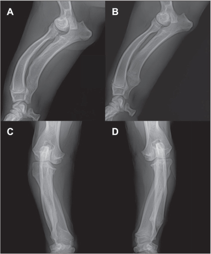
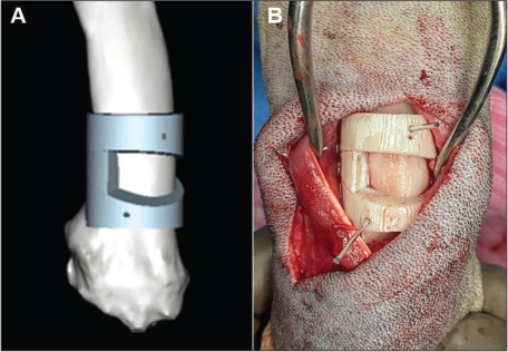
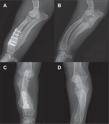
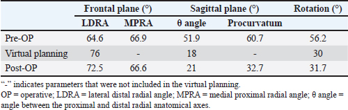
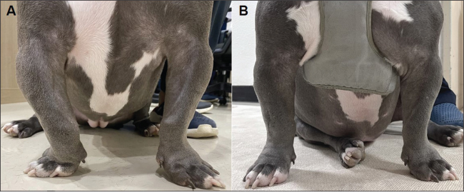
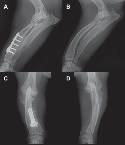
AbstractBackground: Antebrachial deformity is characterized by multiplanar malalignment of the radius and ulna, making accurate surgical correction challenging in dogs. Various patient-specific surgical guides have recently been introduced in veterinary medicine to improve surgical precision. However, their clinical application is often limited by high cost and the requirement for specialized engineering support. This study describes the use of a simple, cost-effective, and clinic-fabricated patient-specific osteotomy guide to correct antebrachial deformities in a dog. Case Description: A 10-month-old neutered female American Bully presented with bilateral antebrachial deformities, with a more severe right-sided deformity. Computed tomography revealed procurvatum, valgus, and torsional deformities of the right radius accompanied by ulnar shortening. As both limbs were affected, correction angles were determined through visual assessment of three-dimensional (3D) reconstructed images with reference to the angles of clinically normal dogs. A single osteotomy guide was designed according to the planned correction and configured in a press-fit manner to allow stable and reproducible positioning by conforming to the anatomical contours of the distal radius. Corrective osteotomy was performed after several simulations using a 3D-printed bone model. The postoperative recovery was uneventful, and no guide-related complications were observed during bone healing. The differences between the planned and achieved correction angles were within 4°. The dog’s bow-legged stance was resolved after surgery. No significant postoperative complications were identified over a 3-year follow-up period. The dog maintained satisfactory forelimb function. Conclusion: This case suggests that this press-fit-type guide can be designed with relative simplicity while enhancing surgical accuracy with minimal surgical exposure. It may be a viable option for the precise correction of complex antebrachial deformities in dogs. Keywords: Antebrachial deformity, Corrective osteotomy, Dog, Patient-specific guide, 3D printing. IntroductionAntebrachial deformity is a condition characterized by angular, torsional, and translational malalignment of the radius and ulna (Fox et al., 2006). This condition is generally caused by a disturbance of the distal physes associated with asynchronous growth of the radius and ulna (Ramadan and Vaughan, 1978; Balfour et al., 2000; Fox et al., 2006; Knapp et al., 2016). Lameness is a common clinical symptom due to altered joint alignment and abnormal load distribution (Theyse et al., 2005; Rovesti et al., 2009). When angular deformities cause incongruity and degenerative disease of the elbow joints, surgical correction is often indicated (Balfour et al., 2000; Theyse et al., 2005; Fox et al., 2006; Rovesti et al., 2009). The center of rotation of angulation (CORA)-based corrective osteotomy, originally developed in human orthopedics, has been widely applied in veterinary medicine to correct angular deformities (Knapp et al., 2016). Surgical planning based solely on two-dimensional radiographic images remains challenging in cases involving torsional or complex multiplanar deformities (Apelt et al., 2005; Piras et al., 2012). Computed tomography (CT)-based three-dimensional (3D) virtual surgical planning has been increasingly adopted in veterinary orthopedics for the assessment and correction of complex deformities (De Armond et al., 2022). In addition, when combined with 3D printing technology, it can be used to fabricate patient-specific surgical guides (Krishnan et al., 2012; Wong, 2016). Recently, various commercial patient-specific surgical guides have been proposed for the correction of antebrachial deformities in veterinary medicine (Worth et al., 2019; Carwardine et al., 2021; De Armond et al., 2022). Although these systems enhance surgical precision, their clinical application may be limited by high production costs, design complexity, and the need for specialized engineering support. Simplified patient-specific guides that focus on osteotomy accuracy with minimal design complexity have been proposed as a more practical alternative (Zhang et al., 2019; Hu et al., 2020; Serra Aguado et al., 2020). However, information regarding the clinical applicability of these systems remains limited, particularly in cases involving complex multiplanar deformities in which a normal contralateral limb is not available as a reference for preoperative planning. In such cases, surgical planning may be more challenging due to the lack of a patient-specific anatomical reference, and there are limited reports describing practical approaches for guide fabrication under these conditions. Therefore, this case report describes a practical workflow for fabricating a clinic-based, patient-specific osteotomy guide with a press-fit design in a dog with bilateral antebrachial deformities and no contralateral reference for surgical planning and reports the associated clinical outcome. Case DetailsA 10-month-old neutered female American Bully weighing 17.5 kg was referred for evaluation of suspected bilateral developmental angular limb deformities of the forelimbs. Upon physical examination, the dog exhibited a bow-legged stance of the right forelimb. Although no obvious lameness was observed, abnormal medial deviation of the forelimb placement, accompanied by external rotation of the forepaws, was noted during both walking and trotting. No signs of pain were identified. Radiographs obtained using a digital radiography system (REX-525R, Listem, Wonju, Korea) revealed bilateral ulnar shortening and radial procurvatum and valgus deformities, resulting in elbow incongruity (Fig. 1). Deformity and incongruity were more pronounced on the right side. Pre-anesthetic thoracic radiographs and blood tests did not reveal any abnormalities. A CT scan was performed using a 16-slice helical CT scanner (Revolution ACT, GE Healthcare, Milwaukee, WI, USA). External rotation of the right radius was also identified on CT. A 3D-reconstructed model was created for the surgical simulation and fabrication of the osteotomy guide (Fig. 2). The forelimb CT data were stored in Digital Imaging and Communications in Medicine format and imported into 3D planning software (Mimics version 17, Materialise, Leuven, Belgium). As both forelimbs were affected, a corrective plan was established based on visual assessment of the 3D-reconstructed images and reference angle ranges reported for clinically normal chondrodystrophic dogs (Duncan et al., 2022; Kwon et al., 2022). The degrees of deformity were assessed using 3D imaging and previously described methods (Duncan et al., 2022; Pulkkinen et al., 2022). Planned correction of the procurvatum was determined based on the angular intersection of the segmental radial axes (θ angle), cranial proximal radial angle (CPRA), and caudal distal radial angle (CDRA). Joint orientation lines were established at the proximal and distal radius in the sagittal plane. The angles formed between these lines and the anatomical axes of the elbow and carpus were measured to determine the CPRA and CDRA, respectively. The anatomical axis was determined by connecting the center points of perpendicular lines drawn at 25%, 50%, and 75% of the radial diaphyseal length. Total radial procurvatum was calculated as θ + (90 − [180 − CPRA]) + (90 − CDRA). The degree of valgus deformity was assessed using the medial proximal radial angle (MPRA) and lateral distal radial angle (LDRA) (Duncan et al., 2022). These angles were measured between the joint orientation lines and the anatomical axes of the elbow and carpus in the frontal plane. Torsional deformity was measured as the angle between the axis of the third digit and the spine (Pulkkinen et al., 2022). The osteotomy guide was designed to conform to the bone curvature and reduce multiplanar deformities using a single-wedge osteotomy. The wedge was configured to correct the procurvatum and valgus deformities. In addition, 2 Kirschner (K-) wire holes were created for temporary fixation and correction of external rotation. A proximal hole was placed in the center of the guide, and a distal hole was positioned according to the planned degree of rotational correction (Fig. 3A). After the wedge osteotomy, the 2 holes were aligned linearly before plate fixation. The design data were saved in STL format and exported to a 3D printer (3DWOX, Sindoh, Seoul, Korea). The osteotomy guide was fabricated from polylactic acid (PLA) with a layer thickness of 0.2 mm and a print speed of 40 mm/s. Subsequently, the printed guide was sterilized using a hydrogen peroxide gas plasma sterilization system, which has been recommended for PLA-based medical devices (Pérez Davila et al., 2021). In addition to the osteotomy guide, a 3D-printed bone model of the dog’s radius was used for surgical simulation. The planned surgery was based on the results of several preoperative simulations. The patient was pre-medicated with midazolam (0.2 mg/kg, IV; Bukwang Pharm, Seoul, Korea). Anesthesia was induced with propofol (6 mg/kg, IV; Provive, Myungmoon Pharm, Seoul, Korea) and maintained with isoflurane (2–2.5%; Terrell, Piramal Critical Care, Bethlehem, PA, USA) in 100% oxygen at 1–2 L/min. Perioperative analgesia was provided with fentanyl (1–8 μg/kg/h; Hana Pharm, Seoul, Korea), followed by a fentanyl transdermal patch (50 μg/h; Durogesic, Janssen Pharmaceutica, Beerse, Belgium). The dog was positioned in lateral recumbency after anesthetic stabilization. After exposure of the radius and ulna, a transverse ulnar osteotomy was performed using an oscillating saw. Thereafter, an osteotomy guide was placed on the radius and temporarily fixed with K-wires (diameter, 1.0 mm) (Fig. 3B). Wedge osteotomy was performed according to the surgical guide. The guide was removed once the bone wedge was excised. The torsional deformity was reduced by aligning the K-wire holes as planned. The osteotomy site was stabilized using a 2.7-mm 6-hole T-plate (Veterinary Instrumentation, Sheffield, UK) with an overall length of 39 mm. A circular external skeletal fixator (CESF) was also applied to enhance stabilization. Based on the degree of radial deformity, only ulnar osteotomy was performed on the left side. The dog’s recovery from anesthesia was uneventful. Cefazolin (22 mg/kg, IV, TID; Chong Kun Dang Pharm, Seoul, Korea) was administered as perioperative antibiotic therapy. The dog was hospitalized for pain management and exercise restriction and was discharged 6 days after surgery. No complications were identified during bone healing, and the CESF was removed 1 month postoperatively (Fig. 4). The complete blood count revealed no abnormalities, and the C-reactive protein concentration was within the reference range at the time of CESF removal. Correction was evaluated using postoperative radiographs, with most angles deviating by less than 4° from the planned values (Table 1). Compared with the preoperative stance, the bow-legged posture resolved following surgery (Fig. 5). The forelimb alignment and external rotation of the forepaws during gait also improved. Despite the lack of significant improvement in elbow incongruity following bilateral ulnar osteotomy, no progressive degenerative changes were observed during the follow-up period. Intermittent discomfort accompanied by mild lameness was effectively managed with short-term analgesic therapy. The dog remained highly active and showed normal limb function without complications over the 3-year follow-up (Fig. 6).
Fig. 1. Ulnar shortening, radial procurvatum and valgus deformities, and elbow incongruity were identified on preoperative radiographs of the right (A: lateral view; C: craniocaudal view) and left (B: lateral view; D: craniocaudal view) forelimbs.
Fig. 2. Three-dimensional reconstruction of the right antebrachium showing angular and torsional deformities. (A) Craniocaudal view and (B) mediolateral view.
Fig. 3. An osteotomy guide was designed to correct the multiplanar deformities of the right radius using three-dimensional planning software (A). The guide was press-fitted onto the distal radius and temporarily secured with Kirschner wires (B). Ethical approvalEthical approval was not required for this case report because it involved a single clinical case without experimental intervention. Written informed consent for the procedures was obtained from the owner. DiscussionIn dogs, antebrachial deformities frequently involve complex 3D malalignments, making accurate surgical correction challenging (Fox et al., 2006). Although various surgical techniques, including CORA-based corrective osteotomy, have been described, the precise determination of correction angles and accurate osteotomy remain difficult, particularly in cases involving multiplanar deformities (Apelt et al., 2005; Piras et al., 2012). In this context, 3D simulation and printing technologies have facilitated the development of patient-specific surgical guides in human and veterinary orthopedics (Krishnan et al., 2012; Wong, 2016). These guides improve the accuracy of osteotomy and reduce the operative time, particularly in cases with complex deformities or individual anatomical variations (Kroner et al., 2017). Most advanced guide systems generally comprise an osteotomy guide and a repositioning guide (De Armond et al., 2022; Tzimtzimis and Rutherford, 2025). These guides enable accurate osteotomy and fixation of the bony fragments. However, designing these guide systems is difficult for individual veterinary surgeons and often requires collaboration with computer-aided design engineers (Carwardine et al., 2021). Furthermore, guides are sometimes abandoned due to soft tissue constraints, as their placement requires a relatively wide bony surface (De Armond et al., 2022). A single osteotomy guide designed to fit the radial surface was used in this study. The guide could be placed with minimal surgical exposure because it was pressed against the bone for fixation. Additional pin holes were created to stabilize the osteotomy guide on the bone. These holes were designed to align after the osteotomy, allowing the planned correction of torsional deformity. Compared with previously reported patient-specific guide systems that incorporate separate repositioning guides and more complex fixation strategies, the present guide design was simplified to facilitate osteotomy and overall alignment correction. Relative to commercial guide systems, the present guide design may be easier to fabricate because it consists of a single osteotomy guide without a repositioning component. This simplified design may facilitate guide fabrication and clinical application by individual veterinary surgeons. In the present case, this approach enabled clinically acceptable correction across multiple planes of deformity with limited surgical exposure. However, direct comparisons regarding cost, surgical time, and correction accuracy were not performed. In planning corrective osteotomy, the correction angle was determined based on the contralateral normal limb when available (Fox et al., 2006). However, as the dog in this study was bilaterally affected, the contralateral limb could not be used for surgical planning. Breed-specific reference values for antebrachial alignment in American Bully dogs have not been well established. Substantial variation in forelimb conformation has been reported among chondrodystrophic dogs (Kwon et al., 2022). Therefore, a correction plan was established by a board-certified orthopedic surgeon based on previously reported ranges of joint orientation angles, clinical judgment, and 3D visual assessment. With computer-assisted 3D surgical planning and the patient-specific osteotomy guide, the achieved correction angles closely approximated the planned values. Joint orientation angles in the frontal plane vary widely among breeds, with LDRA generally greater than MPRA (Duncan et al., 2022; Kwon et al., 2022). However, this case revealed a smaller LDRA than the MPRA. After the planned osteotomy for valgus deformity, the LDRA was corrected to be larger than the MPRA. Postoperative angles for natural procurvatum and rotational alignment were considered acceptable compared with previously reported normal ranges (Fox et al., 2006; Pulkkinen et al., 2022). Despite bilateral ulnar osteotomies, elbow incongruity persisted in both limbs. The lack of improvement may be related to the dog’s age at surgery (10 months), as ulnar ostectomy has been reported to prevent further deformity progression rather than correct established deformities when growth potential is limited (Christopher, 2022). Clinically, the bow-legged stance resolved after surgery, and the dog maintained satisfactory forelimb function throughout the 3-year follow-up period without major complications. This study has several limitations. Since functional outcomes were evaluated based on clinical assessment, the incorporation of additional objective parameters may allow for more robust validation. Furthermore, because only one case was evaluated in this study, additional studies involving more cases are required to further validate this approach. The current guide system could also be improved by optimizing the alignment of bony fragments following osteotomy.
Fig. 4. Radiographs obtained 7 weeks postoperatively demonstrated improvement in radial procurvatum and valgus deformities of the right forelimb (A: lateral view; C: craniocaudal view). Callus formation was evident at the osteotomy site of the left forelimb (B: lateral view; D: craniocaudal view). Table 1. Preoperative, virtual planning, and postoperative measurements of angular limb deformities.
Fig. 5. Frontal views showing the bow-legged stance before surgery (A) and improved limb alignment at 3 months postoperatively (B).
Fig. 6. Radiographs of the right (A: lateral view; C: craniocaudal view) and left (B: lateral view; D: craniocaudal view) forelimbs at 3 years postoperatively did not reveal significant degenerative changes. ConclusionThis case demonstrates that a patient-specific osteotomy guide with a press-fit design can achieve clinically acceptable correction of complex antebrachial deformities. This approach may provide a practical and accessible alternative for veterinary orthopedic practice by reducing design complexity and surgical exposure while maintaining accuracy. As functional outcomes were evaluated primarily based on clinical assessment, further studies incorporating objective outcome measures and larger case series are warranted. AcknowledgmentsThis work was supported by a funding for the academic research program of Chungbuk National University in 2025. Conflict of interestThe authors declare no conflict of interest. FundingThis study received no specific grant. The support acknowledged above was limited to non-monetary institutional support (reduced teaching workload) and did not constitute direct financial support or a research grant. Authors’ ContributionsBA, DK, HA, DC, and GK performed the clinical assessment and designed the study. DK and GK performed the surgery. BA analyzed the data. BA, DK, and GK drafted the manuscript. All authors critically revised the manuscript and approved the final version. Data availabilityAll data supporting the findings of this study are available within the manuscript. ReferencesApelt, D., Kowaleski, M.P. and Dyce, J. 2005. Comparison of computed tomographic and standard radiographic determination of tibial torsion in the dog. Vet. Surg. 34, 457–462. Balfour, R.J., Boudrieau, R.J. and Gores, B.R. 2000. T-plate fixation of distal radial closing wedge osteotomies for treatment of angular limb deformities in 18 dogs. Vet. Surg. 29, 207–217. Carwardine, D.R., Gosling, M.J., Burton, N.J., O’Malley, F.L. and Parsons, K.J. 2021. Three-dimensional-printed patient-specific osteotomy guides, repositioning guides and titanium plates for acute correction of antebrachial limb deformities in dogs. Vet. Comp. Orthop. Traumatol. 34, 43–52. Christopher, S. 2022. Short term outcomes and complications of distal ulnar ostectomy in 23 juvenile dogs with carpal valgus secondary to discordant radial-ulnar physeal growth. Front. Vet. Sci. 9, 971527. De Armond, C.C., Lewis, D.D., Kim, S.E. and Biedrzycki, A.H. 2022. Accuracy of virtual surgical planning and custom three-dimensionally printed osteotomy and reduction guides for acute uni- and biapical correction of antebrachial deformities in dogs. J. Am. Vet. Med. Assoc. 260, 1–9. Duncan, K.L., Mielke, B.A., Phillips, A., Pead, M., Meeson, R. and Kaye, B.M. 2022. Assessment of normal radial joint orientation angles in nonchondrodystrophic small-breed dogs. J. Am. Vet. Med. Assoc. 261, 1–4. Fox, D.B., Tomlinson, J.L., Cook, J.L. and Breshears, L.M. 2006. Principles of uniapical and biapical radial deformity correction using dome osteotomies and the center of rotation of angulation methodology in dogs. Vet. Surg. 35, 67–77. Hu, X., Zhong, M., Lou, Y., Xu, P., Jiang, B., Mao, F., Chen, D. and Zheng, P. 2020. Clinical application of individualized 3D-printed navigation template to children with cubitus varus deformity. J. Orthop. Surg. Res. 15, 111. Knapp, J.L., Tomlinson, J.L. and Fox, D.B. 2016. Classification of angular limb deformities affecting the canine radius and ulna using the center of rotation of angulation method. Vet. Surg. 45, 295–302. Krishnan, S.P., Dawood, A., Richards, R., Henckel, J. and Hart, A.J. 2012. A review of rapid prototyped surgical guides for patient-specific total knee replacement. J. Bone Joint Surg. Br. 94, 1457–1461. Kroner, K., Cooley, K., Hoey, S., Hetzel, S.J. and Bleedorn, J.A. 2017. Assessment of radial torsion using computed tomography in dogs with and without antebrachial limb deformity. Vet. Surg. 46, 24–31. Kwon, M., Kwon, D., Lee, J., Lee, K. and Yoon, H. 2022. Evaluation of the radial procurvatum using the center of rotation of angulation methodology in chondrodystrophic dogs. Front. Vet. Sci. 8, 774993. Pérez Davila, S., González Rodríguez, L., Chiussi, S., Serra, J. and González, P. 2021. How to sterilize polylactic acid based medical devices? Polymers (Basel) 13, 2115. Piras, L.A., Peirone, B. and Fox, D. 2012. Effects of antebrachial torsion on the measurement of angulation in the frontal plane: a cadaveric radiographic analysis. Vet. Comp. Orthop. Traumatol. 25, 89–94. Pulkkinen, H.S.M., Lappalainen, A.K., Junnila, J.J.T., Laitinen-Vapaavuori, O.M. and Hyytiäinen, H.K. 2022. Thoracic limb angular deformity in chondrodystrophic dogs: repeatability of goniometric measurement of external rotation and carpal valgus. VCOT Open 05, e123–e130. Ramadan, R.O. and Vaughan, L.C. 1978. Premature closure of the distal ulnar growth plate in dogs--a review of 58 cases. J. Small Anim. Pract. 19, 647–667. Rovesti, G.L., Schwarz, G. and Bogoni, P. 2009. Treatment of 30 angular limb deformities of the antebrachium and the crus in the dog using circular external fixators. Open Vet. Sci. J. 3, 41–54. Serra Aguado, C.I., Doménech, L., Soriano, J., Fages, A., Moratalla, V. and Soler, C. 2020. Use of a customized device for correction of antebrachial angular deformity in a dog. Am. J. Biomed. Sci. Res. 8, 195–200. Theyse, L.F.H., Voorhout, G. and Hazewinkel, H.A.W. 2005. Prognostic factors in treating antebrachial growth deformities with a lengthening procedure using a circular external skeletal fixation system in dogs. Vet. Surg. 34, 424–435. Tzimtzimis, E. and Rutherford, S. 2025. Long-term outcome and complications of acute correction of canine antebrachial deformities with patient-specific three-dimensional printed osteotomy and reduction guides in 15 dogs. Front. Vet. Sci. 12, 1533805. Wong, K.C. 2016. 3D-printed patient-specific applications in orthopedics. Orthop. Res. Rev. 8, 57–66. Worth, A.J., Crosse, K.R. and Kersley, A. 2019. Computer-assisted surgery using 3D printed saw guides for acute correction of antebrachial angular limb deformities in dogs. Vet. Comp. Orthop. Traumatol. 32, 241–249. Zhang, Y.W., Xiao, X., Gao, W.C., Xiao, Y., Zhang, S.L., Ni, W.Y. and Deng, L. 2019. Efficacy evaluation of three-dimensional printing assisted osteotomy guide plate in accurate osteotomy of adolescent cubitus varus deformity. J. Orthop. Surg. Res. 14, 353. | ||

| How to Cite this Article |
| Pubmed Style An B, Kim D, Ahn H, Chang D, Kim G. Correction of antebrachial deformities using a simple patient-specific surgical guide in an American Bully dog. Open Vet. J.. 2026; 16(8): 5897-5904. doi:10.5455/OVJ.2026.v16.i8.78 Web Style An B, Kim D, Ahn H, Chang D, Kim G. Correction of antebrachial deformities using a simple patient-specific surgical guide in an American Bully dog. https://www.openveterinaryjournal.com/?mno=321580 [Access: September 04, 2026]. doi:10.5455/OVJ.2026.v16.i8.78 AMA (American Medical Association) Style An B, Kim D, Ahn H, Chang D, Kim G. Correction of antebrachial deformities using a simple patient-specific surgical guide in an American Bully dog. Open Vet. J.. 2026; 16(8): 5897-5904. doi:10.5455/OVJ.2026.v16.i8.78 Vancouver/ICMJE Style An B, Kim D, Ahn H, Chang D, Kim G. Correction of antebrachial deformities using a simple patient-specific surgical guide in an American Bully dog. Open Vet. J.. (2026), [cited September 04, 2026]; 16(8): 5897-5904. doi:10.5455/OVJ.2026.v16.i8.78 Harvard Style An, B., Kim, . D., Ahn, . H., Chang, . D. & Kim, . G. (2026) Correction of antebrachial deformities using a simple patient-specific surgical guide in an American Bully dog. Open Vet. J., 16 (8), 5897-5904. doi:10.5455/OVJ.2026.v16.i8.78 Turabian Style An, Byoungho, Dongwook Kim, Heesoo Ahn, Dongwoo Chang, and Gonhyung Kim. 2026. Correction of antebrachial deformities using a simple patient-specific surgical guide in an American Bully dog. Open Veterinary Journal, 16 (8), 5897-5904. doi:10.5455/OVJ.2026.v16.i8.78 Chicago Style An, Byoungho, Dongwook Kim, Heesoo Ahn, Dongwoo Chang, and Gonhyung Kim. "Correction of antebrachial deformities using a simple patient-specific surgical guide in an American Bully dog." Open Veterinary Journal 16 (2026), 5897-5904. doi:10.5455/OVJ.2026.v16.i8.78 MLA (The Modern Language Association) Style An, Byoungho, Dongwook Kim, Heesoo Ahn, Dongwoo Chang, and Gonhyung Kim. "Correction of antebrachial deformities using a simple patient-specific surgical guide in an American Bully dog." Open Veterinary Journal 16.8 (2026), 5897-5904. Print. doi:10.5455/OVJ.2026.v16.i8.78 APA (American Psychological Association) Style An, B., Kim, . D., Ahn, . H., Chang, . D. & Kim, . G. (2026) Correction of antebrachial deformities using a simple patient-specific surgical guide in an American Bully dog. Open Veterinary Journal, 16 (8), 5897-5904. doi:10.5455/OVJ.2026.v16.i8.78 |


