





| Case Report | ||
Open Vet. J.. 2026; 16(8): 5517-5524 !
Open Veterinary Journal, (2026), Vol. 16(8): 5517–5524 Case Report Single second-digit phalangeal fillet reconstruction after total metacarpal pad excision for malignant melanoma in a dogYasuhiro Yamaguchi*, Takashi Higuchi, Sumire Iriyama and Yuji SawatariPETEMO Animal Medical Center Sagamihara, Sagamihara, Japan *Corresponding Author: Yasuhiro Yamaguchi. PETEMO Animal Medical Center Sagamihara, Sagamihara, Japan. Email: yr_yamaguchi [at] aeonpet.co.jp Submitted: 12/05/2026 Revised: 15/06/2026 Accepted: 24/06/2026 Published: 08/08/2026 © 2025 Open Veterinary Journal
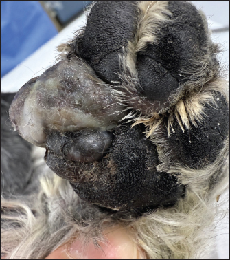
AbstractBackground: Malignant melanoma of the canine metacarpal pad presents a surgical challenge because removal of tumor-bearing pad tissue must be balanced with reconstruction of a durable weight-bearing surface. Although second-digit pad transfer has been described for selected metacarpal pad defects, reconstruction completed with the second digit alone after total metacarpal pad excision for canine metacarpal pad malignant melanoma, while preserving the fifth digit, has not been documented to the authors’ knowledge. Case Description: A 12-year-old male Miniature Schnauzer was referred for a bleeding mass contiguous with the left forelimb metacarpal pad. Punch biopsy confirmed malignant melanoma, and cytology of the left superficial cervical and axillary lymph nodes suggested reactive lymphadenopathy. Twelve days after initial examination, the tumor-bearing metacarpal pad was excised en bloc, the left superficial cervical lymph node and a pigmented right upper lip lesion were excised, and the metacarpal pad defect was reconstructed using a phalangeal fillet technique. Reconstruction using both the second and fifth digits had been anticipated preoperatively, but temporary transposition of the filleted second digit suggested adequate central weight-bearing coverage and contour. Therefore, reconstruction was completed with the second digit alone, and the fifth digit was preserved. Histopathology confirmed malignant melanoma of the metacarpal pad, with no neoplastic cells identified at the surgical margins, and no neoplastic lesion in the left superficial cervical lymph node. The right upper lip lesion was also malignant melanoma; subsequent wider excision and right mandibular lymph node biopsy revealed no residual or nodal neoplastic lesion. Retrospective two-dimensional planimetry showed that the contralateral second digital pad area was 26.9% of the contralateral metacarpal pad area. Postoperatively, localized incomplete wound healing occurred without infection. Weight-bearing was possible by postoperative day 14, and no visually apparent left forelimb lameness was observed during ordinary in-hospital walking approximately 4 weeks after surgery. Conclusion: This case suggests that, in selected dogs, short-term weight-bearing reconstruction after total metacarpal pad excision for malignant melanoma may be achieved using the second digit alone while preserving the fifth digit. Keywords: Digital pad transposition, Malignant melanoma, Metacarpal pad, Phalangeal fillet technique, Weight-bearing reconstruction. IntroductionThe paw pads of dogs and cats are specialized weight-bearing structures composed of thick cornified epithelium and specialized subcutaneous tissue. These structures provide cushioning, abrasion resistance, and traction, and loss of pad tissue may substantially compromise limb function. The metacarpal and metatarsal pads, together with the third and fourth digital pads, are considered major load-bearing structures during walking in dogs (Besancon et al., 2004; Neat and Smeak, 2007). Digital pad transposition and phalangeal fillet techniques have been described as reconstructive options for metacarpal or metatarsal pad defects (Olsen et al., 1997; Danielsson, 1998; Neat and Smeak, 2007; Shaw et al., 2014). Danielsson (1998) reported transposition of the second digital pad after excision of a metacarpal pad lymphangioma in a dog, with eventual full weight-bearing and marked adaptation of the transposed pad. Digital pad transposition has also been summarized in a case series and review, with generally favorable outcomes when performed after tumor excision (Neat and Smeak, 2007). More recently, reconstruction of a metatarsal pad defect using the second digital pad has been reported in Japan (Morishima and Yamamura, 2025). Canine foot pad malignant melanoma is uncommon but may show aggressive biological behavior. In a multi-institutional retrospective study of canine foot pad malignant melanomas, regional lymph node metastasis at diagnosis, subsequent regional or distant metastasis, and relatively short survival were reported (Jeon et al., 2022). In addition, the diagnosis and prognostic interpretation of canine melanocytic neoplasms should consider anatomic site, histopathologic features, and clinical stage (Smedley et al., 2022). Therefore, in metacarpal pad malignant melanoma, local tumor control and restoration of a durable weight-bearing surface must both be considered. Second-digit reconstruction alone has been described for selected metacarpal pad defects, and digital pad transposition has been used for partial or complete metacarpal or metatarsal pad defects (Danielsson, 1998; Neat and Smeak, 2007). In addition, bilateral phalangeal fillet reconstruction using the second and fifth digits has been reported after complete metacarpal pad excision for malignant melanoma in a dog (Shaw et al., 2014). Therefore, the uniqueness of the present case does not lie in the use of the second digit alone per se, but rather in the combination of canine metacarpal pad malignant melanoma, total excision of the tumor-bearing metacarpal pad without retaining pad tissue for functional preservation, reconstruction completed with the second digit alone, and preservation of the fifth digit. To the authors’ knowledge, reconstruction completed with a single second-digit phalangeal fillet technique after total metacarpal pad excision for canine metacarpal pad malignant melanoma while preserving the fifth digit has not been documented. This report aims to describe a dog in which total metacarpal pad excision for malignant melanoma was followed by short-term weight-bearing reconstruction using the second digit alone while preserving the fifth digit. Case DetailsA 12-year-old male Miniature Schnauzer weighing 6.2 kg was referred 6 days after the owner noticed bleeding from the left forelimb metacarpal pad. At initial examination, a 1.8 × 1.5 cm mass contiguous with the left forelimb metacarpal pad and enlargement of the left superficial cervical lymph node were identified (Fig. 1). The owner did not wish to pursue limb amputation. Initial diagnostic workup included hematologic and biochemical testing, left forelimb and thoracic radiography, abdominal ultrasonography, plain and contrast-enhanced computed tomography, punch biopsy of the left metacarpal pad lesion, and fine-needle aspiration of the left superficial cervical and left axillary lymph nodes. Clinicopathologic abnormalities included mild anemia, increased alanine aminotransferase activity, and hyperfibrinogenemia. Left forelimb radiographs showed no obvious osseous abnormality adjacent to the mass, and thoracic radiographs showed no obvious abnormality. Abdominal ultrasonography identified multiple choleliths but no clinically relevant abnormalities in the liver, spleen, kidneys, adrenal glands, or abdominal lymph nodes. Computed tomography showed a solitary mass contiguous with the left forelimb metacarpal pad. On radiology review, the mass measured 15.2 × 13.0 × 16.2 mm, was isoattenuating on precontrast images, and showed poor contrast enhancement. No obvious abnormality was identified in the adjacent bones. The left ventral superficial cervical lymph node was severely enlarged, and the left axillary lymph node was mildly enlarged. Lymph node metastasis and reactive hyperplasia were considered differential diagnoses.
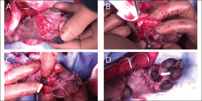
Fig. 1. Preoperative appearance of the left forelimb metacarpal pad lesion at initial presentation. A mass contiguous with the metacarpal pad is visible. The arrow indicates the lesion. Histopathologic examination of the punch biopsy specimen from the left metacarpal pad showed ulceration of the superficial epithelium and nested proliferation of atypical non-epithelial cells, predominantly short spindle cells, extending into deeper tissue. The neoplastic cells had mildly eosinophilic cytoplasm, round to oval nuclei, and moderate anisokaryosis. Abundant melanin pigment was present in the cytoplasm of a subset of cells. Mitotic count was 1 per 10 high-power fields. The lesion was diagnosed as malignant melanoma. Fine-needle aspirates from the left superficial cervical and left axillary lymph nodes consisted predominantly of small lymphocytes, with no obvious atypical cells, and were interpreted as suggestive of reactive lymphadenopathy. The mass enlarged progressively, and the dog became reluctant to bear weight on the affected limb. In addition, multiple small black lesions appeared on the mucosal surface of the right upper lip. At the time of surgery, the left metacarpal pad lesion involved approximately one-half of the metacarpal pad. Because a previous report of reconstruction after complete metacarpal pad excision for malignant melanoma used both the second and fifth digits, reconstruction using both digits was anticipated preoperatively in the present dog (Shaw et al., 2014). Twelve days after initial examination, total excision of the left metacarpal pad melanoma, excisional biopsy of the left superficial cervical lymph node, excisional biopsy of the pigmented right upper lip lesion, and reconstruction of the metacarpal pad defect using a phalangeal fillet technique were performed. Before anesthesia, atropine (0.05 mg/kg), midazolam (0.2 mg/kg), maropitant (1 mg/kg), meloxicam (0.2 mg/kg), and ampicillin (20 mg/kg) were administered. General anesthesia was induced with propofol (5 mg/kg) and maintained with isoflurane in oxygen. No local anesthetic block was performed. Intraoperative analgesia was provided with a fentanyl continuous-rate infusion at 10 μg/kg/hour, which was reduced to 5 μg/kg/hour after surgery and continued until the following morning. No obvious adjacent bone involvement was identified on preoperative imaging. The tumor-bearing metacarpal pad and surrounding skin were excised en bloc along the contour of the pad. The deep plane of excision was selected to remove all identifiable metacarpal pad tissue. Thus, the metacarpal pad was not partially preserved for functional purposes; rather, the entire metacarpal pad was removed as an anatomic unit. After total metacarpal pad excision, the second digit was prepared using a phalangeal fillet technique. The proximal, middle, and distal phalanges were removed. Soft tissue was carefully separated from the bones to preserve the second digital pad and the surrounding soft tissue as much as possible. Particular attention was given to preservation of the vascular supply. Vessels that could not be controlled by compression were coagulated using bipolar electrocautery. The filleted second digital pad was temporarily transposed into the metacarpal pad defect. Based on gross intraoperative assessment, the second digit alone appeared to provide adequate central weight-bearing coverage and acceptable contour. Therefore, the fifth digit was preserved and was not incorporated into the reconstruction. Accordingly, the decision to complete the reconstruction with the second digit alone was based on intraoperative gross assessment after temporary transposition of the filleted pad, rather than on preoperative or intraoperative area measurements. The post-excision defect, preparation of the second-digit fillet, temporary transposition of the second digital pad, and immediate postoperative appearance after preservation of the fifth digit are shown in Figure 2. After the second digital pad was positioned within the metacarpal pad defect, the dermis was apposed with closely spaced simple interrupted sutures using 5–0 polydioxanone. This was intended to minimize wound-edge step-off and increase support at the reconstruction site. The skin was then closed with simple interrupted sutures using 4–0 nylon. Histopathologic examination of the initial surgical specimens identified malignant melanoma in both the left forelimb metacarpal pad and the right upper lip. In the metacarpal pad lesion, atypical non-epithelial cells, predominantly short spindle cells containing abundant melanin pigment, proliferated from the dermis into the subcutis. The metacarpal pad tumor formed a polypoid exophytic mass, and no neoplastic cells were identified at the surgical margins of the metacarpal pad lesion. The left superficial cervical lymph node retained its architecture, and no neoplastic lesion was detected.
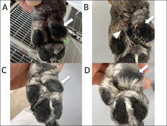
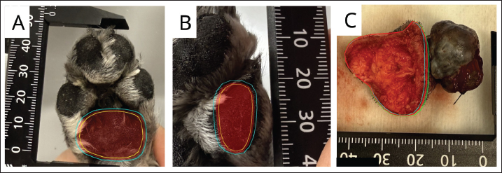
Fig. 2. Intraoperative findings during total metacarpal pad excision and reconstruction using a single second-digit phalangeal fillet technique. (A) Defect after complete metacarpal pad excision. (B) Preparation of the second digit using the phalangeal fillet technique. (C) Temporary transposition of the second digital pad into the defect. (D) Immediate postoperative appearance after reconstruction with preservation of the fifth digit. Arrows in panels C and D indicate the transposed second digital pad. In the right upper lip, a similar neoplastic population was present immediately beneath the mucosa. The right upper lip lesion had initially been removed with minimal margins, and neoplastic cells were close to the horizontal margin. Postoperatively, the left forelimb was managed with a thick Robert Jones-type protective bandage to protect the reconstruction site and reduce early loading that could place tensile or distraction forces on the wound. Additional padding was applied around the distal paw to prevent direct contact between the paw and the floor during early weight-bearing. Ampicillin was administered perioperatively, and oral amoxicillin was used after oral administration became possible. After oral administration became possible, postoperative nonsteroidal anti-inflammatory therapy was continued with oral firocoxib instead of meloxicam and was administered until postoperative day 5. The protective bandage was maintained until postoperative day 14, with scheduled bandage changes and wound inspection. Formal physiotherapy or objective rehabilitation training was not performed. Postoperative management consisted of bandage protection, wound monitoring, activity restriction, and gradual return to ordinary walking after removal of the protective bandage. At the bandage change on postoperative day 4, there was no evidence of infection or dehiscence. On postoperative day 7, a localized area of incomplete wound healing was present at the proximal aspect of the reconstruction, but no evidence of infection was observed, and the dog was discharged for home care. Figure 3 shows the position of the transposed second digital pad and serial postoperative wound changes. On postoperative day 10, localized incomplete healing was noted at the proximal aspect of the reconstruction. Area measurements were performed retrospectively after the surgical decision had been made. Because exophytic tumor growth distorted the excised specimen and the native contour of the metacarpal pad could not be reliably reconstructed from the specimen, the contralateral normal metacarpal pad was used as a surrogate estimate of the original metacarpal pad area. Retrospective two-dimensional digital planimetry was performed on available clinical photographs that included a scale. For regions in which the pad boundary was not completely unequivocal, conservative, central, and liberal contours were generated to assess boundary uncertainty. Reported values and the pad replacement ratio were calculated from the central contour.
Fig. 3. Serial postoperative changes in the surgical wound. (A) Postoperative day 4. (B) Postoperative day 10. (C) Postoperative day 14. (D) Postoperative day 21. Arrows in panels A–D indicate the location of the transposed second digital pad. The arrowhead in panel B indicates a localized area of incomplete healing at the proximal aspect of the reconstruction. The contralateral metacarpal pad area was 421.3 mm2, and the contralateral second digital pad area was 113.3 mm2. The pad replacement ratio, calculated as second digital pad area/metacarpal pad area × 100, was 26.9%. The two-dimensional projected area of the excised metacarpal pad specimen was 474.0 mm2 and was recorded as a reference value only. These measurements were not used to determine the reconstructive approach; rather, they were collected as descriptive data to characterize the conditions under which single second-digit reconstruction was feasible in this dog (Fig. 4). By postoperative day 14, no infection or dehiscence was observed, and the protective bandage was removed. Walking was initiated in the hospital. Mild lameness was observed during ordinary walking, but the dog was able to bear weight on the affected limb. At approximately 3 weeks postoperatively, mild lameness persisted but became less noticeable as walking speed increased. The surgical site had healed adequately, and all sutures were removed. Representative images showing clinical weight-bearing on the operated left forelimb are shown in Figure 5. Approximately 4 weeks after the metacarpal pad surgery, the dog was admitted for planned wider excision of the right upper lip malignant melanoma and right mandibular lymph node biopsy. During the preoperative in-hospital walking assessment at that visit, no visually apparent left forelimb lameness was observed during ordinary walking. Histopathologic examination of the subsequent wider excision specimen from the right upper lip showed granulation tissue at the previous excision site with mild inflammatory and melanophage infiltration, but no residual neoplastic lesion. No neoplastic lesion was identified in the right mandibular lymph node. Gait assessment was based on visual evaluation during ordinary walking in the hospital. Trotting evaluation, numerical lameness scoring, visual analog scale assessment, force-plate analysis, and pressure-distribution analysis were not performed. Ethical approvalThis case report describes diagnostic and therapeutic procedures performed as part of the routine clinical management of a client-owned dog. No experimental procedures were performed. DiscussionThe main clinical significance of this case is that the tumor-bearing metacarpal pad was removed in its entirety, without retaining metacarpal pad tissue for functional preservation, and reconstruction was completed using a single second-digit phalangeal fillet technique. Histopathologically, no neoplastic cells were identified at the surgical margins of the metacarpal pad lesion. Second-digit reconstruction alone is not new in itself. Danielsson (1998) reported transposition of the second digital pad after resection of a metacarpal pad lymphangioma in a dog, and Neat and Smeak (2007)included cases in which digital pad transposition was used for partial or complete metacarpal or metatarsal pad defects. The distinctive feature of the present case is the combination of canine metacarpal pad malignant melanoma, total metacarpal pad excision without retaining pad tissue for functional preservation, and reconstruction completed with a single second-digit phalangeal fillet technique, allowing preservation of the fifth digit. In addition, no neoplastic cells were identified at the surgical margins of the metacarpal pad lesion.
Fig. 4. Retrospective two-dimensional pad area measurements. (A) Contralateral metacarpal pad. (B) Contralateral second digital pad. (C) Excised metacarpal pad specimen, inner view. Multiple outlines indicate plausible contours used to assess uncertainty in boundary selection. In panels A and B, the yellow, red, and cyan outlines indicate conservative, central, and liberal contours, respectively. In panel C, the cyan, red, and green outlines indicate conservative, central, and liberal contours, respectively. Reported values and the pad replacement ratio were calculated from the central contour, shown in red in all panels. The contralateral metacarpal pad area was 421.3 mm2, and the contralateral second digital pad area was 113.3 mm2, yielding a pad replacement ratio of 26.9%. The projected area of the excised metacarpal pad specimen was 474.0 mm2 and was recorded as a reference value only. These measurements represent two-dimensional projected areas and do not represent true three-dimensional pad surface area or dynamic ground-contact area.
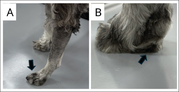
Fig. 5. Representative postoperative clinical weight-bearing at approximately 3 weeks after surgery. (A) Lateral view of the dog standing on the examination table. The arrow indicates the operated left forelimb. (B) Caudal view of the operated paw during weight bearing. The arrow indicates the transposed second digital pad contacting the supporting surface. These images represent clinical observation only and do not indicate quantitative gait analysis, load measurement, or pressure-distribution assessment. In the Neat and Smeak (2007) series, the dog with metacarpal pad melanoma reconstructed with the second digit alone underwent 33% metacarpal pad excision, not total metacarpal pad excision. The case involving 100% metacarpal pad excision and second-digit reconstruction alone was a cat with a basal cell tumor, not a dog with malignant melanoma. A dog with metacarpal pad malignant melanoma reconstructed after complete metacarpal pad excision using bilateral second- and fifth-digit phalangeal fillets has been reported, but that reconstruction was not completed with the second digit alone (Shaw et al., 2014). Therefore, although previous reports indicate that digital pad transposition and phalangeal fillet techniques can be used for large weight-bearing defects, the present case appears to represent a distinct scenario in which total metacarpal pad excision for canine malignant melanoma was reconstructed using the second digit alone. Canine foot pad malignant melanoma is uncommon, and its biological behavior remains incompletely defined, but it can be aggressive. In a multi-institutional study of 20 canine foot pad malignant melanomas, regional lymph node metastasis was identified at diagnosis in 20% of cases, and the overall rate of regional or distant metastasis was 55% (Jeon et al., 2022). Therefore, long-term oncologic outcome in the present case should be interpreted cautiously. In addition, melanocytic neoplasms in dogs require diagnostic and prognostic interpretation based on anatomic site, histopathologic features, and clinical stage (Smedley et al., 2022). The oncologic interpretation of this case is limited by the concurrent right upper lip malignant melanoma identified at the time of the initial surgery. Subsequent wider excision of the right upper lip lesion did not identify residual neoplastic tissue, and no neoplastic lesion was detected in the right mandibular lymph node. However, based on the available clinical and histopathologic information, it was not possible to determine whether the right upper lip melanoma represented a separate primary lesion or was related to the metacarpal pad melanoma. Therefore, this report should not be interpreted as demonstrating long-term oncologic control; its principal focus is the short-term feasibility of weight-bearing reconstruction after total metacarpal pad excision. Compared with the most analogous previous report, in which bilateral second- and fifth-digit phalangeal fillets were used, the present reconstruction was completed with the second digit alone, allowing preservation of the fifth digit (Shaw et al., 2014). This intraoperative decision was based on gross assessment after temporary transposition of the filleted second digital pad, rather than on the retrospective pad replacement ratio. The pad replacement ratio should, therefore, be interpreted as descriptive, hypothesis-generating information from a single case. The use of conservative, central, and liberal contours allowed assessment of boundary-selection uncertainty; however, the reported values were derived from the central contour and were not calculated as averages of multiple contours. These measurements represented two-dimensional projected areas from scale-containing clinical photographs and did not represent true three-dimensional pad surface area or dynamic ground-contact area. The ratio of the contralateral second digital pad area to the contralateral metacarpal pad area was 26.9%. This value does not define an indication for single second-digit reconstruction. Rather, it provides one measurable descriptor that may be useful for comparison if similar cases are accumulated. Postoperative protection was considered important because the reconstruction site was located on a weight-bearing surface and early loading could place tensile or distraction forces on the wound. Therefore, the limb was protected for 14 days using a thick Robert Jones-type protective bandage. In addition, closely spaced simple interrupted dermal sutures with 5–0 polydioxanone were used before skin closure with 4–0 nylon to minimize wound-edge step-off and increase support at the reconstruction site. Formal physiotherapy was not performed; instead, postoperative management consisted of bandage protection, wound monitoring, activity restriction, analgesic and anti-inflammatory medication, and gradual return to ordinary walking after bandage removal. Neat and Smeak (2007) described the use of a protective bandage after digital pad transposition until wound healing was considered adequate. Danielsson (1998)also reported splint protection after the second digital pad transposition. Thus, the postoperative protection used in the present case was broadly consistent with previous management strategies for digital pad transfer. A localized wound-healing complication was observed postoperatively, but there was no evidence of infection. By postoperative day 14, the surgical site was stable enough to remove the protective bandage and initiate walking. The dog was able to bear weight despite mild lameness. At approximately 3 weeks, mild lameness persisted but was improving, and at approximately 4 weeks, no visually apparent left forelimb lameness was observed during ordinary in-hospital walking. These findings suggest that the reconstructed surface functioned as a weight-bearing surface in the short term. Representative postoperative images obtained at approximately 3 weeks showed clinical weight bearing on the operated limb, including contact of the transposed second digital pad with the supporting surface. However, these images represent clinical observation only, and objective gait, load, or pressure-distribution analyses were not performed. This report has several limitations. First, oncologic follow-up was short; local recurrence, regional lymph node metastasis, and distant metastasis could not be fully evaluated. Second, malignant melanoma was also present in the right upper lip, complicating the interpretation of the overall oncologic course. Third, the gait assessment was based only on visual evaluation during ordinary walking in the hospital. Trotting evaluation, numerical lameness scoring, visual analog scale assessment, force-plate analysis, and pressure-distribution analysis were not performed. Therefore, this report does not prove normalization of gait; it documents short-term clinical weight bearing. Fourth, the postoperative area of the transposed second digital pad could not be quantified from standardized images. Future cases should include standardized photographs with a scale and, ideally, an objective functional assessment. In summary, this case suggests that, in selected dogs, total metacarpal pad excision for malignant melanoma may be followed by short-term weight-bearing reconstruction using a single second-digit phalangeal fillet technique. Further case accumulation and standardized functional assessment are needed to clarify the indications for reconstruction completed with the second digit alone while preserving the fifth digit. ConclusionA dog with malignant melanoma of the metacarpal pad underwent total metacarpal pad excision without partial preservation of the pad for functional purposes. No neoplastic cells were identified at the surgical margins of the metacarpal pad lesion, and reconstruction was completed using a single second-digit phalangeal fillet technique. Although a localized wound-healing complication occurred, no infection was observed, weight-bearing was possible by postoperative day 14, and no visually apparent left forelimb lameness was observed during ordinary in-hospital walking approximately 4 weeks after surgery. This case suggests that single second-digit reconstruction may be feasible after total metacarpal pad excision in selected dogs. AcknowledgmentsNone. Conflict of interestThe authors declare that there is no conflict of interest. FundingThis report received no specific grant from any funding agency in the public, commercial, or not-for-profit sectors. Authors’ contributionsYasuhiro Yamaguchi performed the surgical procedure, managed the case, collected clinical data, drafted the manuscript, and approved the final manuscript. Takashi Higuchi contributed to diagnostic evaluation, oncologic case management, interpretation of clinical data, critical revision of the manuscript, and approval of the final manuscript. Sumire Iriyama contributed to case management, data organization, manuscript revision, and approval of the final manuscript. Yuji Sawatari contributed to case management, manuscript revision, and approval of the final manuscript. Data availabilityThe anonymized clinical data supporting the findings of this case report are available from the corresponding author upon reasonable request. ReferencesBesancon, M.F., Conzemius, M.G., Evans, R.B. and Ritter, M.J. 2004. Distribution of vertical forces in the pads of greyhounds and labrador retrievers during walking. Am. J. Vet. Res. 65, 1497–1501; doi:10.2460/ajvr.2004.65.1497 Danielsson, F. 1998. Lymphangioma in the metacarpal pad of a dog. J. Small Anim. Pract. 39, 295–298; doi:10.1111/j.1748-5827.1998.tb03654.x Jeon, M.D., Leeper, H.J., Cook, M.R., McMillan, S.K., Bennett, T., Murray, C.A., Tripp, C.D. and Curran, K.M. 2022. Multi-institutional retrospective study of canine foot pad malignant melanomas: 20 cases. Vet. Comp. Oncol. 20, 854–861; doi:10.1111/vco.12846 Morishima, T. and Yamamura, T. 2025. Surgical reconstruction of the metatarsal pad using 2nd digital pad in a dog. Jpn. J. Vet. Anesth. Surg. 55, 51–55; doi:10.2327/jjvas.55.51 Neat, B.C. and Smeak, D.D. 2007. Reconstructing weight-bearing surfaces: digital pad transposition. Compend. Contin. Educ. Vet. 29, 39–46. Olsen, D., Straw, R.C., Withrow, S.J. and Basher, A.W. 1997. Digital pad transposition for replacement of the metacarpal or metatarsal pad in dogs. J. Am. Anim. Hosp. Assoc. 33, 337–341; doi:10.5326/15473317-33-4-337 Shaw, T., James, F., Beierer, L. and Hosgood, G. 2014. Bilateral phalangeal fillet technique for metacarpal pad reconstruction in a dog. Can. Vet. J. 55, 955–960. Smedley, R.C., Bongiovanni, L., Bacmeister, C., Clifford, C.A., Christensen, N., Dreyfus, J.M., Gary, J.M., Pavuk, A., Rowland, P.H., Swanson, C., Tripp, C., Woods, J.P. and Bergman, P.J. 2022. Diagnosis and histopathologic prognostication of canine melanocytic neoplasms: a consensus of the Oncology-Pathology Working Group. Vet. Comp. Oncol. 20, 739–751; doi:10.1111/vco.12827 | ||
| How to Cite this Article |
| Pubmed Style Yamaguchi Y, Higuchi T, Iriyama S, Sawatari Y. Single second-digit phalangeal fillet reconstruction after total metacarpal pad excision for malignant melanoma in a dog. Open Vet. J.. 2026; 16(8): 5517-5524. doi:10.5455/OVJ.2026.v16.i8.44 Web Style Yamaguchi Y, Higuchi T, Iriyama S, Sawatari Y. Single second-digit phalangeal fillet reconstruction after total metacarpal pad excision for malignant melanoma in a dog. https://www.openveterinaryjournal.com/?mno=320639 [Access: August 08, 2026]. doi:10.5455/OVJ.2026.v16.i8.44 AMA (American Medical Association) Style Yamaguchi Y, Higuchi T, Iriyama S, Sawatari Y. Single second-digit phalangeal fillet reconstruction after total metacarpal pad excision for malignant melanoma in a dog. Open Vet. J.. 2026; 16(8): 5517-5524. doi:10.5455/OVJ.2026.v16.i8.44 Vancouver/ICMJE Style Yamaguchi Y, Higuchi T, Iriyama S, Sawatari Y. Single second-digit phalangeal fillet reconstruction after total metacarpal pad excision for malignant melanoma in a dog. Open Vet. J.. (2026), [cited August 08, 2026]; 16(8): 5517-5524. doi:10.5455/OVJ.2026.v16.i8.44 Harvard Style Yamaguchi, Y., Higuchi, . T., Iriyama, . S. & Sawatari, . Y. (2026) Single second-digit phalangeal fillet reconstruction after total metacarpal pad excision for malignant melanoma in a dog. Open Vet. J., 16 (8), 5517-5524. doi:10.5455/OVJ.2026.v16.i8.44 Turabian Style Yamaguchi, Yasuhiro, Takashi Higuchi, Sumire Iriyama, and Yuji Sawatari. 2026. Single second-digit phalangeal fillet reconstruction after total metacarpal pad excision for malignant melanoma in a dog. Open Veterinary Journal, 16 (8), 5517-5524. doi:10.5455/OVJ.2026.v16.i8.44 Chicago Style Yamaguchi, Yasuhiro, Takashi Higuchi, Sumire Iriyama, and Yuji Sawatari. "Single second-digit phalangeal fillet reconstruction after total metacarpal pad excision for malignant melanoma in a dog." Open Veterinary Journal 16 (2026), 5517-5524. doi:10.5455/OVJ.2026.v16.i8.44 MLA (The Modern Language Association) Style Yamaguchi, Yasuhiro, Takashi Higuchi, Sumire Iriyama, and Yuji Sawatari. "Single second-digit phalangeal fillet reconstruction after total metacarpal pad excision for malignant melanoma in a dog." Open Veterinary Journal 16.8 (2026), 5517-5524. Print. doi:10.5455/OVJ.2026.v16.i8.44 APA (American Psychological Association) Style Yamaguchi, Y., Higuchi, . T., Iriyama, . S. & Sawatari, . Y. (2026) Single second-digit phalangeal fillet reconstruction after total metacarpal pad excision for malignant melanoma in a dog. Open Veterinary Journal, 16 (8), 5517-5524. doi:10.5455/OVJ.2026.v16.i8.44 |


