





| Case Report | ||
Open Vet. J.. 2026; 16(8): 5881-5887
Open Veterinary Journal, (2026), Vol. 16(8): 5881–5887 Case Report Calcium carbonate urolithiasis with ureteral obstruction in an adult wether sheep: A case reportChihiro Kanno*, Shogo Sato, Izumi Yamauchi, Mai Shimamura, Yosuke Maeda and Fumiaki TakahashiDepartment of Veterinary Medicine, School of Veterinary Medicine, Kitasato University, Towada, Aomori, Japan *Corresponding Author: Chihiro Kanno. Department of Veterinary Medicine, School of Veterinary Medicine, Kitasato University, Towada, Aomori, Japan. Email: c-kanno [at] vmas.kitasato-u.ac.jp Submitted: 11/05/2026 Revised: 15/07/2026 Accepted: 30/07/2026 Published: 20/08/2026 © 2025 Open Veterinary Journal
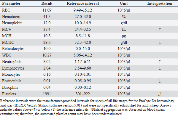
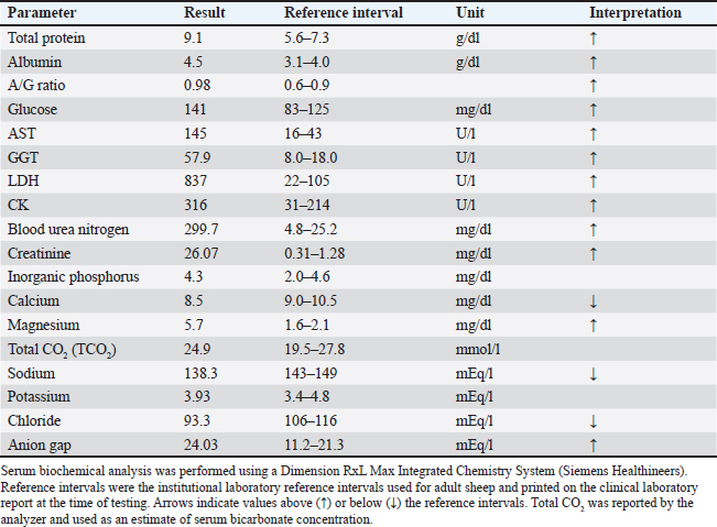
AbstractBackground: Obstructive urolithiasis is an important urinary tract disorder in male small ruminants and is usually recognized as lower urinary tract obstruction. Upper urinary tract involvement may be overlooked when gastrointestinal or systemic signs predominate. Case Description: A three-year-two-month-old Corriedale wether sheep was presented with anorexia, lethargy, foul-smelling diarrhea, and pale mucous membranes. Parasitic enteritis was initially suspected. Hematology and serum biochemistry revealed marked azotemia, hypermagnesemia, hypocalcemia, hypochloremia, and an increased anion gap. Despite supportive treatment, the sheep deteriorated and was euthanized for animal welfare reasons. Gross necropsy revealed right hydronephrosis, a calculus obstructing the right ureter, ureteral mucosal ulceration, bilateral renal pelvic hemorrhage, small uroliths and urinary sediment in the urinary bladder, and bladder mucosal petechiae. No urinary bladder rupture was observed. Infrared spectrophotometric analysis identified calcium carbonate as the predominant component of the right ureteral calculus. Conclusion: Calcium carbonate urolithiasis in adult wether sheep may involve the ureter and contribute to severe renal dysfunction without typical signs of distal urethral obstruction. Urolithiasis should be considered in adult sheep with non-specific systemic deterioration, especially when urination cannot be confirmed. Keywords: Azotemia, Calcium carbonate, Sheep, Ureteral obstruction, Urolithiasis. IntroductionObstructive urolithiasis is one of the most important urinary tract disorders in small ruminants, particularly in male and castrated animals (Videla and van Amstel, 2016; Scully, 2021; Cook, 2023). Clinical attention is usually directed toward lower urinary tract obstruction because anatomical narrowing at the sigmoid flexure and urethral process predisposes males to distal obstruction (Videla and van Amstel, 2016; Riedi et al., 2018a; Scully, 2021; Cook, 2023). Affected animals may show dysuria, stranguria, abdominal pain, anorexia, depression, azotemia, and systemic deterioration; however, clinical signs vary, and urinary disease may be difficult to recognize when typical urinary signs are absent (Videla and van Amstel, 2016; Riedi et al., 2018a; Cook, 2023). Urolith composition is clinically relevant because it can influence prevention and herd management (Videla and van Amstel, 2016; Jones et al., 2017; Cook, 2023). In sheep and goats, amorphous magnesium calcium phosphate, struvite, and calcium carbonate are important urolith components (Jones et al., 2017). Calcium carbonate represented a substantial proportion of uroliths in a previous analysis of sheep and goat calculi (Jones et al., 2017). In goats, calcium carbonate urolithiasis has been associated with male sex, neuter status, age, geographic location, and season (Nwaokorie et al., 2015), and recent data in goats also support calcium carbonate as an important urolith type (Chigerwe et al., 2025). Experimental work in goats showed that calcium carbonate urolithiasis can be induced under high-calcium dietary conditions (Jones et al., 2018). In lambs fed high-concentrate diets, urinary acidifying additives have been investigated as preventive measures, reflecting the clinical relevance of diet and urinary pH (Buarque et al., 2023). This report describes an adult Corriedale wether sheep with calcium carbonate urolithiasis associated with right ureteral obstruction, severe renal dysfunction, and non-specific clinical signs initially suggestive of gastrointestinal disease. The clinical significance of this case lies in the combination of ureteral obstruction in an adult wether, the absence of recognized typical signs of distal urethral obstruction, and the predominance of gastrointestinal and systemic signs that obscured recognition of urinary tract disease. Case DetailsA three-year-two-month-old castrated male Corriedale sheep, weighing approximately 80 kg, was examined at the university farm by veterinarians from the Large Animal Medical Center of Kitasato University Veterinary Teaching Hospital. The sheep was an institutionally maintained animal and was managed as a clinical case. It was housed in a group and received 300 g/day of a commercially available formulated feed for sheep and goats and 550 g/day of timothy hay. According to the guaranteed analysis provided on the feed label, the formulated feed contained ≥12.0% crude protein, ≥2.0% crude fat, ≤8.0% crude fiber, ≤7.0% crude ash, ≥0.35% calcium, ≥0.30% phosphorus, and ≥74.0% total digestible nutrients. The feed consisted primarily of cereals (73%), milling by-products (12%), plant-derived oilseed meals (10%), and other ingredients (5%). Tap water and mineral salt were available ad libitum; the detailed composition of the mineral salt was unavailable. The sheep developed anorexia, lethargy, and diarrhea eight days before the first veterinary examination. The animal was isolated, and oral treatment intended to improve ruminal function was administered, but feed intake gradually decreased. At the first examination, the sheep showed anorexia, depression, a rectal temperature of 39.4°C, heart rate of 120 beats/min, respiratory rate of 100 breaths/min, foul-smelling scant diarrhea, and pale mucous membranes. Parasitic enteritis was initially suspected based on the presence of diarrhea, pale mucous membranes, and poor general condition. Fecal examination was not performed because an adequate fecal sample could not be obtained. Hematological analysis was performed using an automated veterinary hematology analyzer (ProCyte Dx, IDEXX Laboratories, Inc., Westbrook, ME, USA). Compared with the manufacturer-provided reference intervals for sheep of all life stages, mean corpuscular volume and absolute neutrophil count were increased, whereas mean corpuscular hemoglobin concentration, absolute lymphocyte and eosinophil counts, and the automated platelet count were decreased (Table 1). Red blood cell count, hematocrit, hemoglobin concentration, mean corpuscular hemoglobin, reticulocyte count, and total white blood cell count were within the reference intervals. Blood smear examination revealed numerous acanthocytic and echinocytic erythrocytes. Platelet aggregation was also observed, indicating that the automated platelet count may have been underestimated. Serum biochemical analysis was performed using an automated chemistry analyzer (Dimension RxL Max Integrated Chemistry System, Siemens Healthineers, Erlangen, Germany). Based on institutional laboratory reference intervals used for adult sheep, serum biochemistry revealed increased total protein, albumin, albumin-to-globulin ratio, glucose, aspartate aminotransferase, gamma-glutamyl transferase, lactate dehydrogenase, creatine kinase, blood urea nitrogen, creatinine, magnesium, and anion gap, together with decreased calcium, sodium, and chloride concentrations (Table 2). The marked increases in blood urea nitrogen and creatinine were consistent with severe renal dysfunction. Table 1. Hematological findings at presentation in an adult Corriedale wether sheep with calcium carbonate urolithiasis and right ureteral obstruction.
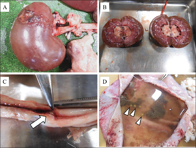
Ivermectin and meloxicam were administered at the first examination. The following day, no clinical improvement was observed. Fluid therapy was administered with sodium bicarbonate, vitamin supplementation, a methionine preparation, and flunixin meglumine. Similar treatment was continued on the next day. The sheep remained at the university farm and was examined there without hospitalization. During the four-day observation period, farm staff checked the bedding for evidence of urination twice daily during feeding and bedding replacement. During veterinary examinations, the hair surrounding the preputial orifice was inspected for wetness, and gentle preputial massage was performed to assess whether urine could be expressed. No evidence of urination was identified during this period; however, continuous observation and objective measurement of urine output were not performed. Because the general condition progressively deteriorated and the prognosis was considered poor, humane euthanasia was elected on the fourth day for animal welfare reasons. The sheep was sedated with xylazine (0.2 mg/kg, intravenously), anesthetized with pentobarbital sodium (20 mg/kg, intravenously), and then exsanguinated according to the institutional euthanasia method specified by the Animal Experimentation Committee of Kitasato University School of Veterinary Medicine. A gross necropsy was performed. Gross examination revealed mild enlargement and hydronephrosis of the right kidney. A focal dark discoloration was present on the surface of the right kidney (Fig. 1A). Petechial hemorrhages were observed in the renal pelvis of both kidneys (Fig. 1B). The left kidney had no other gross abnormalities. A calculus was identified within the right ureter and obstructed the lumen. The ureteral mucosa at the obstructed site was ulcerated, consistent with pressure injury caused by the calculus (Fig. 1C). The left ureter was opened longitudinally, and no calculus or gross obstructive lesion was identified. The urinary bladder was opened and contained a large volume of urine, urinary sediment, and multiple small spherical uroliths (Fig. 1D). Petechial hemorrhages were present on the bladder mucosa, but urinary bladder rupture was not identified. The bladder neck, urethral process, sigmoid flexure, and pelvic urethra were not systematically dissected and examined. Gross gastrointestinal lesions included focal abomasal erosion and congestion or petechial hemorrhages of the large intestinal mucosa. Table 2. Serum biochemical findings at presentation in an adult Corriedale wether sheep with calcium carbonate urolithiasis and right ureteral obstruction.
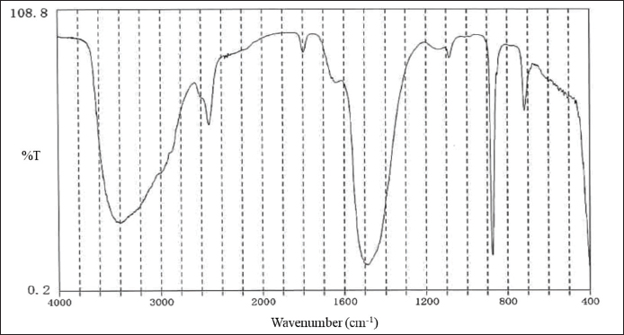
Fig. 1. Gross necropsy findings of the urinary tract in an adult Corriedale wether sheep with calcium carbonate urolithiasis. (A) Right kidney showing focal dark discoloration on the renal surface. (B) Cut surfaces of the kidneys showing renal pelvic hemorrhage. (C) Right ureter showing the obstructed site with mucosal ulceration associated with an intraluminal calculus (arrow). (D) Urinary bladder containing urinary sediment and multiple small spherical uroliths (arrowheads). Infrared spectrophotometric analysis was performed on the calculus removed from the obstructed right ureter because urolith composition cannot be reliably inferred from gross appearance alone (Videla and van Amstel, 2016; Jones et al., 2017; Cook, 2023). The central portion of the calculus was mixed with potassium bromide, ground, compressed into a pellet, and analyzed using infrared absorption spectroscopy. Calcium carbonate was identified as the predominant component, accounting for ≥98% of the calculus composition (Fig. 2). Ethical approvalThis case involved an institutionally maintained sheep that was managed as a clinical case at the Large Animal Medical Center of Kitasato University Veterinary Teaching Hospital. No experimental procedures were performed. Humane euthanasia was performed for animal welfare reasons because the sheep was judged to have a poor prognosis and was considered unsalvageable on clinical grounds. Euthanasia was performed according to the institutional euthanasia method specified by the Animal Experimentation Committee of Kitasato University School of Veterinary Medicine. Briefly, the sheep was sedated with xylazine (0.2 mg/kg, intravenously), anesthetized with pentobarbital sodium (20 mg/kg, intravenously), and then exsanguinated. Ethics approval was not required because the procedures were performed as clinical management and post-mortem investigation of a clinical case, not as an experimental study. DiscussionThis case highlights calcium carbonate urolithiasis with right ureteral obstruction in an adult wether sheep in which gastrointestinal and systemic signs predominated and typical signs of distal urethral obstruction were not recognized clinically. The initial signs, including anorexia, lethargy, foul-smelling diarrhea, and pale mucous membranes, were non-specific and led to suspicion of parasitic enteritis. Necropsy revealed severe urinary tract lesions, including right ureteral obstruction, hydronephrosis, bilateral renal pelvic hemorrhage, bladder uroliths, and bladder mucosal hemorrhage. The combination of adult age, upper urinary tract obstruction, and gastrointestinal-predominant presentation represents the principal clinical value of this case.
Fig. 2. Infrared absorption spectrum of the right ureteral calculus. Infrared spectrophotometric analysis identified calcium carbonate as the predominant component of the right ureteral calculus (≥98%). Most discussions of obstructive urolithiasis in small ruminants emphasize lower urinary tract obstruction (Videla and van Amstel, 2016; Riedi et al., 2018a; Scully, 2021; Cook, 2023). In the present case, ante-mortem ultrasonography or radiography was not performed because urinary tract disease was not initially suspected. This is an important limitation, because ultrasonography was useful in a large retrospective study (Riedi et al., 2018a), and plain radiography has been reported to aid diagnosis and surgical management in goats and sheep (Kinsley et al., 2013). In retrospect, diagnostic imaging may have helped identify urinary tract distension or uroliths before euthanasia. Nevertheless, detection of a ureteral calculus may be limited by its size, location, and radiographic visibility, and indirect findings such as hydronephrosis or ureteral dilation may be more readily recognized than the calculus itself (Kinsley et al., 2013; Riedi et al., 2018a). The predominance of calcium carbonate was clinically important. Urolith composition varies substantially among small ruminants and cannot be determined accurately from gross appearance alone (Videla and van Amstel, 2016; Jones et al., 2017; Cook, 2023). Jones et al. (2017) showed that calcium carbonate represented an important urolith type in sheep and goats. More recent data in goats also support calcium carbonate as an important urolith type, although extrapolation to sheep should be cautious because species-specific and management-related factors may influence urolith composition (Chigerwe et al., 2025). These studies support the clinical relevance of calcium carbonate urolithiasis in small ruminants, but they do not establish the cause of urolith formation in the present sheep. Risk factors and pathogenesis should be interpreted cautiously in this case. In goats, calcium carbonate urolithiasis has been associated with male sex, neuter status, age, geographic location, and season (Nwaokorie et al., 2015). Experimental work in goats has shown that calcium carbonate urolithiasis can be induced under high-calcium dietary conditions (Jones et al., 2018). In lambs fed high-concentrate diets, urinary acidifying additives have been investigated for prevention of urolithiasis (Buarque et al., 2023). In the present sheep, the type, amount, and guaranteed composition stated on the feed label were documented. However, the actual feed batch, timothy hay, mineral salt, drinking water, urine, and total ration were not chemically analyzed, and urine pH and urine mineral concentrations were unavailable. Therefore, the etiologic factors responsible for calcium carbonate urolith formation could not be determined, and no causal association can be made between the urolithiasis and any specific feed product or dietary component. The marked azotemia was likely associated with urinary tract disease and renal dysfunction, but unilateral ureteral obstruction alone was unlikely to fully account for its severity. Possible additional contributors included prerenal azotemia associated with dehydration and reduced intake, concurrent intrinsic renal dysfunction that was not detectable by gross examination, and impaired lower urinary outflow that could not be completely excluded because the bladder neck and urethra were not systematically dissected. Although the left kidney showed no gross abnormality other than renal pelvic petechial hemorrhages and no gross obstructive lesion was identified in the left ureter, histopathological examination of both kidneys was not performed. Therefore, the presence and extent of intrinsic renal injury could not be determined. Treatment decisions in small ruminant urinary obstruction depend on patient condition, obstruction site, urinary tract integrity, and available surgical options (Mejia et al., 2022). Although surgical options, including tube cystostomy, have been reported (Ewoldt et al., 2006), their applicability depends on obstruction site and systemic condition. Variables at initial examination and clinical management have also been associated with survival in small ruminants with obstructive urolithiasis (Riedi et al., 2018b). In this case, delayed recognition of urinary tract disease, severe azotemia, and confirmed ureteral obstruction would have complicated therapeutic decision-making. This case has several limitations. Ante-mortem urinary imaging, histopathology, urinalysis, urine pH, bacterial culture, urine mineral analysis, and independent chemical analysis of the actual feed batch, hay, mineral salt, drinking water, and total ration were not performed. Continuous observation and objective measurement of urine output were unavailable. In addition, the bladder neck, urethral process, sigmoid flexure, and pelvic urethra were not systematically dissected; therefore, distal urethral obstruction could not be completely excluded. The contribution of gross gastrointestinal lesions, possible parasitism, or uremia-associated gastrointestinal dysfunction to the clinical signs could not be determined because fecal examination, histopathology, and detailed gastrointestinal evaluation were unavailable. Therefore, concomitant gastrointestinal disease or parasitism could not be completely excluded. Nevertheless, the severity of azotemia and the gross urinary tract lesions indicated that calcium carbonate urolithiasis with right ureteral obstruction was a major contributor to renal dysfunction and systemic deterioration. ConclusionCalcium carbonate urolithiasis in adult wether sheep may involve the ureter and present with non-specific systemic and gastrointestinal signs rather than typical signs of distal urethral obstruction. In adult sheep with anorexia, depression, diarrhea, azotemia, or unconfirmed urination, urolithiasis should remain an important differential diagnosis. AcknowledgmentsThe authors thank the staff of the Large Animal Medical Center of Kitasato University Veterinary Teaching Hospital for their assistance with clinical care and sample handling. Conflict of interestThe authors declare that there is no conflict of interest. FundingThis research received no specific grant. Authors’ contributionsCK conceptualized the case report, interpreted the clinical and necropsy findings, and drafted the manuscript. SS managed the clinical case and contributed to interpretation of clinical findings. IY organized the initial case information and contributed to data curation. MS contributed to validation of case interpretation, discussion of limitations, and critical revision of the manuscript. YM contributed to interpretation of clinical findings and manuscript revision. FT supervised the case report preparation and critically reviewed the manuscript. CK, IY, and MS prepared the figures and tables. All authors reviewed and approved the final manuscript. Data availabilityAll data supporting the findings of this report are included in the manuscript. Additional clinical records are available from the corresponding author upon reasonable request. ReferencesBuarque, V.L.M., Bezerra, H.V.A., Rissi, G.P., Oliveira, R.Í.G., Silva, N.M., Cônsolo, N.R.B., et al. 2023. The use of additives to prevent urolithiasis in lambs fed diets with a high proportion of concentrate. Vet. Sci. 10, 617; doi:10.3390/vetsci10100617. Chigerwe, M., Killilea, D.W., Hardgrove, C.N., Mann, U., Stoller, M.L. and Westropp, J.L. 2025. Trace element content is associated with urolith type in goats and pigs diagnosed with urolithiasis. Am. J. Vet. Res. 86, ajvr.25.01.0011; doi:10.2460/ajvr.25.01.0011. Cook, M.J. 2023. Urinary calculi of small ruminants. Vet. Clin. North Am. Food Anim. Pract. 39, 355–370; doi:10.1016/j.cvfa.2023.02.006. Ewoldt, J.M., Anderson, D.E., Miesner, M.D. and Saville, W.J. 2006. Short- and long-term outcome and factors predicting survival after surgical tube cystostomy for treatment of obstructive urolithiasis in small ruminants. Vet. Surg. 35, 417–422; doi:10.1111/j.1532-950X.2006.00169.x. Jones, M.L., Gibbons, P.M., Roussel, A.J. and Dominguez, B.J. 2017. Mineral composition of uroliths obtained from sheep and goats with obstructive urolithiasis. J. Vet. Intern. Med. 31, 1202–1208; doi:10.1111/jvim.14743. Jones, M.L., Dominguez, B.J. and Deveau, M.A. 2018. An experimental model for calcium carbonate urolithiasis in goats. J. Vet. Intern. Med. 32, 1268–1273; doi:10.1111/jvim.15061. Kinsley, M.A., Semevolos, S., Parker, J.E., Duesterdieck-Zellmer, K. and Huber, M. 2013. Use of plain radiography in the diagnosis, surgical management, and postoperative treatment of obstructive urolithiasis in 25 goats and 2 sheep. Vet. Surg. 42, 663–668; doi:10.1111/j.1532-950X.2013.12021.x. Mejia, S., McOnie, R.C., Nelligan, K.L. and Fubini, S.L. 2022. Small ruminant urinary obstruction: decision trees for treatment. J. Am. Vet. Med. Assoc. 260, S64–S71; doi:10.2460/javma.22.02.0071. Nwaokorie, E.E., Osborne, C.A., Lulich, J.P., Fletcher, T.F., Ulrich, L.K., Koehler, L.A., et al. 2015. Risk factors for calcium carbonate urolithiasis in goats. J. Am. Vet. Med. Assoc. 247, 293–299; doi:10.2460/javma.247.3.293. Riedi, A.K., Knubben-Schweizer, G. and Meylan, M. 2018a. Clinical findings and diagnostic procedures in 270 small ruminants with obstructive urolithiasis. J. Vet. Intern. Med. 32, 1274–1282; doi:10.1111/jvim.15128. Riedi, A.K., Nathues, C., Knubben-Schweizer, G., Nuss, K. and Meylan, M. 2018b. Variables of initial examination and clinical management associated with survival in small ruminants with obstructive urolithiasis. J. Vet. Intern. Med. 32, 2105–2114; doi:10.1111/jvim.15336. Scully, C.M. 2021. Management of urologic conditions in small ruminants. Vet. Clin. North Am. Food Anim. Pract. 37, 93–104; doi:10.1016/j.cvfa.2020.10.003. Videla, R. and van Amstel, S. 2016. Urolithiasis. Vet. Clin. North Am. Food Anim. Pract. 32, 687–700; doi:10.1016/j.cvfa.2016.05.010. | ||
| How to Cite this Article |
| Pubmed Style Kanno C, Sato S, Yamauchi I, Shimamura M, Maeda Y, Takahashi F. Calcium carbonate urolithiasis with ureteral obstruction in an adult wether sheep: A case report. Open Vet. J.. 2026; 16(8): 5881-5887. doi:10.5455/OVJ.2026.v16.i8.76 Web Style Kanno C, Sato S, Yamauchi I, Shimamura M, Maeda Y, Takahashi F. Calcium carbonate urolithiasis with ureteral obstruction in an adult wether sheep: A case report. https://www.openveterinaryjournal.com/?mno=320512 [Access: September 04, 2026]. doi:10.5455/OVJ.2026.v16.i8.76 AMA (American Medical Association) Style Kanno C, Sato S, Yamauchi I, Shimamura M, Maeda Y, Takahashi F. Calcium carbonate urolithiasis with ureteral obstruction in an adult wether sheep: A case report. Open Vet. J.. 2026; 16(8): 5881-5887. doi:10.5455/OVJ.2026.v16.i8.76 Vancouver/ICMJE Style Kanno C, Sato S, Yamauchi I, Shimamura M, Maeda Y, Takahashi F. Calcium carbonate urolithiasis with ureteral obstruction in an adult wether sheep: A case report. Open Vet. J.. (2026), [cited September 04, 2026]; 16(8): 5881-5887. doi:10.5455/OVJ.2026.v16.i8.76 Harvard Style Kanno, C., Sato, . S., Yamauchi, . I., Shimamura, . M., Maeda, . Y. & Takahashi, . F. (2026) Calcium carbonate urolithiasis with ureteral obstruction in an adult wether sheep: A case report. Open Vet. J., 16 (8), 5881-5887. doi:10.5455/OVJ.2026.v16.i8.76 Turabian Style Kanno, Chihiro, Shogo Sato, Izumi Yamauchi, Mai Shimamura, Yosuke Maeda, and Fumiaki Takahashi. 2026. Calcium carbonate urolithiasis with ureteral obstruction in an adult wether sheep: A case report. Open Veterinary Journal, 16 (8), 5881-5887. doi:10.5455/OVJ.2026.v16.i8.76 Chicago Style Kanno, Chihiro, Shogo Sato, Izumi Yamauchi, Mai Shimamura, Yosuke Maeda, and Fumiaki Takahashi. "Calcium carbonate urolithiasis with ureteral obstruction in an adult wether sheep: A case report." Open Veterinary Journal 16 (2026), 5881-5887. doi:10.5455/OVJ.2026.v16.i8.76 MLA (The Modern Language Association) Style Kanno, Chihiro, Shogo Sato, Izumi Yamauchi, Mai Shimamura, Yosuke Maeda, and Fumiaki Takahashi. "Calcium carbonate urolithiasis with ureteral obstruction in an adult wether sheep: A case report." Open Veterinary Journal 16.8 (2026), 5881-5887. Print. doi:10.5455/OVJ.2026.v16.i8.76 APA (American Psychological Association) Style Kanno, C., Sato, . S., Yamauchi, . I., Shimamura, . M., Maeda, . Y. & Takahashi, . F. (2026) Calcium carbonate urolithiasis with ureteral obstruction in an adult wether sheep: A case report. Open Veterinary Journal, 16 (8), 5881-5887. doi:10.5455/OVJ.2026.v16.i8.76 |


