





| Research Article | ||
Open Vet. J.. 2026; 16(8): 5114-5124 !
Open Veterinary Journal, (2026), Vol. 16(8): 5114–5124 Research Article Clinical and pathological characterization of chromatophoromas in captive leopard geckos: A retrospective case seriesHeyi Zhao1, Haoyu Xuanyuan2, Chi Zhang3, Yi Zhou1, Zixiang Lin1, Fuqiang Qiao1 and Hua Yao1*1College of Veterinary Medicine, Beijing University of Agriculture, Beijing, China 2Faculty of Science, The University of Sydney, Sydney, Australia 3Beijing Ornamental Animal Hospital, Haidian, Beijing, China *Corresponding Author: Hua Yao. College of Veterinary Medicine, Beijing University of Agriculture, Beijing, China. Email: vetyao [at] 163.com Submitted: 07/05/2026 Revised:11/06/2026 Accepted: 18/06/2026 Published: 08/08/2026 © 2025 Open Veterinary Journal
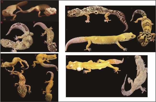
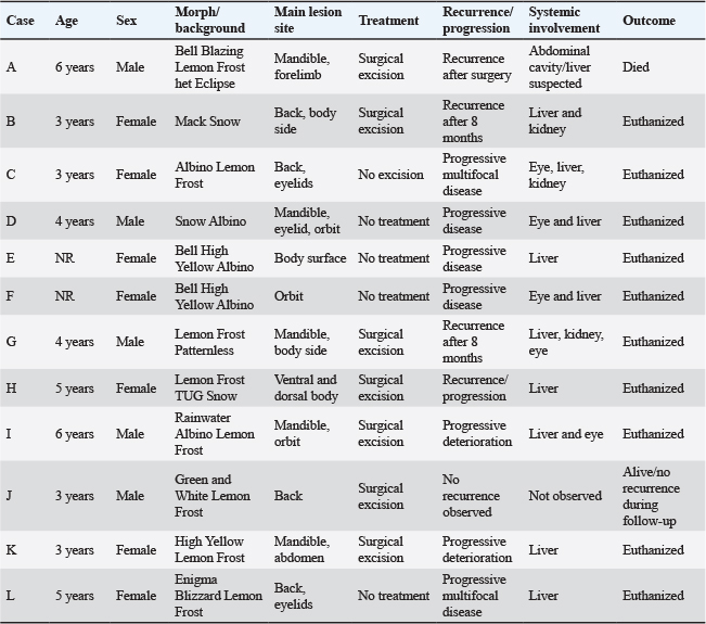
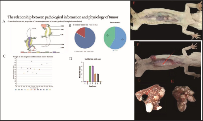
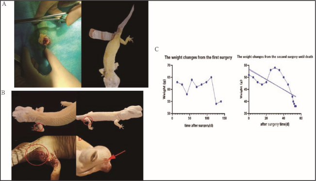
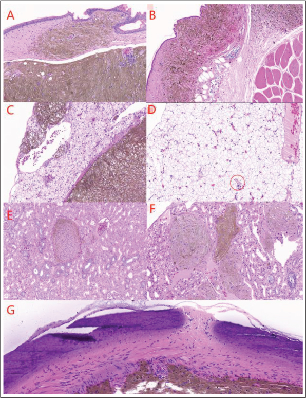
AbstractBackground: Chromatophoromas are pigment cell tumors reported in reptiles; however, their clinical behavior, pathological characteristics, and prognosis in leopard geckos (Eublepharis macularius) remain insufficiently described. Aim: This study aimed to characterize the clinical, cytological, histopathological, immunohistochemical, and outcome features of chromatophoromas in captive leopard geckos. Methods: A retrospective case series was conducted, reviewing 12 cases examined between 2023 and 2024. Clinical records, cytological smears, hematoxylin and eosin-stained sections, and immunohistochemical staining results were analyzed. Results: Affected geckos were predominantly Lemon Frost-related morphs aged 3–6 years. Cutaneous masses most frequently involved the mandible, back, eyelids, ventral body, and limbs. Cytology revealed spindle-shaped to stellate pigmented cells. Histopathology demonstrated invasive, proliferative neoplasms with variable pigmentation; advanced cases exhibited metastasis to the liver, kidneys, and ocular tissues. Immunohistochemistry was positive for HMB45, Melan-A, S100, and PNL2. Despite surgical intervention, recurrence and systemic involvement were frequent, with only one case remaining recurrence-free during follow-up. Conclusion: Chromatophoromas in captive leopard geckos often exhibit aggressive biological behavior. Early diagnosis, histopathological confirmation, and long-term monitoring are recommended for affected animals. Keywords: Leopard gecko (Eublepharis macularius), Chromatophoroma, Iridophoroma, Immunohistochemistry, Reptile oncology. IntroductionReptile neoplasia has been increasingly recognized in captive species, partly because of improved husbandry, longer survival, increased clinical attention, and more frequent diagnostic submissions (Sykes and Trupkiewicz, 2006). Although tumors in reptiles were once considered uncommon, retrospective studies have shown that neoplastic diseases occur in a wide range of reptile species and may represent an important clinical problem in captive populations (Hernandez -Divers and Garner, 2003; Garner et al., 2004). Among pet reptiles, leopard geckos (Eublepharis macularius) are commonly kept because of their manageable size, diverse color morphs, and relatively well-established husbandry requirements (Kubiak et al., 2020). Chromatophoromas are neoplasms derived from pigment-bearing or light-reflecting cells in reptiles, amphibians, and fish. In reptiles, pigment cells are mainly located in the dermis and include melanophores, xanthophores, and iridophores (Heckers et al., 2012; Monahan et al., 2022; ). Accordingly, pigment cell tumors may be classified as melanophoromas, xanthophoromas, iridophoromas, or mixed chromatophoromas, depending on the predominant cell lineage and histological features (Monahan et al., 2022). These tumors may show variable biological behavior, ranging from localized cutaneous masses to invasive or metastatic disease (Heckers et al., 2012). However, species-specific information on their clinical presentation, pathological diagnosis, treatment outcome, and prognosis remains limited. Leopard geckos with Lemon Frost-related morphs have attracted particular attention because of the reported association between this morph and iridophoroma development. Szydłowski first described the clinical and histopathological features of malignant iridophoroma in three privately owned Lemon Frost leopard geckos, although diagnostic imaging did not reveal obvious abnormalities in those cases (Szydłowski et al., 2020). Subsequent genetic mapping linked the Lemon Frost phenotype and iridophoroma susceptibility to a genomic region containing SPINT1, a candidate gene associated with epithelial proliferation and tumor-suppressive regulation (Guo et al., 2021). More recently, Boulanger et al. (2025) described four adult Lemon Frost or Super Lemon Frost leopard geckos with iridophoroma and reported clinical findings, including weight loss, submandibular masses, oral plaques, hepatic lesions, and pigment-containing neoplastic cells on fine-needle aspiration (Boulanger et al., 2025; Ikeda et al., 2026). Nevertheless, larger case series integrating lesion distribution, surgical outcome, recurrence, cytology, histopathology, immunohistochemistry, post-mortem findings, and prognosis remain limited. The diagnosis of chromatophoromas in reptiles usually requires integration of clinical findings, cytological examination, histopathological assessment, and, in selected cases, immunohistochemistry. Histopathology remains the primary diagnostic method for evaluating cellular morphology, tissue invasion, mitotic activity, and metastasis, while immunohistochemistry and electron microscopy may provide additional information in poorly differentiated or poorly pigmented tumors (Brot et al., 2015). Immunohistochemical markers, such as Melan-A, S100, HMB45, and PNL2, may provide supportive diagnostic information, although their cross-reactivity and diagnostic reliability in reptile tissues require cautious interpretation and further validation (Brot et al., 2015; Monahan et al., 2022). Therefore, the present study retrospectively describes 12 cases of chromatophoromas in captive leopard geckos examined between 2023 and 2024. The aim was to summarize their clinical characteristics, anatomical distribution, cytological and histopathological findings, immunohistochemical profiles, surgical outcomes, recurrence patterns, and prognosis, thereby providing additional clinical and pathological information for the diagnosis and management of pigment cell tumors in leopard geckos. Materials and MethodsStudy design and case selectionThis study was designed as a retrospective clinical case series. Medical records and diagnostic samples from 12 captive leopard geckos (Eublepharis macularius) with suspected or confirmed pigment cell tumors examined between 2023 and 2024 were reviewed. Cases were included when clinical information, gross lesion records, cytological findings, histopathological sections, immunohistochemical results, surgical records, post-mortem findings, or follow-up information were available. All tissue samples were obtained during routine veterinary diagnostic procedures, surgical excision, fine-needle aspiration, biopsy, or post-mortem examination. No experimental tumor induction or additional invasive procedure was performed specifically for this study. Clinical data collectionFor each case, available clinical information was collected, including age, sex, morph type, breeding history, body weight, lesion location, lesion size, gross appearance, clinical progression, treatment method, recurrence, evidence of systemic involvement, and outcome. Lesion distribution was recorded according to anatomical site, including the mandible, eyelids, back, ventral body region, limbs, and other affected regions. Clinical outcomes were classified descriptively as no recurrence during follow-up, recurrence after surgery, progressive disease without surgery, euthanasia, or death. Follow-up information was obtained from clinical records and owner-reported observations when available. Surgical procedures and postoperative managementSurgical excision was performed in selected cases based on clinical condition, lesion location, tumor burden, and owner consent. Before surgery, each gecko underwent clinical assessment to evaluate general condition and tumor distribution. Animals were fasted for approximately 24 hours before surgery when appropriate. Anesthesia was induced using isoflurane inhalation, followed by analgesic support according to clinical judgment. Tumor excision was planned according to lesion size and anatomical location. When possible, grossly visible tumor tissue was removed with a surgical margin while avoiding major vessels, nerves, and functionally important structures. Hemostasis was achieved during surgery, and the incision was closed using absorbable suture material suitable for reptile skin. After surgery, geckos were maintained under appropriate environmental temperature and monitored for recovery from anesthesia, wound healing, food and water intake, activity level, recurrence, and systemic deterioration. Excised tissues were submitted for cytological and/or histopathological examination when available. Cytological examinationFine-needle aspiration or impression smears were prepared from suspicious masses or excised tumor tissues. Smears were stained with Wright–Giemsa staining and examined by light microscopy. Cytological evaluation focused on cellular morphology, cell arrangement, nuclear characteristics, cytoplasmic features, pigment granules, inflammatory cells, and background material. A cytological diagnosis of pigment cell tumor was considered when neoplastic cells with spindle-shaped, stellate, or round morphology, and variable pigment granules were observed. Histopathological examinationExcised tumor tissues and post-mortem tissue samples were fixed in 4% neutral buffered formaldehyde. After fixation, tissues were processed routinely, dehydrated through graded alcohols, cleared, embedded in paraffin, sectioned, and stained with hematoxylin and eosin. Histopathological evaluation included tumor cell morphology, cellular arrangement, pigment deposition, degree of pleomorphism, mitotic activity when identifiable, inflammatory changes, tissue invasion, and involvement of internal organs. Skin, liver, kidneys, and ocular tissues were examined when available. Evidence of systemic involvement was assessed based on gross post-mortem findings and histopathological changes. ImmunohistochemistryImmunohistochemical staining was performed on selected paraffin-embedded tissue sections to further characterize tumor cells. After deparaffinization, rehydration, antigen retrieval, and blocking of non-specific binding, sections were incubated with primary antibodies against HMB45, Melan-A, S100, and PNL2. After washing, enzyme-labeled secondary antibodies were applied, and immunoreactivity was visualized using a chromogenic substrate. Sections were counterstained and examined by light microscopy. Immunohistochemical results were interpreted descriptively according to the presence and distribution of cytoplasmic or cellular immunoreactivity in tumor tissues. Because antibody validation in reptile tissues remains limited, positive staining was considered supportive diagnostic information rather than definitive evidence of tumor lineage. Data presentation and statistical analysisBecause of the small sample size and retrospective nature of this case series, data were summarized descriptively. Categorical variables, including sex, morph type, lesion location, treatment, recurrence, systemic involvement, and outcome, were presented as numbers and percentages when appropriate. Continuous variables, including age and body weight, were reported as ranges or individual case values. No inferential statistical comparisons were performed. Statistical testing, such as one-way ANOVA, was not applied because the study was not designed as a controlled experimental study and did not include sufficiently powered comparison groups. Ethical approvalThis retrospective study used clinical records and diagnostic tissue samples obtained during routine veterinary care, surgical treatment, fine-needle aspiration, biopsy, or post-mortem examination with owner consent. No experimental procedures were performed specifically for this study. Therefore, institutional animal ethics approval was not required according to the retrospective and clinical diagnostic nature of the study. ResultsSignalment and clinical presentationA total of 12 captive leopard geckos (Eublepharis macularius) with suspected or confirmed chromatophoromas were included in this retrospective case series. The affected geckos included 5 males and 7 females, with ages ranging from 3 to 6 years. Most animals were Lemon Frost-related morphs or morphs with a Lemon Frost background. Available clinical information showed that most cases had a history of breeding, except for Case A. Cutaneous masses were observed at multiple anatomical sites. The most frequently affected regions were the mandible, back, and eyelids, followed by the ventral body region and limbs. Lesions were grossly described as white to pale, flat or nodular masses of variable size. In several cases, the masses showed progressive enlargement, rupture, or multifocal distribution. Advanced cases presented with reduced body condition, decreased activity, severe weight loss, or ocular involvement (Fig. 1). A summary of the clinical characteristics, lesion distribution, treatment, recurrence, systemic involvement, and outcome of all cases is provided in Table 1. Gross findings and lesion distributionGrossly, the tumors appeared as white to pale nodular or flat cutaneous masses. In surgically excised cases, the cut surface of the masses was usually white or caseous. In most cases, the masses were not grossly attached to the underlying muscle at the time of initial excision. However, recurrent or advanced cases showed more extensive lesion distribution and systemic deterioration. Post-mortem examination revealed internal organ involvement in several cases. The liver was the most frequently affected internal organ. Gross hepatic changes included enlargement, multifocal white nodules, abnormal coloration, and loss of normal architecture. Ascites was observed in Case A. In some cases, nodular lesions or pigment deposition were also observed in the kidneys and ocular tissues, suggesting systemic involvement (Fig. 2). Surgical treatment and clinical outcomeSurgical excision was performed in selected cases when the tumor burden and anatomical location allowed removal. Cases A, B, G, H, I, J, and K underwent surgical excision. Temporary postoperative recovery was observed in several cases. However, recurrence or clinical deterioration developed in most surgically treated animals during follow-up. Case J had a single small dorsal mass and showed no recurrence during the available follow-up period. On the contrary, most other surgically treated cases developed recurrent masses, weight loss, systemic involvement, or poor clinical outcomes. Non-surgically treated cases generally showed progressive disease, multifocal tumor development, severe body condition loss, or ocular/internal organ involvement. Euthanasia was elected in several advanced cases because of severe clinical deterioration (Fig. 3). Cytological findingsCytological smears from affected masses showed numerous neoplastic cells arranged individually or in clusters. The tumor cells were predominantly spindle-shaped or stellate, with occasional round cells. Cytoplasmic borders were often indistinct, and the cytoplasm ranged from pale blue to lightly stained. The nuclei were round to oval, with variable size, coarse chromatin, and small nucleoli. Variable amounts of pigment granules were observed within or around the neoplastic cells. Scattered inflammatory cells and erythrocytes were also present in the background. These cytological findings were consistent with a pigment cell tumor and supported a diagnosis of chromatophoroma (Fig. 4).
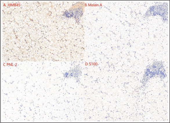
Fig. 1. Representative clinical presentation of chromatophoromas in 12 captive leopard geckos. (A–L) Gross appearance of the 12 leopard geckos included in this retrospective case series. The affected animals showed single or multifocal white to pale cutaneous masses at different anatomical sites, including the mandible, back, eyelids, ventral body region, and limbs. (A) A 6-year-old male Bell Blazing Lemon Frost het Eclipse leopard gecko showing a mandibular mass indicated by the red arrow. (B) A 3-year-old female Mack Snow leopard gecko with multiple dorsal cutaneous nodules. (C) A 3-year-old female Albino Lemon Frost leopard gecko with multiple dorsal and periocular lesions. (D–L) Additional affected leopard geckos showing variable lesion distribution and tumor burden. All samples used in this study were obtained from surgical biopsies, excised tissues, or post-mortem specimens collected during routine clinical diagnostic evaluation. Histopathological findingsHistopathological examination was performed on selected skin and internal organ tissues. In cutaneous lesions, neoplastic cells were densely proliferative and arranged in nests, sheets, or poorly organized patterns. Tumor cells showed variable morphology, including spindle-shaped, epithelioid, and round forms. Pigment granules were observed in the cytoplasm or extracellularly. Cellular pleomorphism, variable nuclear size, prominent nucleoli, and mitotic figures were observed in some sections. Inflammatory and vacuolar changes were also present in affected skin tissues. In advanced cases, internal organ involvement was identified. Liver sections showed replacement or disruption of normal hepatic architecture by neoplastic tissue, fibrous proliferation, pigment deposition, and multifocal tumor-like nodules. Kidney sections showed nodular pigment deposition, tissue destruction, fibrosis, and necrosis in affected regions. In ocular tissues, neoplastic changes involved the iris and adjacent ocular structures, with pigment deposition and spindle-shaped neoplastic cells. These histopathological findings supported invasive and systemic involvement in advanced cases (Fig. 5). Immunohistochemical findingsImmunohistochemical staining was performed on selected tumor tissues, including skin and liver samples. Tumor cells showed positive immunoreactivity for HMB45, Melan-A, S100, and PNL2. HMB45 showed strong cytoplasmic staining in the examined tumor tissues. Melan-A and S100 also showed positive immunoreactivity in tumor cells. PNL2 staining was positive but appeared less extensive than HMB45 in the examined sections. Table 1. Clinical summary of 12 leopard geckos with chromatophoromas.
The immunohistochemical findings were consistent with pigment cell tumor differentiation and provided supportive diagnostic information for chromatophoromas in leopard geckos. Because these markers have not been fully validated across reptile species, the staining results were interpreted as supportive rather than definitive evidence of cell lineage (Fig. 6). DiscussionThis retrospective case series describes the clinical, cytological, histopathological, immunohistochemical, and outcome features of chromatophoromas in 12 captive leopard geckos. The main findings were that cutaneous masses were commonly observed on the mandible, back, eyelids, ventral body region, and limbs; several cases showed recurrence or progressive disease after surgical excision; and advanced cases showed evidence of internal organ involvement, particularly in the liver, kidneys, and ocular tissues. These observations suggest that chromatophoromas in leopard geckos may have clinically aggressive behavior in some cases, although the biological behavior likely varies among individuals. Previous studies have reported neoplasia in captive reptiles, and improved clinical recognition and diagnostic submission may have increased the detection of such diseases (Hernandez-Divers and Garner, 2003). Chromatophoromas are derived from pigment cells and may include melanophoromas (Sykesand Trupkiewicz, 2006), xanthophoromas, iridophoromas, or mixed forms depending on the predominant pigment cell lineage (Garner et al., 2004). In leopard geckos, Lemon Frost-related morphs have received particular attention because of the reported association with iridophoroma development. In the present study, most affected geckos were Lemon Frost-related morphs or had a Lemon Frost background. However, because no genetic testing was performed, this study cannot confirm the genetic basis of tumor development in these cases. Therefore, the relationship between morph background and tumor occurrence should be interpreted as a clinical observation rather than evidence of causation (Kubiak et al., 2020).
Fig. 2. Lesion distribution and gross post-mortem findings in leopard geckos with chromatophoromas. (A) Anatomical distribution of gross cutaneous lesions among the 12 affected leopard geckos. (B) Distribution of morph background and sex among the included cases. (C) Relationship between body weight at first diagnosis and maximum tumor diameter. (D) Age distribution of affected geckos. (E and F) Representative gross abdominal findings in advanced cases, showing internal discoloration or masses visible through the body wall and during post-mortem examination. (G and H) Representative gross lesions involving internal organs, including multifocal pale nodules and abnormal tissue architecture. Arrows indicate representative lesions. The anatomical distribution of lesions in this case series was relatively broad. Masses were observed on the mandible, back, eyelids, ventral body region, and limbs (Boulanger et al., 2025). Some cases had multifocal lesions at first presentation, whereas others initially presented with localized masses and later developed recurrence or systemic deterioration (Heckers et al., 2012). These findings indicate that careful whole-body examination is important when evaluating leopard geckos with suspected pigment cell tumors. Ocular or periocular involvement may be clinically important because it can interfere with vision, feeding behavior, and general condition (Gardhouse et al., 2014; Wiggans et al., 2018). In addition, the presence of internal organ lesions in advanced cases highlights the need for further staging when feasible (Zborilova et al., 2023). The clinical findings in this study are partly consistent with those of Boulanger et al. (2025) who described hyporexia, lethargy, weight loss, submandibular masses, oral plaques, hepatic nodules, and pigment-containing neoplastic cells in Lemon Frost or Super Lemon Frost leopard geckos with iridophoroma (Boulanger et al., 2025). Similarly, several cases in the present series showed mandibular or periocular masses, weight loss, cytological evidence of pigment cell tumors, and hepatic involvement in advanced disease. However, the present study includes a larger number of cases and further summarizes surgical treatment, recurrence, immunohistochemical findings, and clinical outcomes, thereby expanding the available clinicopathological information. Cytological examination provided useful preliminary diagnostic information (Boulanger et al., 2025). In this study, smears showed neoplastic cells with spindle-shaped to stellate morphology, variable nuclear features, and pigment granules. These findings supported the diagnosis of a pigment cell tumor. However, cytology alone may not be sufficient to determine the specific subtype or biological behavior of chromatophoromas (Heckers et al., 2012). Histopathology remains essential for evaluating tissue architecture (Irizarry-Rovira et al., 2008), cellular morphology, invasion, mitotic activity, and involvement of adjacent or internal tissues (Brot et al., 2015).
Fig. 3. Surgical excision, recurrence, and postoperative body-weight changes in a representative leopard gecko with chromatophoroma. (A) Representative surgical excision of a cutaneous mass and postoperative wound management. (B) Recurrent or progressive cutaneous masses observed during follow-up; circles and arrows indicate representative lesions. (C) Descriptive body-weight changes after the first surgery and after the second surgery until death in Case A. Body-weight data are presented descriptively for an individual case, and no inferential statistical analysis was performed.
Fig. 4. Cytological findings of chromatophoromas in leopard geckos. Wright–Giemsa-stained cytological smears showed numerous neoplastic cells arranged individually or in clusters. Tumor cells were predominantly spindle-shaped to stellate, with indistinct cytoplasmic borders, variable nuclear morphology, and pigment granules. Arrows indicate representative neoplastic pigment cells. Histopathological findings in the present cases included dense proliferation of neoplastic cells, variable cell morphology, pigment deposition, inflammatory changes, tissue invasion, and, in advanced cases, involvement of the liver, kidneys, and ocular tissues. These findings support the view that some chromatophoromas in leopard geckos can behave as locally invasive or systemically disseminated tumors (Szydłowski et al., 2020). Nevertheless, because post-mortem examination and histopathology were not equally available for all cases, the true frequency of metastasis or systemic involvement cannot be determined from this case series (Zborilova et al., 2023).
Fig. 5. Histopathological findings in skin and internal organs of leopard geckos with chromatophoromas. (A and B) Representative skin tumor sections from Cases A and C showing dense proliferation of neoplastic cells, variable cellular morphology, pigment deposition, and disruption of normal tissue architecture. (C and D) Liver sections from Cases A and C showing abnormal hepatic architecture, fibrous proliferation, pigment deposition, and tumor-like cellular infiltration. (E and F) Kidney sections from Cases A and C showing renal tissue disruption, pigment deposition, fibrosis, necrosis, or abnormal cellular infiltration. (G) Ocular tissue section from Case C showing neoplastic involvement of ocular structures. Hematoxylin and eosin staining. Scale bars should be indicated in each panel.
Fig. 6. Immunohistochemical staining of liver tissue in leopard geckos with chromatophoroma. Representative liver tissue sections showing immunohistochemical staining for HMB45, Melan-A, S100, and PNL2. (A) HMB45, (B) Melan-A, (C) PNL2, (D) S100. Positive immunoreactivity (brown staining) was observed in tumor-associated areas and was interpreted as supportive diagnostic information. Images were captured at 10× magnification. Immunohistochemistry was used as an adjunctive diagnostic tool. Tumor cells showed positive immunoreactivity for HMB45, Melan-A, S100, and PNL2 in the examined tissues (Monahan et al., 2022). Previous reviews have suggested that immunohistochemistry and electron microscopy may be useful for poorly differentiated or poorly pigmented chromatophoromas, but histopathology remains the primary diagnostic approach (Monahan et al., 2022). These results suggest that the markers used in the present study may provide supportive diagnostic information for leopard gecko chromatophoromas, especially in poorly pigmented or diagnostically challenging tumors (Zborilova et al., 2023). Surgical excision remains one of the main practical treatment options for localized cutaneous masses in reptiles (Rivera et al., 2015). In the present case series, several geckos underwent surgical removal of visible masses, and short-term recovery was observed in some cases (Futema et al., 2020). However, recurrence or clinical deterioration occurred in most surgically treated cases, whereas only one case showed no recurrence during the available follow-up period. These findings suggest that complete surgical control may be difficult when tumors are multifocal, invasive, or already associated with systemic involvement (Rivera et al., 2015). Early detection and removal of small, localized lesions may improve local control, but this assumption requires further confirmation in larger studies (Kent, 2016). The poor outcomes observed in several advanced cases may be related to tumor burden, lesion distribution, systemic involvement, nutritional decline (Zborilova et al., 2023), and the limited treatment options currently available for reptile pigment cell tumors (Wen et al., 2025). Although chemotherapy, radiotherapy, immunotherapy, and other adjunctive approaches have been explored in other species or tumor types, there is currently insufficient evidence to recommend specific systemic therapies for chromatophoromas in leopard geckos (Ritter et al., 2009; A et al., 2011). Any extrapolation from mammalian melanoma or other animal tumors should be made cautiously because of major differences in species biology, tumor classification, drug metabolism, and clinical response (Irizarry-Rovira et al., 2008; Mineshige et al., 2024). This study has several limitations. First, the sample size was small, and the retrospective design limited the completeness and consistency of clinical records. Second, not all cases had the same diagnostic work-up, and cytology, histopathology, immunohistochemistry, post-mortem examination, and follow-up information were not uniformly available for every animal. Third, no molecular testing, electron microscopy, or standardized immunohistochemical validation was performed (Mineshige et al., 2024). Therefore, the precise pigment cell lineage, genetic background, and mechanisms of tumor development could not be confirmed (Pinto et al., 2023). Fourth, treatment outcomes could not be compared statistically because there was no controlled treatment group and the cases differed in lesion burden, disease stage, and clinical condition (McLean and Vickaryous, 2011). Despite these limitations, this case series provides additional clinical and pathological information on chromatophoromas in captive leopard geckos (Heckers et al., 2012). The findings emphasize the importance of early clinical recognition, histopathological confirmation, cautious interpretation of immunohistochemistry, and long-term follow-up after surgical excision (Divers, 2010). Future studies with larger case numbers, standardized diagnostic protocols (Rivera et al., 2015; Pinto et al., 2023), molecular characterization, electron microscopy, and prospective follow-up are needed to better define tumor subtype, biological behavior, prognostic indicators, and treatment strategies in leopard geckos. ConclusionThis retrospective case series provides clinical and pathological information on chromatophoromas in 12 captive leopard geckos. The tumors commonly involved the mandible, back, eyelids, ventral body region, and limbs, and several cases showed recurrence or systemic involvement after clinical progression. Cytology and histopathology were central for diagnosis, while HMB45, Melan-A, S100, and PNL2 immunohistochemistry provided supportive diagnostic information. The findings suggest that chromatophoromas in leopard geckos may show aggressive behavior in advanced cases, but interpretation is limited by the small sample size, retrospective design, and lack of molecular validation. Early diagnosis, careful surgical planning, histopathological confirmation, and long-term follow-up are recommended. Larger prospective studies are needed to clarify tumor classification, prognostic factors, and effective treatment strategies. AcknowledgmentsThe authors thank Beijing Ornamental Animal Hospital for providing the clinical facilities and diagnostic resources for this study. Conflicts of interestThe authors declare no conflicts of interest. FundingThis research received no external funding. Authors’ contributionsHeyi Zhao: Writing–review & editing, Investigation, Original draft, Software, Methodology, Formal analysis, Data curation. Haoyu Xuanyuan: Writing–review & editing, Original draft, Investigation, Funding acquisition. Chi Zhang: Diagnosis and case summarization. Yi Zhou: Writing–review & editing. Zixiang Lin: Writing–review & editing, Funding acquisition, Conceptualization. Fuqiang Qiao: Conceptualization. Hua Yao: Methodology, Conceptualization. Data availabilityThe data presented in this study are available from the corresponding author upon reasonable request. ReferencesBoulanger, M., Lee, Y. and Keller, J. 2025. Iridophoroma in leopard geckos (Eublepharis macularius): clinical complications and histopathology. J. Am. Assoc. For. Lab. Anim. Sci. 64(1), 1–7; doi:10.30802/AALAS-JAALAS-25-095 Cardona, J.A.C., Conley, K.J., Wellehan, J.F.X., Farina, L.L., Origgi, F.C. and Wamsley, H.L. 2011. Incomplete ovariosalpingectomy and subsequent malignant granulosa cell tumor in a female green iguana (Iguana iguana). J. Am. Vet. Med. Assoc. 239(2), 237–242; doi:10.2460/javma.239.2.237 De Brot, S., Sydler, T., Nufer, L. and Ruetten, M. 2015. Histologic, immunohistochemical, and electron microscopic characterization of a malignant iridophoroma in a dwarf bearded dragon (Pogona henrylawsoni). J. Zoo. Wildlife. Med. 46(3), 583–587; doi:10.1638/2013-0113.1 Divers, S.J. 2010. Reptile diagnostic endoscopy and endosurgery. Vet. Clinics. North. Amer. Exotic. Anim. Pract. 13(2), 217–242; doi:10.1016/j.cvex.2010.01.004 Futema, F., De Carvalho, F.M. and Werneck, M.R. 2020. Spinal anesthesia in green sea turtles (Chelonia mydas) undergoing surgical removal of cutaneous fibropapillomas. J. Zoo. Wildlife. Med. 51(2), 357–362; doi:10.1638/2015-0084 Gardhouse, S., Eshar, D., Lee-Chow, B., Foster, R.A., Ingrao, J.C. and Poirier, V.J. 2014. Diagnosis and treatment of a periocular myxosarcoma in a bearded dragon (Pogona vitticeps). Can. Vet. J. 55(7), 663–666. Garner, M.M., Hernandez-Divers, S.M. and Raymond, J.T. 2004. Reptile neoplasia: a retrospective study of case submissions to a specialty diagnostic service. Vet. Clinics. North. Amer. Exotic. Anim. Pract. 7(3), 653–671; doi:10.1016/j.cvex.2004.04.002 Guo, L., Bloom, J., Sykes, S., Huang, E., Kashif, Z., Pham, E., Ho, K., Alcaraz, A., Xiao, X.G., Duarte-Vogel, S. and Kruglyak, L. 2021. Genetics of white color and iridophoroma in “Lemon Frost” leopard geckos. PLos Genet. 17(6), e1009580; doi:10.1371/journal.pgen.1009580 Heckers, K.O., Aupperle, H., Schmidt, V. and Pees, M. 2012. Melanophoromas and iridophoromas in reptiles. J. Comparative Pathol. 146(2–3), 258–268; doi:10.1016/j.jcpa.2011.07.003 Hernandez-Divers, S.M. and Garner, M.M. 2003. Neoplasia of reptiles with an emphasis on lizards. Vet. Clinics. North. Amer. Exotic. Anim. Pract. 6(1), 251–273; doi:10.1016/S1094-9194(02)00028-2 Ikeda, M., Kondo, H., Takekubo, A., Tsunesumi, N. and Shibuya, H. 2026. Localized oral histiocytic sarcoma in a leopard gecko (Eublepharis macularius). J. Vet. Med. Sci. 88(4), 665–669; doi:10.1292/jvms.25-0581 Irizarry-Rovira, A.R., Lennox, A.M. and Ramos-Vara, J.A. 2008. Granular cell tumor in the testis of a rabbit: cytologic, histologic, immunohistochemical, and electron microscopic characterization. Vet. Pathol. 45(1), 73–77; doi:10.1354/vp.45-1-73 Kent, M.S. 2016. Principles and applications of radiation therapy in exotic animals. Vet. Clinics. North. Amer. Exotic. Anim. Pract. 20(1), 255–270; doi:10.1016/j.cvex.2016.07.008 Kubiak, M., Denk, D. and Stidworthy, M.F. 2020. Retrospective review of neoplasms of captive lizards in the United Kingdom. Vet. Rec. 186(1), 28; doi:10.1136/vr.105308 Mclean, K.E. and Vickaryous, M.K. 2011. A novel amniote model of epimorphic regeneration: the leopard gecko, Eublepharis macularius. BMC. Develop. Biol. 11, 50; doi:10.1186/1471-213X-11-50 Mineshige, T., Tanaka, Y., Watanabe, K., Tagawa, M., Tomihari, M. and Kobayashi, Y. 2024. Histologic, immunohistochemical, and in situ hybridization study of myxoid stroma in feline oral squamous cell carcinoma. J. Vet. Med. Sci. 86(3), 258–265; doi:10.1292/jvms.23-0356 Monahan, C.F., Garner, M.M. and Kiupel, M. 2022. Chromatophoromas in reptiles. Vet. Sci. 9(3), 115; doi:10.3390/vetsci9030115 Pinto, B.J., Gamble, T., Smith, C.H., Keating, S.E., Havird, J.C. and Chiari, Y. 2023. The revised reference genome of the leopard gecko (Eublepharis macularius) provides insight into the considerations of genome phasing and assembly. J. Heredity 114(5), 513–520; doi:10.1093/jhered/esad016 Ritter, J.M., Garner, M.M., Chilton, J.A., Jacobson, E.R. and Kiupel, M. 2009. Gastric neuroendocrine carcinomas in bearded dragons (Pogona vitticeps). Vet. Pathol. 46(6), 1109–1116; doi:10.1354/vp.09-VP-0019-K-FL Rivera, S., Crane, M.M., Mcmanamon, R. and Gregory, C.R. 2015. Surgical treatment of pulmonary melanophoroma in a beaded lizard (Heloderma horridum exasperatum). J. Zoo Wildlife Med. 46(2), 397–399; doi:10.1638/2014-0188R2.1 Sykes, J.M. and Trupkiewicz, J.G. 2006. Reptile neoplasia at the Philadelphia Zoological Garden, 1901–2002. J. Zoo Wildlife Med. 37(1), 11–19; doi:10.1638/04-112.1 Szydłowski, P., Madej, J.P., Duda, M., Madej, J.A., Sikorska-Kopyłowicz, A., Chełmońska-Soyta, A., Ilnicka, L. and Duda, P. 2020. Iridophoroma associated with the Lemon Frost colour morph of the leopard gecko (Eublepharis macularius). Scientific. Rep. 10(1), 5734; doi:10.1038/s41598-020-62717-8 Wen, A., Sandiford, T., Nakashima, A., Sojka, P.A., Freer, S. and Mishra, N. 2025. Diagnosis and management of pericardial mesothelioma in a leopard gecko (Eublepharus macularius). Vet. Radiol. Ultrasound. 66(5), e70062; doi:10.1111/vru.70062 Wiggans, K.T., Sanchez-migallon Guzman, D., Reilly, C.M., Vergneau-Grosset, C., Kass, P.H. and Hollingsworth, S.R. 2018. Diagnosis, treatment, and outcome of and risk factors for ophthalmic disease in leopard geckos (Eublepharis macularius) at a veterinary teaching hospital: 52 cases (1985–2013). J. Am. Vet. Med. Assoc. 252(3), 316–323; doi:10.2460/javma.252.3.316 Zborilova, H., Dietz, J., Heckers, K., Dvorakova, R., Knotek, Z. and Cermakova, E. 2023. Malignant ovarian dysgerminoma in a 16-year-old leopard gecko (Eublepharis macularius). Veterinární. Medicína. 68(3), 129–134; doi:10.17221/107/2022-VETMED | ||

| How to Cite this Article |
| Pubmed Style Zhao H, Xuanyuan H, Zhang C, Zhou Y, Lin Z, Qiao F, Yao H. Clinical and pathological characterization of chromatophoromas in captive leopard geckos: A retrospective case series. Open Vet. J.. 2026; 16(8): 5114-5124. doi:10.5455/OVJ.2026.v16.i8.7 Web Style Zhao H, Xuanyuan H, Zhang C, Zhou Y, Lin Z, Qiao F, Yao H. Clinical and pathological characterization of chromatophoromas in captive leopard geckos: A retrospective case series. https://www.openveterinaryjournal.com/?mno=319985 [Access: August 08, 2026]. doi:10.5455/OVJ.2026.v16.i8.7 AMA (American Medical Association) Style Zhao H, Xuanyuan H, Zhang C, Zhou Y, Lin Z, Qiao F, Yao H. Clinical and pathological characterization of chromatophoromas in captive leopard geckos: A retrospective case series. Open Vet. J.. 2026; 16(8): 5114-5124. doi:10.5455/OVJ.2026.v16.i8.7 Vancouver/ICMJE Style Zhao H, Xuanyuan H, Zhang C, Zhou Y, Lin Z, Qiao F, Yao H. Clinical and pathological characterization of chromatophoromas in captive leopard geckos: A retrospective case series. Open Vet. J.. (2026), [cited August 08, 2026]; 16(8): 5114-5124. doi:10.5455/OVJ.2026.v16.i8.7 Harvard Style Zhao, H., Xuanyuan, . H., Zhang, . C., Zhou, . Y., Lin, . Z., Qiao, . F. & Yao, . H. (2026) Clinical and pathological characterization of chromatophoromas in captive leopard geckos: A retrospective case series. Open Vet. J., 16 (8), 5114-5124. doi:10.5455/OVJ.2026.v16.i8.7 Turabian Style Zhao, Heyi, Haoyu Xuanyuan, Chi Zhang, Yi Zhou, Zixiang Lin, Fuqiang Qiao, and Hua Yao. 2026. Clinical and pathological characterization of chromatophoromas in captive leopard geckos: A retrospective case series. Open Veterinary Journal, 16 (8), 5114-5124. doi:10.5455/OVJ.2026.v16.i8.7 Chicago Style Zhao, Heyi, Haoyu Xuanyuan, Chi Zhang, Yi Zhou, Zixiang Lin, Fuqiang Qiao, and Hua Yao. "Clinical and pathological characterization of chromatophoromas in captive leopard geckos: A retrospective case series." Open Veterinary Journal 16 (2026), 5114-5124. doi:10.5455/OVJ.2026.v16.i8.7 MLA (The Modern Language Association) Style Zhao, Heyi, Haoyu Xuanyuan, Chi Zhang, Yi Zhou, Zixiang Lin, Fuqiang Qiao, and Hua Yao. "Clinical and pathological characterization of chromatophoromas in captive leopard geckos: A retrospective case series." Open Veterinary Journal 16.8 (2026), 5114-5124. Print. doi:10.5455/OVJ.2026.v16.i8.7 APA (American Psychological Association) Style Zhao, H., Xuanyuan, . H., Zhang, . C., Zhou, . Y., Lin, . Z., Qiao, . F. & Yao, . H. (2026) Clinical and pathological characterization of chromatophoromas in captive leopard geckos: A retrospective case series. Open Veterinary Journal, 16 (8), 5114-5124. doi:10.5455/OVJ.2026.v16.i8.7 |


