





| Research Article | ||
Open Vet. J.. 2026; 16(8): 5821-5829 !
Open Veterinary Journal, (2026), Vol. 16(8): 5821–5829 Research Article Beta-adrenergic receptor expression in canine splenic hemangiosarcoma versus non-neoplastic nodular splenic lesions: A comparative immunohistochemical studyKiyeon Son1, Youngmi Youk2, Mungyu Song2, Hyoyoung Jung2* and Joonghyun Song11Department of Veterinary Internal Medicine, College of Veterinary Medicine, Chungnam National University, Daejeon, South Korea 2Department of Veterinary Medicine and Institute of Veterinary Science, Chungnam National University, Daejeon, South Korea *Corresponding Author: Hyoyoung JUNG. Department of Veterinary Medicine and Institute of Veterinary Science, Chungnam National University, Daejeon 34134, South Korea. Tel: +82-10-6310-6384 Fax: +82-42-821-6769 E-mail: hyjung [at] cnu.ac.kr Submitted: 03/05/2026 Revised: 15/07/2026 Accepted: 24/07/2026 Published: 20/08/2026 © 2025 Open Veterinary Journal
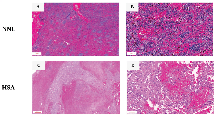
AbstractBackground: Hemangiosarcoma (HSA) is a highly aggressive malignant neoplasm of vascular endothelial origin in dogs, exhibiting pathophysiological and molecular features analogous to human angiosarcoma. β-adrenergic receptors (β-ARs) are significantly upregulated in human angiosarcoma, and β-AR antagonists are under investigation as potential therapeutics. Aim: This study aimed to quantify β1- and β2-AR expression in canine splenic HSA tissue relative to non-neoplastic splenic tissue. Methods: Splenic tissue samples were obtained from ten client-owned dogs—four with splenic HSA and six with non-neoplastic nodular lesions (NNL), including nodular hyperplasia (NH) and nodular lymphoid hyperplasia—and subjected to immunohistochemical (IHC) analysis using primary antibodies targeting β1- and β2-AR. Quantification of IHC expression was conducted using the histochemical score (H-score) and immunoreactive score (IRS). Results: Compared with NNL tissues, HSA specimens showed higher β1-AR immunoreactivity (H-score: 30.74 vs. 1.78, respectively, p < 0.01; IRS: 2.45 vs. 0.52, respectively, p < 0.05) and β2-AR immunoreactivity (H-score: 100.50 vs. 1.69, respectively, p < 0.01; IRS: 7.36 vs. 0.50, respectively, p < 0.01). Within HSA tissues, β2-AR expression was higher than β1-AR (H-score: 100.50 vs. 30.74, respectively, p < 0.05; IRS: 7.36 vs. 2.45, respectively, p < 0.05). Conclusion: These findings demonstrate marked β-AR overexpression, especially β2-AR, in canine splenic HSA and provide foundational tissue-level evidence for further investigation into the pathophysiological role and therapeutic relevance of β-adrenergic signaling in HSA. Keywords: Angiosarcoma, Beta-adrenergic receptor, Dogs, Hemangiosarcoma, Spleen. IntroductionCanine splenic hemangiosarcoma (HSA) is a highly aggressive, malignant tumor that originates from vascular endothelial cells (Faulhaber et al., 2021; De Nardi et al., 2023). It is recognized as the most common splenic malignancy in dogs, characterized by its high metastatic potential and poor prognosis (Dickerson and Bryan, 2015; Wendelburg et al., 2015; De Nardi et al., 2023). The current standard treatment for splenic HSA, which involves splenectomy with or without adjunctive chemotherapy, provides only limited therapeutic outcomes (De Nardi et al., 2023). In affected dogs, median survival time (MST) with surgery alone is only 42–86 d (Faulhaber et al., 2021), and adjuvant chemotherapy may extend MST to 4–6 m; however, the one-year survival rate for stage II or III disease remains <16% (Wendelburg et al., 2015; Faulhaber et al., 2021). These limitations underscore the urgent need for novel and more effective therapeutic strategies. From a comparative oncology perspective, canine HSA shares substantial histopathological, molecular, and clinical characteristics with human angiosarcoma (Dickerson and Bryan, 2015; Wong et al., 2021; De Nardi et al., 2023). Both neoplasms originate from vascular endothelial cells, are highly vascularized, and exhibit aggressive biological behavior with marked metastatic potential. In human angiosarcoma, β1- and β2-adrenergic receptors (β1- and β2-AR) are markedly overexpressed at both mRNA and protein levels in angiosarcoma cell lines and tissues compared with normal vascular endothelium (Stiles et al., 2013; Porcelli et al., 2020).Experimental studies demonstrate that β-AR activation functionally drives tumor-related processes through cyclic adenosine monophosphate (cAMP)-dependent signaling pathways, thereby promoting enhanced cellular proliferation, angiogenesis, and resistance to apoptosis (Cole and Sood, 2012; Dickerson and Bryan, 2015). Specifically, functional assays have shown that β-AR stimulation increases cAMP–protein kinase A (PKA) signaling, upregulates pro-angiogenic and pro-inflammatory mediators, such as vascular endothelial growth factor (VEGF) and interleukin-6 (IL-6), and modulates B-cell lymphoma 2 (Bcl-2) family proteins and anoikis resistance in cancer models (Madden et al., 2011; Cole and Sood, 2012; Stiles et al., 2013). Based on these findings, the nonselective β-blocker, propranolol, has been proposed as a potential therapeutic option for angiosarcoma. Propranolol has demonstrated antitumor effects, including reduced tumor cell viability, induction of apoptosis, and inhibition of angiogenesis and cellular invasiveness in both experimental and clinical settings (Stiles et al., 2013; Amaya et al., 2018; Saha et al., 2020). In combination with chemotherapy, propranolol has been shown to substantially prolong progression-free survival (PFS) and overall survival (OS) in patients with human angiosarcoma (Banavali et al., 2015; Porcelli et al., 2020). These findings suggest that β-AR signaling contributes to the development and progression of human angiosarcoma and may represent a promising therapeutic target (Stiles et al., 2013; Dickerson and Bryan, 2015). In contrast, investigations of β-AR expression in canine splenic HSA have been largely limited to in vitro cell line studies or transcriptomic analyses, and their expression in clinical tissue specimens has been poorly characterized. Therefore, the present study aimed to evaluate β1- and β2-AR protein expression in canine splenic HSA using immunohistochemistry (IHC) and to compare these findings with those in nodular hyperplasia (NH) and nodular lymphoid hyperplasia (NLH), the two most common non-neoplastic splenic lesions detected in dogs via ultrasound (Ko et al., 2023). We hypothesized that both β1- and β2-ARs would be markedly overexpressed in HSA tissues compared with NH/NLH tissues. This protein-level analysis using clinical tissue specimens is expected to provide foundational insight into the role of β-AR signaling in the pathogenesis of canine splenic HSA and to establish a molecular rationale for the future evaluation of β-blockers as adjunctive therapeutic strategies in this tumor. Materials and MethodsSubjects and tissue samplesSplenic tissue samples were collected from 10 client-owned dogs that underwent splenectomy at Chungnam National University Veterinary Medicine Teaching Hospital. Splenectomy was performed for diagnostic or therapeutic purposes following ultrasonographic detection of splenic lesions, such as nodules ≥2 cm in diameter, multiple nodules, or splenomegaly with mixed echogenicity. All splenic specimens were stained with hematoxylin and eosin (H&E) and histopathologically evaluated at the Department of Veterinary Medicine and Institute of Veterinary Science, Chungnam National University. All histopathological diagnoses were confirmed by board-certified veterinary anatomic pathologists at IDEXX Reference Laboratories. Specimens were classified into two groups: splenic HSA (HSA group, n=4) and non-neoplastic nodular lesions (NNL group, n=6), including NH and NLH. Normal splenic tissue from healthy dogs was not included as a control because splenectomy is not ethically justifiable in clinically normal animals. Instead, NH and NLH were used as comparative non-neoplastic controls because these lesions are commonly encountered in clinical practice and are frequently excised for diagnostic purposes. Representative H&E-stained photomicrographs of NNL and HSA are shown in Figure 1. Informed consent was obtained from the owners, and the study protocol was approved by the University’s Ethics committee (protocol no.: 202502-CNU-047). Immunohistochemical stainingFormalin-fixed, paraffin-embedded splenic tissues were sectioned at a thickness of 4 μm using a rotary microtome (Leica RM2235, Leica Microsystems, Wetzlar, Germany) and mounted on silane-coated glass slides. Serial sections were prepared from each paraffin block. One section was stained with H&E for histopathological evaluation, and adjacent sections were used for IHC to assess β1-AR and β2-AR immunoreactivity in corresponding regions of interest. After deparaffinization and rehydration, antigen retrieval was conducted in citrate buffer (pH 6.0) at 115°C for 15 minutes. Endogenous peroxidase activity was blocked with 0.3% hydrogen peroxide in phosphate-buffered saline for 30 minutes, and nonspecific binding was prevented using 5% normal goat serum. The slides were incubated with rabbit polyclonal anti-β1-AR antibody (1:200 dilution, Abbiotec, Escondido, CA, USA; Cat. No. 250919) and rabbit polyclonal anti-β2-AR (1:300 dilution, Abbiotec, Escondido, CA, USA; Cat. No. 251604) at 4°C for 48 hours. According to the manufacturer’s datasheets, both primary antibodies are reactive in dogs and suitable for IHC; specifically, their use has been validated in peer-reviewed studies, including immunoblotting data in canine cell lines (Saha et al., 2020). Thereafter, the slides were sequentially incubated with goat anti-rabbit IgG (Vector Laboratories, Burlingame, CA) and VECTASTAIN Elite ABC system at 25°C for 2 hours. The immunoreactions were visualized using 3,3-diaminobenzidine tetrachloride (DAB; Sigma-Aldrich, St. Louis, MO, USA; Cat. No. 12750-5G-F) as a chromogen. The DAB working solution was freshly prepared by mixing 20 mL of phosphate-buffered saline (PBS), 20 μL of 30% hydrogen peroxide, and 500 μL of DAB substrate solution. Sections were developed for 30–60 s at 25 °C under microscopic control and then rinsed in distilled water to stop the reaction. The slides were counterstained with Harris hematoxylin for 1 minute 10 seconds, rinsed in running tap water for 15 minutes to develop the blue hematoxylin color, dehydrated through graded ethanol, cleared in xylene, and mounted. For both β1-AR and β2-AR, membranous and/or cytoplasmic staining was considered positive. Negative control sections were processed in parallel by omission of the primary antibody, with all other staining conditions identical to those of the test sections.
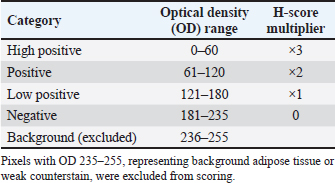
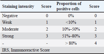
Fig. 1. Representative hematoxylin and eosin (H&E)-stained photomicrographs of canine splenic tissues. Digital image analysis and immunohistochemical scoringImmunostained slides were initially examined at × 200 magnification to identify evaluable tissue regions and exclude areas containing staining artifacts, tissue folds, or insufficient tissue for analysis. Within these predefined evaluable regions, 10 non-overlapping × 400 high-power fields per sample were randomly selected. The same field-selection criteria were applied to both the NNL and HSA groups to ensure consistent assessment of immunostaining. The selected × 400 fields were digitized as high-resolution JPEG files and subjected to quantitative image analysis using ImageJ software (version 1.54; National Institutes of Health, Bethesda, MD, USA) with the IHC Profiler plugin, according to previously published protocols (Varghese et al., 2014). IHC Profiler was used to obtain a semi-quantitative, optical density-based measure of staining intensity, with the aim of reducing the subjectivity and inter-observer variability associated with conventional manual scoring. In each field, DAB-stained images were subjected to color deconvolution using the “H DAB” vector to separate the DAB (brown; immunopositive signal) and hematoxylin (blue; counterstain) channels. The IHC Profiler automatically classified pixels into four intensity categories: high positive, positive, low positive, and negative, based on calibrated optical density (OD) thresholds. The proportional area (%) for each category was then determined (Table 1) (Varghese et al., 2014). The histochemical score (H-score) was calculated as a weighted sum of the percentages of high positive (×3), positive (×2), and low positive (×1) pixels, yielding a continuous value between 0 and 300: The immunoreactive score (IRS) was calculated by multiplying the staining intensity score (0–3) by the score based on the proportion of positive cells (0–4), yielding a composite score ranging from 0 to 12. The scoring criteria for both intensity and proportion are summarized in Table 2 (Remmele and Stegner, 1987). All image analyses and scoring procedures were independently conducted by two observers blinded to the sample identities. Statistical analysisStatistical analyses were conducted using GraphPad Prism, version 10.0 (GraphPad Software, San Diego, CA, USA). Comparisons of IHC scores between the HSA and NNL groups, as well as β1- and β2-AR expression within the HSA group, were analyzed using the two-sided Mann–Whitney U test. This nonparametric test was selected due to the small sample size and to avoid assumptions about data distribution. For all analyses, p < 0.05 was considered statistically significant. Table 1. Immunohistochemical (IHC) Profiler scoring criteria.
Table 2. Immunohistochemistry scoring protocol (IRS system).
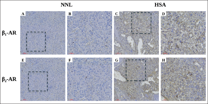
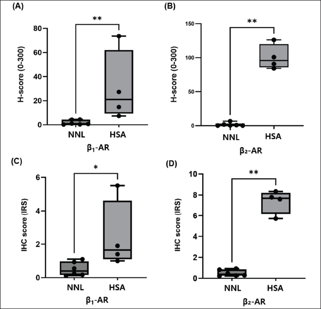
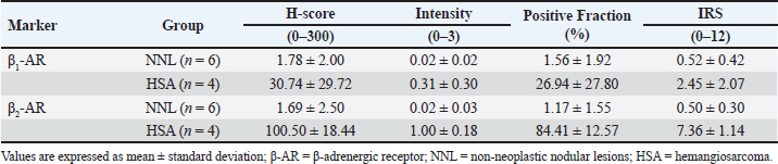
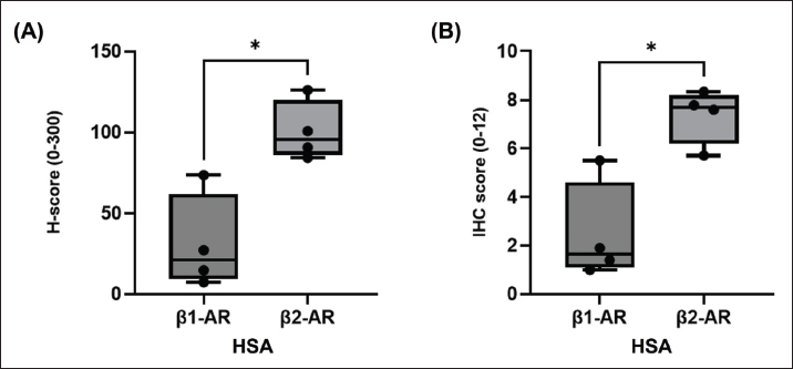
Ethics approvalInformed consent was obtained from the owners, and the study protocol was approved by the University’s Ethics committee (protocol no.: 202502-CNU-047). ResultsTissue samples and clinical dataSplenic tissue samples were collected from 10 client-owned dogs that underwent splenectomy for splenic abnormalities detected on ultrasound examinations. The cohort included seven spayed female dogs and three male neutered dogs, with the following breed distribution: Maltese (n=3), Poodle (n=2), Pomeranian (n=1), Shih Tzu (n=1), Golden Retriever (n=1), Beagle (n=1), and Coton de Tulear (n=1). The overall median age was 10.1 years (range, 5–12 years), and the median body weight was 8.78 kg (range, 3–33.1 kg). Histopathological examination classified four samples as HSA group (n=4) and six as NNL group (n=6), including NLH (n=4) and NH with hematoma (n=2). In the HSA group, the median age was 12 years (range not provided as all cases were 12 years), and the median body weight was 13.96 kg (range, 5.0–33.1 kg). In the NNL group, the median age was 8.8 years (range, 5–12 years), and the median body weight was 3.6 kg (range, 3.0–5.4 kg). Immunohistochemical detection of β-adrenergic receptorsImmunohistochemical analysis showed different β1- and β2-AR staining patterns between the two tissue types. No staining above background was observed in any negative control sections, supporting the specificity of the immunohistochemical staining reactions. NNL tissues exhibited minimal to faint immunoreactivity for β1-AR (Fig. 2A, B) and β2-AR (Fig. 2E, F), with only scattered, weakly-stained cells observed. In contrast, splenic HSA tissues demonstrated diffuse membranous and cytoplasmic immunoreactivity for β1-AR (Fig. 2C, D) and β2-AR (Fig. 2G, H), predominantly in neoplastic endothelial cells lining irregular, blood-filled vascular channels. Increased β-AR immunoreactivity in hemangiosarcomaQuantitative analysis using H-score and IRS indicated higher β1-AR and β2-AR immunoreactivity in HSA tissues compared with NNL controls (Fig. 3, Table 3). For β1-AR, the mean H-score was 30.74 in HSA versus 1.78 in NNL (p=0.0095), and the mean IRS was 2.45 versus 0.52 (p=0.0190). For β2-AR the mean H-score was 100.50 in HSA versus 1.69 in NNL (p=0.0095), and the mean IRS was 7.36 versus 0.50 (p=0.0095). These findings confirm marked overexpression of both receptor and subtypes in HSA tissues. Comparison of β1-AR and β2-AR expression within HSA tissuesWithin HSA, β2-AR expression was higher than β1-AR expression. The mean H-score for β2-AR was 100.50, compared with 30.74 for β1-AR (p= 0.0286), and the mean IRS for β2-AR was 7.36, compared with 2.45 for β1-AR (p=0.0286) (Fig. 4). These results suggest that β2-AR may represent the more prominently-immunoreactive β-AR subtype in canine splenic HSA. DiscussionThis study demonstrated that both β1- and β2-AR expressions were markedly increased in canine splenic HSA tissue compared with non-neoplastic nodular splenic tissues, including NH and NLH. This β-AR overexpression pattern is consistent with profiles previously reported in various human malignancies, including breast, pancreatic, gastric, and lung cancers ( Dickerson and Bryan, 2015; Rains et al., 2017; Liu et al., 2018; Kaira et al., 2019). Increased expression of β1- and β2-ARs has also been documented in human angiosarcoma, a malignant vascular tumor that shares biological characteristics with canine HSA, at both mRNA and protein levels compared with normal vascular endothelium (Grimes et al., 2016; De Nardi et al., 2023). Sustained overexpression of β-ARs may lead to chronic activation of β-AR signaling pathways, even in the absence of markedly elevated catecholamine concentrations (Dickerson and Bryan, 2015; Mravec et al., 2020). Such persistent β-AR signaling has been shown to activate downstream pathways associated with tumor-related processes, including cell proliferation, survival, angiogenesis, invasion, and immune modulation (Cole and Sood, 2012; Dickerson and Bryan, 2015). Consequently, chronic β-AR activation may contribute to tumor progression and metastatic potential in aggressive vascular tumors such as HSA.
Fig. 2. Representative immunohistochemical staining of β1- and β2-adrenergic receptors in canine splenic tissues. In the present study, β2-AR expression was markedly higher than β1-AR expression within the HSA group. This finding aligns with reports from various human malignancies, including gastric, non-small cell lung, and breast cancers, in which β2-AR has been shown to be preferentially overexpressed or functionally-dominant compared with other β-AR subtypes (Dickerson and Bryan, 2015; Rains et al., 2017; Liu et al., 2018; Kaira et al., 2019). Elevated β2-AR expression in these tumors has been strongly associated with high tumor grade, increased metastatic potential, and poor clinical prognosis (Coelho et al., 2019; Zhang et al., 2019). The predominance of β2-AR may be attributed to its preferential expression in endothelial and immune cell compartments, thereby contributing to angiogenesis, extracellular matrix remodeling, and immunosuppressive pathways within the tumor microenvironment, including suppression of cytotoxic CD8+ T-cell activity (Dickerson and Bryan, 2015; Zhang et al., 2025a; Zhang et al., 2025b). In contrast, the role of β1-AR in tumor biology is relatively underexplored and is thought to be primarily related to cardiovascular and metabolic regulation rather than direct oncogenic processes (Dickerson and Bryan, 2015; Zhang et al., 2025a; Zhang et al., 2025b). Therefore, the predominant β2-AR expression observed in canine splenic HSA tissues in this study supports its established role in promoting invasive and aggressive tumor behavior. Collectively, these findings extend prior in vitro and transcriptomic observations of β-AR signaling in canine HSA to clinical tissue-level protein expression, providing a foundation for future pathophysiological investigations and exploration of β-adrenergic–targeted therapeutic strategies. β-AR expression in canine splenic tissue may be influenced by various factors, including cell type composition, local microenvironment, blood flow, and inflammatory status, potentially inducing non-specific immunoreactivity (Dickerson and Bryan, 2015). In particular, β2-AR is predominantly expressed in endothelial and immune cells, and hemorrhagic lesions such as hematomas can disrupt local hemodynamics and oxygenation, potentially leading to the upregulation of β-AR transcription and protein stability (Cheong et al., 2016; Corvera et al., 2024). Similarly, NNL (NH/NLH) may impair marginal sinus blood flow, resulting in parenchymal blood pooling and hematoma formation, a phenomenon frequently observed in clinical settings (Corvera et al., 2024). Accordingly, the increased βAR immunoreactivity in this study cannot be entirely excluded as a nonspecific response associated with hemorrhagic or vascular disturbances. However, in the NNL group, including cases with co-existing hematomas, β1- and β2-ARs both exhibited consistently low immunoreactivity across H-score and IRS assessments, with limited data point distributions (Fig. 3). In contrast, both receptors showed substantially elevated immunoreactivity in the HSA group. This distinct expression pattern suggests that β-AR overexpression in HSA tissues is unlikely to be explained by nonspecific immunoreactivity alone and more plausibly reflects tumor-specific alterations. Given the limited sample size and the heterogeneous composition of the non-neoplastic control group, future studies incorporating isolated hematoma or hemangioma cohorts are warranted to further validate the specificity of β-AR expression in canine splenic tumors.
Fig. 3. Quantitative analysis of β-adrenergic receptor expression in canine splenic tissues. Table 3. Quantitative summary of β-AR expression in NNL spleen and HSA tissues.
Fig. 4. Comparison of β1- and β2-adrenergic receptor expression in canine splenic HSA tissues. This study had several limitations. The HSA group included a small number of cases (n=4), which may limit the generalizability of the findings. Protein expression was assessed exclusively using IHC. Although semi-quantitative scoring systems such as H-score and IRS were used to reduce subjectivity, a formal concordance analysis between the ImageJ-based digital scoring method and conventional pathologist-based manual scoring was not performed. In addition, variability in tissue fixation, processing, and antibody specificity could affect the accuracy and reproducibility of the results. Therefore, complementing IHC with other quantitative methods and incorporating pathologist-based manual scoring in future studies would further strengthen the reliability of the findings. The study did not evaluate correlations between β-AR expression and clinical parameters such as tumor stage, metastasis, or survival. Future research should address these aspects to clarify the prognostic and therapeutic implications of β-AR expression in canine splenic HSA. ConclusionThis study demonstrated marked overexpression of β1- and β2-AR in canine splenic HSA compared with non-neoplastic splenic tissue, with β2-AR showing predominant expression. Adrenergic signaling, particularly mediated through β2-AR, may therefore be involved in the pathophysiology of canine splenic HSA. This study extends previous molecular and transcriptomic observations by providing tissue-level protein expression data derived from clinical splenic specimens. Thus, these results provide foundational data for future investigations into the pathophysiological role and potential therapeutic implications of β-adrenergic signaling in canine splenic HSA. AcknowledgmentsThis work was supported by the National Research Foundation of Korea (NRF) grantfunded by the Korea government (MSIT) (RS-2026-25483764). Conflict of InterestThe Authors declare that there is no conflict of interest. FundingThis work was supported by the Basic Science Research Program through the National Research Foundation of Korea, funded by the Ministry of Education, under Grant RS-2023-00219710314820926400. Authors’ contributionsConceptualization: Son KY, Song JH; Data curation: Son KY, Youk YM, and Song MG; Formal analysis: Son KY and Jung HY; Investigation: Son KY; Methodology: Son KY, Jung HY, and Song JH; Project administration: Son KY and Song JH; Resources: Youk YM, Song MG, and Song JH; Software: Son KY, Youk YM, and Song MG; Supervision: Jung HY and Song JH; Validation: Jung HY and Song JH; Visualization: Son KY, Youk YM, and Song MG; Writing - original draft: Son KY; Writing - review & editing: all authors. Data availabilityThe datasets used and/or analyzed during the current study are available from the corresponding author upon reasonable request. ReferencesAmaya, C.N., Perkins, M., Belmont, A., Herrera, C., Nasrazadani, A., Vargas, A., Khayou, T., Montoya, A., Ballou, Y., Galvan, D., Rivas, A., Rains, S., Patel, L., Ortega, V., Lopez, C., Chow, W., Dickerson, E.B. and Bryan, B.A. 2018. Non-selective beta blockers inhibit angiosarcoma cell viability and increase progression free- and overall-survival in patients diagnosed with metastatic angiosarcoma. Oncoscience 5(3–4), 109–119. Banavali, S., Pasquier, E. and Andre, N. 2015. Targeted therapy with propranolol and metronomic chemotherapy combination: sustained complete response of a relapsing metastatic angiosarcoma. Ecancermedicalscience 9, 499. Cheong, H.I., Asosingh, K., Stephens, O.R., Queisser, K.A., Xu, W., Willard, B., Hu, B., Dermawan, J.K.T., Stark, G.R., Naga Prasad, S.V. and Erzurum, S.C. 2016. Hypoxia sensing through β-adrenergic receptors. JCI Insight 1(21), e90240. Coelho, M., Imperatori, A., Chiaravalli, A.M., Franzi, F., Castiglioni, M., Rasini, E., Luini, A., Legnaro, M., Marino, F., Ribeiro, L. and Cosentino, M. 2019. Beta1- and Beta2-adrenoceptors expression patterns in human non-small cell lung cancer: relationship with cancer histology. J. Neuroimmune Pharmacol. 14(4), 697–708. Cole, S.W. and Sood, A.K. 2012. Molecular pathways: beta-adrenergic signaling in cancer. Clin. Cancer Res. 18(5), 1201–1206. Corvera, G., Alegría-Morán, R., Cifuentes, F.F. and Torres, C.G. 2024. Pathological characterization and risk factors of splenic nodular lesions in dogs (Canis lupus familiaris). Animals (Basel), 14(5), 802. De Nardi, A.B., de Oliveira Massoco Salles Gomes, C., Fonseca-Alves, C.E., de Paiva, F.N., Linhares, L.C.M., Carra, G.J.U., Dos Santos Horta, R., Ruiz Sueiro, F.A., Jark, P.C., Nishiya, A.T., de Carvalho Vasconcellos, C.H., Ubukata, R., Batschinski, K., Sobral, R.A., Fernandes, S.C., Biondi, L.R., De Francisco Strefezzi, R., Matera, J.M., Rangel, M.M.M., Dos Anjos, D.S., Brunner, C.H.M., Laufer-Amorim, R., Cadrobbi, K.G., Cirillo, J.V., Martins, M.C., de Paula Reis Filho, N., Silva Lessa, D.F., Portela, R., Scarpa Carneiro, C., Ricci Lucas, S.R., Fukumasu, H., Feliciano, M.A.R., Gomes Quitzan, J. and Dagli, M.L.Z. 2023. Diagnosis, prognosis, and treatment of canine hemangiosarcoma: a review based on a consensus organized by the brazilian association of veterinary oncology, ABROVET. Cancers (Basel), 15(7), 2025. Dickerson, E.B. and Bryan, B.A. 2015. Beta adrenergic signaling: a targetable regulator of angiosarcoma and hemangiosarcoma. Vet. Sci. 2(3), 270–292. Faulhaber, E.A., Janik, E. and Thamm, D.H. 2021. Adjuvant carboplatin for treatment of splenic hemangiosarcoma in dogs: retrospective evaluation of 18 cases (2011-2016) and comparison with doxorubicin-based chemotherapy. J. Vet. Intern. Med. 35(4), 1929–1934. Grimes, J.A., Prasad, N., Levy, S., Cattley, R., Lindley, S., Boothe, H.W., Henderson, R.A. and Smith, B.F. 2016. A comparison of microRNA expression profiles from splenic hemangiosarcoma, splenic nodular hyperplasia, and normal spleens of dogs. BMC Vet. Res. 12(1), 272. Kaira, K., Kamiyoshihara, M., Kawashima, O., Endoh, H., Imaizumi, K., Sugano, M., Tanaka, S., Fujita, A., Kogure, Y., Shimizu, A., Oyama, T., Asao, T., Shimizu, K. and Mogi, A. 2019. Prognostic impact of β2 adrenergic receptor expression in surgically resected pulmonary pleomorphic carcinoma. Anticancer Res. 39(1), 395–403. Ko, Y.U., Bae, M.K., Sur, J.H. and Choe, N.H. 2023. Analysis of the prevalence of canine splenic mass lesions in Republic of Korea via histopathological diagnosis with immunohistochemistry. Vet. Sci. 10(4), 247. Liu, H., Wang, C., Xie, N., Zhuang, Z., Liu, X., Hou, J. and Huang, H. 2018. Activation of adrenergic receptor β2 promotes tumor progression and epithelial mesenchymal transition in tongue squamous cell carcinoma. Int. J. Mol. Med. 41(1), 147–154. Madden, K.S., Szpunar, M.J. and Brown, E.B. 2011. β-Adrenergic receptors (β-AR) regulate VEGF and IL-6 production by divergent pathways in high β-AR-expressing breast cancer cell lines. Breast Cancer Res. Treat. 130(3), 747–758. Mravec, B., Horvathova, L. and Hunakova, L. 2020. Neurobiology of cancer: the role of β-Adrenergic receptor signaling in various tumor environments. Int. J. Mol. Sci. 21(21), 7958. Porcelli, L., Garofoli, M., Di Fonte, R., Fucci, L., Volpicella, M., Strippoli, S., Guida, M. and Azzariti, A. 2020. The beta-adrenergic receptor antagonist propranolol offsets resistance mechanisms to chemotherapeutics in diverse sarcoma subtypes: a pilot study. Sci. Rep. 10(1), 10465. Rains, S.L., Amaya, C.N. and Bryan, B.A. 2017. Beta-adrenergic receptors are expressed across diverse cancers. Oncoscience, 4(7–8), 95–105. Remmele, W. and Stegner, H.E. 1987. [Recommendation for uniform definition of an immunoreactive score (IRS) for immunohistochemical estrogen receptor detection (ER-ICA) in breast cancer tissue]. Pathologe 8(3), 138–140. Saha, J., Kim, J.H., Amaya, C.N., Witcher, C., Khammanivong, A., Korpela, D.M., Brown, D.R., Taylor, J., Bryan, B.A. and Dickerson, E.B. 2020. Propranolol sensitizes vascular sarcoma cells to doxorubicin by altering lysosomal drug sequestration and drug efflux. Front. Oncol. 10, e614288. Stiles, J.M., Amaya, C., Rains, S., Diaz, D., Pham, R., Battiste, J., Modiano, J.F., Kokta, V., Boucheron, L.E., Mitchell, D.C. and Bryan, B.A. 2013. Targeting of beta adrenergic receptors results in therapeutic efficacy against models of hemangioendothelioma and angiosarcoma. PLoS One 8(3), e60021. Varghese, F., Bukhari, A.B., Malhotra, R. and De, A. 2014. IHC profiler: an open source plugin for the quantitative evaluation and automated scoring of immunohistochemistry images of human tissue samples. PLoS One, 9(5), e96801. Wendelburg, K.M., Price, L.L., Burgess, K.E., Lyons, J.A., Lew, F.H., and Berg, J. 2015. Survival time of dogs with splenic hemangiosarcoma treated by splenectomy with or without adjuvant chemotherapy: 208 cases (2001-2012). J. Am. Vet. Med. Assoc. 247(4), 393–403. Wong, K., Ludwig, L., Krijgsman, O., Adams, D.J., Wood, G.A. and van der Weyden, L. 2021. Comparison of the oncogenomic landscape of canine and feline hemangiosarcoma shows novel parallels with human angiosarcoma. Dis. Model Mech. 14(7), dmm049044. Zhang, F., Wang, Y., Liu, F., Li, Y., Liu, X., Ren, X. and Yuan, X. 2025a. Impact of beta blockers on cancer neuroimmunology: a systematic review and meta-analysis of survival outcomes and immune modulation. Front. Immunol. 16, 1635331. Zhang, X., Zhang, Y., He, Z., Yin, K., Li, B., Zhang, L. and Xu, Z. 2019. Chronic stress promotes gastric cancer progression and metastasis: an essential role for ADRB2. Cell Death Dis. 10(11), 788. Zhang, Y., Yu, F., Ouyang, J., Liu, P., Dai, Y., Wang, Y., Yi, H., Wang, S., Liu, D., Song, K., Pei, W., Hong, Z., Zhang, W., Huang, W., Zhou, G., Cao, S., McLeod, H., Peng, C., Chen, L. and He, Y. 2025b. ADRB2 inhibition suppresses cancer immune evasion by regulating tumor SOX10-PD-L1 axis and T cell function. J. Immunother. Cancer 13(6), e011611. | ||
| How to Cite this Article |
| Pubmed Style Son K, Youk Y, Song M, Jung H, Song J. Beta-adrenergic receptor expression in canine splenic hemangiosarcoma versus non-neoplastic nodular splenic lesions: A comparative immunohistochemical study. Open Vet. J.. 2026; 16(8): 5821-5829. doi:10.5455/OVJ.2026.v16.i8.70 Web Style Son K, Youk Y, Song M, Jung H, Song J. Beta-adrenergic receptor expression in canine splenic hemangiosarcoma versus non-neoplastic nodular splenic lesions: A comparative immunohistochemical study. https://www.openveterinaryjournal.com/?mno=319555 [Access: September 04, 2026]. doi:10.5455/OVJ.2026.v16.i8.70 AMA (American Medical Association) Style Son K, Youk Y, Song M, Jung H, Song J. Beta-adrenergic receptor expression in canine splenic hemangiosarcoma versus non-neoplastic nodular splenic lesions: A comparative immunohistochemical study. Open Vet. J.. 2026; 16(8): 5821-5829. doi:10.5455/OVJ.2026.v16.i8.70 Vancouver/ICMJE Style Son K, Youk Y, Song M, Jung H, Song J. Beta-adrenergic receptor expression in canine splenic hemangiosarcoma versus non-neoplastic nodular splenic lesions: A comparative immunohistochemical study. Open Vet. J.. (2026), [cited September 04, 2026]; 16(8): 5821-5829. doi:10.5455/OVJ.2026.v16.i8.70 Harvard Style Son, K., Youk, . Y., Song, . M., Jung, . H. & Song, . J. (2026) Beta-adrenergic receptor expression in canine splenic hemangiosarcoma versus non-neoplastic nodular splenic lesions: A comparative immunohistochemical study. Open Vet. J., 16 (8), 5821-5829. doi:10.5455/OVJ.2026.v16.i8.70 Turabian Style Son, Kiyeon, Youngmi Youk, Mungyu Song, Hyoyoung Jung, and Joonghyun Song. 2026. Beta-adrenergic receptor expression in canine splenic hemangiosarcoma versus non-neoplastic nodular splenic lesions: A comparative immunohistochemical study. Open Veterinary Journal, 16 (8), 5821-5829. doi:10.5455/OVJ.2026.v16.i8.70 Chicago Style Son, Kiyeon, Youngmi Youk, Mungyu Song, Hyoyoung Jung, and Joonghyun Song. "Beta-adrenergic receptor expression in canine splenic hemangiosarcoma versus non-neoplastic nodular splenic lesions: A comparative immunohistochemical study." Open Veterinary Journal 16 (2026), 5821-5829. doi:10.5455/OVJ.2026.v16.i8.70 MLA (The Modern Language Association) Style Son, Kiyeon, Youngmi Youk, Mungyu Song, Hyoyoung Jung, and Joonghyun Song. "Beta-adrenergic receptor expression in canine splenic hemangiosarcoma versus non-neoplastic nodular splenic lesions: A comparative immunohistochemical study." Open Veterinary Journal 16.8 (2026), 5821-5829. Print. doi:10.5455/OVJ.2026.v16.i8.70 APA (American Psychological Association) Style Son, K., Youk, . Y., Song, . M., Jung, . H. & Song, . J. (2026) Beta-adrenergic receptor expression in canine splenic hemangiosarcoma versus non-neoplastic nodular splenic lesions: A comparative immunohistochemical study. Open Veterinary Journal, 16 (8), 5821-5829. doi:10.5455/OVJ.2026.v16.i8.70 |


