





| Research Article | ||
Open Vet. J.. 2026; 16(8): 5479-5485 !
Open Veterinary Journal, (2026), Vol. 16(8): 5479–5485 Research Article Zearalenone drives apoptosis and oxidative damage in bovine mammary epithelial cellsXinyu Chen, Qi Kong, Haixu Xu and Nannan Zhao*School of Food and Biology Engineering, Xuzhou University of Technology, Xuzhou, People’s Republic of China *Corresponding Author: Nannan Zhao. School of Food and Biology Engineering, Xuzhou University of Technology, Xuzhou, Republic of China. Email: zhaonannan512 [at] 126.com Submitted: 24/04/2026 Revised: 07/07/2026 Accepted: 16/07/2026 Published: 08/08/2026 © 2025 Open Veterinary Journal
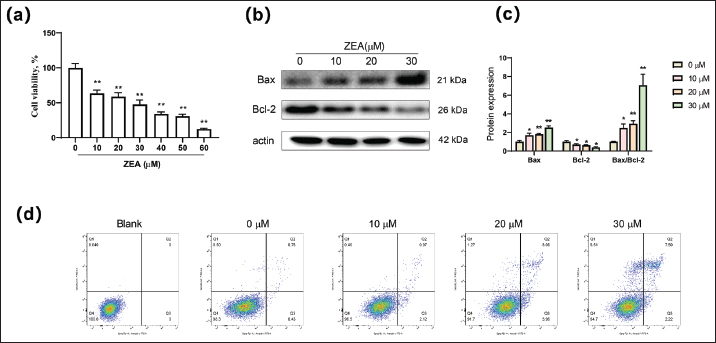
AbstractBackground: Zearalenone (ZEA) is a mycotoxin produced by Fusarium fungi that is widely present in animal and human food. Aim: This study aimed to systematically evaluate the toxicological effects of ZEA on bovine mammary epithelial cells (MAC-T). Methods: To assess cell viability and apoptosis, MAC-T cells were exposed to various concentrations of ZEA for 24 hours, and the optimal ZEA dose was determined for subsequent experiments. Results: A dose-dependent decrease in cell viability and a simultaneous increase in apoptotic proportion were observed by flow cytometry. Western blot analysis revealed that ZEA markedly decreased the level of the antiapoptotic protein Bcl-2 and increased the level of the proapoptotic protein Bax. Cellular reactive oxygen species (ROS) and malondialdehyde concentrations were significantly increased, while glutathione peroxidase (GSH-Px) activity and mitochondrial membrane potential were notably reduced. Gene Ontology and Kyoto Encyclopedia of Genes and Genomes enrichment analyses revealed that ZEA exposure disrupted endoplasmic reticulum function, amino acid metabolism, and glutathione metabolism, among other pathways. Conclusion: ZEA induces apoptosis in MAC-T cells by disrupting mitochondrial dysfunction, increasing ROS production, and triggering oxidative stress. These findings offer experimental evidence that may inform strategies for mitigating ZEA-induced milk production losses and mammary health issues in dairy farming. Keywords: Zearalenone, Bovine mammary epithelial cells, Apoptosis, Oxidative damage. IntroductionExposure to mycotoxins can result in gastrointestinal and reproductive dysfunction in both livestock and humans (Xue et al., 2022). Zearalenone (ZEA), a nonsteroidal mycotoxin with estrogenic properties, is produced by Fusarium (Alshannaq and Yu, 2017). It exhibits reproductive toxicity, immunotoxicity, genotoxicity, and carcinogenicity. Surveys indicate that 40%–50% of agricultural product samples worldwide are contaminated with ZEA (Eskola et al., 2020), thereby posing substantial risks to agricultural production and public health (Wang et al., 2022; Dai et al., 2024). The compound feed for dairy cows typically comprises silage, concentrated feed, and green feed, serving as a potential source of various mycotoxins (Debevere et al., 2019). Rumen microorganisms can degrade some mycotoxins in ruminants, but reports of subclinical health issues in high-yielding dairy cows due to mycotoxins still exist (Valenti et al., 2023). Notably, complete detoxification of ZEA in the rumen may not always occur (Gruber-Dorninger et al., 2021), potentially allowing its residues to contaminate dairy products through milk, posing risks to consumer health. Consumption of ZEA-contaminated feed may lead to toxin residues in breast tissue, potentially elevating intracellular redox levels and reducing milk production (Fusco et al., 2020), leading to toxin accumulation in breast tissue. This accumulation elevates intracellular redox levels, hastening cellular aging and apoptosis, subsequently increasing the predisposition to mastitis in virtuosis (Abuzar et al., 2025). Moreover, mounting evidence suggests that ZEA can prompt ER stress-mediated apoptosis (Fu et al., 2019) or impact the autophagy pathway by modulating reactive oxygen species (ROS) levels, thereby increasing susceptibility to mastitis in vitro (He vitroet al., 2023). However, the molecular mechanism of ZEA in bovine mammary epithelial cells (MAC-T) is poorly understood, especially regarding oxidative stress–apoptosis crosstalk. The aim of this study is to elucidate the molecular mechanisms underlying the effects of ZEA on MAC-T and offer a theoretical basis for mitigating the potential health risks to dairy cows and ensuring the safety of dairy products. Materials and MethodsMaterialsThe MAC-T bovine mammary epithelial cell line was obtained from Shanghai Qingqi Biotechnology Development Co., Ltd. ZEA powder with a purity of ≥98% was procured from Shanghai Macklin Biochemical Technology Co., Ltd. Malondialdehyde (MDA) and glutathione peroxidase (GSH-Px) kits, along with mitochondrial membrane potential (MMP) detection kits, were sourced from Wuhan Elabscience Biotechnology Co., Ltd. The cell counting kit-8 (CCK-8) kit, Color Gel Rapid Kit, Tris–glycine SDS electrophoresis buffer, rapid transfer solution, rapid blocking solution, and hypersensitive ECL chemiluminescence kit were purchased from Suzhou NCM Biotechnology Co., Ltd. Antibodies for Bax, Bcl-2, and actin were acquired from Wuhan Proteintech Biotechnology Co., Ltd. Additionally, supplies such as penicillin-streptomycin solution, pancreatic enzyme cell digestion solution, radio immunoprecipitation assay (RIPA) lysis solution, cell reactive oxygen species kit, and apoptosis detection kit were obtained from Shanghai Beyotime Biotechnology Co., Ltd. Cell cultureMAC-T cells were cultured in Dulbecco’s modified Eagle’s medium/F12 medium supplemented with 10% fetal bovine serum (FBS) and 1% penicillin-streptomycin at 37° in a 5% CO2 atmosphere. Upon reaching 90% confluence, the cells were washed twice with warm PBS, detached using 0.25% trypsin, and subcultured every 2–3 days. Detection of cell viability5,000 cells were seeded in each well of a 96-well cell plate. Subsequently, ZEA solutions at varying concentrations (0, 10, 20, 30, 40, 50, and 60 μM) were individually applied after cell adhesion and incubated for 24 hours. Following this, CCK-8 solution was introduced and incubated for 1 hour before measuring the absorbance at 450 nm. Western blotting (WB)Following the protocol, RIPA lysis buffer was added to the cell samples for lysis. The mixture was centrifuged to obtain the supernatant (12,000 × g, 10 min, 4°), which was then thoroughly mixed with the protein loading buffer to facilitate denaturation. Store the resulting solution at −80° for future use. A volume of 10 μL of denatured protein (30 μg) was subjected to electrophoresis using 12% sodium dodecyl sulfate-polyacrylamide gel electrophoresis (SDS-PAGE). Subsequently, the proteins were transferred to a polyvinylidene difluoride membrane under constant current conditions (400 mA for 25 minutes) and sealed for 10 minutes. The membrane was then sequentially incubated with the primary (overnight at 4°C) and secondary (1 hour at room temperature) antibodies, followed by color imaging using a chemiluminescence imaging device. Apoptosis detectionFollowing stimulation with a ZEA solution, trypsinize the sample for digestion, centrifuge at 1,000 × g for 5 minutes, discard the supernatant, and perform two washes with phosphate-buffered saline. Proceed with flow cytometry analysis according to the manufacturer’s guidelines. Detection of reactive oxygen species and mitochondrial membrane potentialFollowing a 24-hours ZEA treatment, two PBS washes were performed, the 2′,7′-Dichlorodihydrofluorescein diacetate (DCFH-DA) (Ex/Em=488/525 nm) and JC-1 (Ex/Em=514/529 nm for green, 585/590 nm for red) fluorescent probes were applied separately, and the samples were incubated at 37° for 20 minutes. Subsequently, ROS and membrane potential levels were assessed using either an inverted fluorescence microscope or a flow cytometer. Transcriptome sequencingThe collected cells were treated with TRIzol Reagent and submitted to Shanghai Biozeron Co., Ltd for library construction using the Illumina TruSeq™ RNA sample prep kit. DEG analysis between the two groups and samples was performed using edgeR software, with DEGs identified based on the criteria of FDR < 0.05 and fold change (FC) ≥ 1.5. Subsequently, Gene Ontology (GO) function and Kyoto Encyclopedia of Genes and Genomes (KEGG) pathway enrichment analyses for the DEGs were conducted on the WeChat platform. Statistical analysisData are presented as mean ± standard error of the mean. We analyzed the data using one-way analysis of variance or two-tailed Student’s t-test followed by Tukey’s test in GraphPad Prism 7.0. Statistical significance was set at p < 0.05. Ethical approvalNot necessary for this manuscript ResultsZEA-induced apoptosis in MAC-T cellsZEA exhibited a significant inhibitory effect on the viability of MAC-T cells within the concentration range of 10~60 μM, compared with the control group (p < 0.05), and this effect was concentration dependent (Fig. 1a). A negative correlation was observed between ZEA concentration and cell viability. As the concentration of ZEA increased, the levels of the proapoptotic protein Bax were upregulated, while the antiapoptotic protein Bcl-2 was downregulated, resulting in a significant increase in the Bax/Bcl-2 ratio (Fig. 1b and c). Further analysis using flow cytometry demonstrated that the proportions of both early and late apoptotic cells increased significantly with higher ZEA concentrations. Notably, at a ZEA concentration of 30 μM, there was a sharp rise in the proportion of necrotic cells (Fig. 1d), suggesting that 30 μM serves as a critical toxicity threshold beyond which ZEA induces a shift from predominantly apoptotic cell death to necrotic cell death in MAC-T cells. Therefore, CCK-8 assay showed a dose-dependent decrease in cell viability, while flow cytometry and Western blot further confirmed apoptosis. These findings suggest that ZEA is associated with apoptosis induction in MAC-T cells.
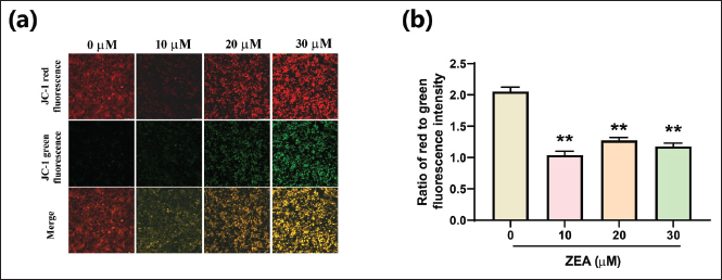
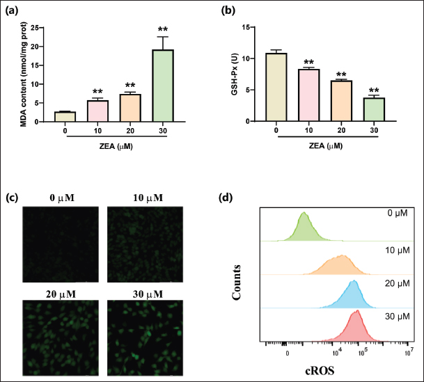
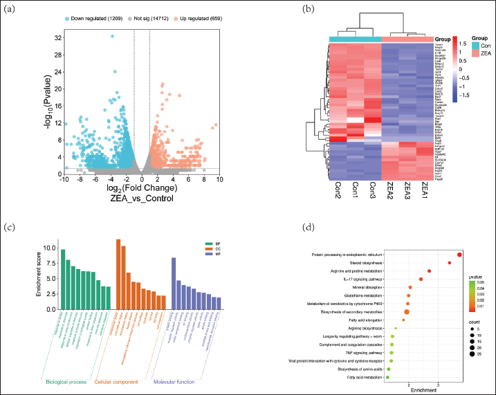
Fig. 1. ZEA induced apoptosis in MAC-T cells (a) Viability of MAC-T cells under different concentrations of ZEA stimulation for 24 hours. (b) Representative Western blot bands for the Bax and Bcl-2 proteins. MAC-T cells were treated with 0, 10, 20, or 30 μM of ZEA for 24 hours. (c) Quantitation of Bax and Bcl-2 proteins. (d) Flow cytometry determination of apoptosis in MAC-T cells after ZEA stimulation. Data are presented as mean ± SEM. *p < 0.05, **p < 0.01 compared with the control group, n=3. ZEA disrupted MMP in MAC-T cells. ZEA disrupted the mitochondrial membrane potential of MAC-T cellsThe JC-1 fluorescent probe was used to monitor the MMP in real-time. The high membrane potential of JC-1 leads to polymer formation within the matrix, resulting in red fluorescence. Conversely, a decrease in membrane potential prevents probe aggregation, causing monomer distribution in the cytoplasm and exhibiting green fluorescence. The ZEA treatment group displayed a notably increased and more intense green fluorescence area than the control group (Fig. 2a). Additionally, the corresponding red-green fluorescence intensity ratio was significantly reduced (Fig. 2b, p < 0.05), indicating that ZEA potentially reduces the MMP of MAC-T cells, leading to apoptosis. ZEA induces oxidative stress in MAC-T cellsAs shown in Figure 3, an increase in ZEA concentration corresponded with a stepwise increase in MDA levels in MAC-T cells, while GSH-PX activity exhibited a concentration-dependent decline. Fluorescence staining was conducted for each treatment group using DCFH-DA. The results indicated that ZEA treatment markedly increased intracellular fluorescence intensity, reflecting a significant increase in ROS levels (P < 0.05). Flow cytometry results corroborated these findings, suggesting that ZEA induces oxidative damage in MAC-T cells. RNA sequencing revealed ZEA-induced changes in MAC-T cellsA concentration of 20 μM ZEA was selected for transcriptomic analysis because it induced significant apoptosis and oxidative stress without causing excessive necrosis, allowing clearer delineation of early signaling events. In the ZEA group (20 μM), 659 genes were upregulated and 1,209 genes were downregulated compared with the control group. Clustering heat maps were generated for the 50 genes with the most significant differences (Fig. 4a and b). GO enrichment analysis identified that these differentially expressed genes (DEGs) primarily function in stress response, immune response, protein folding, and tolerance induction at the BP level. Regarding cell components, DEGs were mainly associated with the extracellular matrix or organelles, such as the ER chaperone complex. In terms of MF, DEGs were predominantly engaged in substance binding (e.g., proteins, compounds, and ions) and enzyme activity (Fig. 4c). KEGG pathway analysis highlighted the affected pathways, including endoplasmic reticulum protein processing, steroid biosynthesis, amino acid metabolism and biosynthesis, glutathione metabolism, interleukin-17 (IL-17), and tumor necrosis factor (TNF) signaling pathways. For instance, alterations in glutathione metabolism and ER processing are consistent with the observed ROS accumulation and apoptotic changes.
Fig. 2. ZEA disrupted MMP in MAC-T cells MAC-T cells were treated with 0, 10, 20, or 30 μM of ZEA for 24 hours. (a) Fluorescence images of JC-1 probe staining in MAC-T cells in response to ZEA stimulation. (b) Quantification of the MMP levels. Data are presented as mean ± SEM. *p < 0.05, **p < 0.01 compared with the control group, n=3.
Fig. 3. ZEA aggravated oxidative stress in MAC-T cells (a) MDA levels in cells after ZEA exposure. (b) GSH-Px activity measurement in MAC-T cells. (c) Representative fluorescence images of DCFH-DA probe staining in ZEA-treated MAC-T cells. (d) Flow cytometry determination of cellular reactive oxygen species in MAC-T cells. Data are presented as mean ± SEM. *p < 0.05, **p < 0.01 compared with the control group, n=3.
Fig. 4. RNA sequencing revealed ZEA-induced changes in MAC-T cells. (a) Volcano plot of distribution trends for differentially expressed genes (DEGs). (b) Heatmap of DEGs in the control and ZEA groups. (c) Gene ontology (GO) pathway enrichment analysis of DEGs in biological processes, cellular components, and molecular functions. (d) KEGG pathway enrichment analysis of DEGs. DiscussionMAC-T, the primary agents of lactation, are highly susceptible to oxidative stress (Tufarelli et al., 2023). Exposure to oxidative insults may result in reduced cell numbers and a surge in apoptosis rates. ZEA concentrations ranging from 7.5 to 240 μM notably impede MAC-T cell viability and disturb intracellular redox balance, culminating in cellular demise and endoplasmic reticulum stress (Fu et al., 2021). These findings align with the outcomes of our investigation, demonstrating a marked decline in cell viability with increasing ZEA concentrations. ZEA-induced abnormalities primarily stem from intracellular oxidative stress and elevated levels of ROS. ZEA cytotoxicity can harm IPEC-J2 cells by triggering the Wnt/FRZB/β-catenin signaling pathway (Cai et al., 2024). This investigation further confirmed that escalating ZEA concentrations heightened intracellular ROS levels and MDA content in MAC-T cells, suppressed GSH-Px activity, and prompted apoptosis. The findings of this study demonstrated that ZEA treatment reduced Bcl-2 protein expression while increasing Bax expression levels, aligning with the findings of a previous study (Ma et al., 2024). Mitochondria serve as both primary energy producers and signal amplifiers in cells, particularly during cell apoptosis. MMP is a crucial indicator of mitochondrial function integrity, directly influencing apoptosis regulation and antioxidant capacity. Research findings indicate that elevated ZEA concentrations lead to a significant decrease in MMP, accompanied by notable increases in early and late apoptosis rates and a substantial rise in the proportion of necrotic cells. This phenomenon is likely attributed to the phosphorylation of mixed lineage kinase domain-like (MLKL) by RIP, resulting in the formation of a necrotic signaling complex that compromises cell membrane integrity (Xuan et al., 2022), subsequently triggering endoplasmic reticulum stress injury (Fu et al., 2021). Consequently, dysregulation of the mTOR-PPAR-ACSL4 pathway obstructs milk fat and milk protein synthesis (Ma et al., 2024), which is consistent with the results of our transcriptomic analysis. Compromised endoplasmic reticulum function after ZEA treatment leads to reduced protein processing and amino acid metabolism capabilities. Recent investigations have demonstrated that ZEA induces Ca2+ overload and increased ROS production through MLKL phosphorylation, thereby inducing mitochondrial dysfunction and promoting apoptosis in goat endometrial cells (Yi et al., 2022). The limitations of this study include the use of an in vitro cell line model, which does not fully replicate the complex rumen and systemic metabolism of ZEA in live animals. Future research should investigate the protective effects of feed additives against ZEA-induced mammary injury and validate these findings in in vivo models. ConclusionIn conclusion, ZEA treatment inhibits cell viability, promotes cell apoptosis, induces mitochondrial dysfunction, and elevates ROS levels, thereby triggering oxidative stress damage. These findings provide a crucial reference for elucidating the mechanism of ZEA toxicity to MAC-T and establish a foundation for assessing feed safety. AcknowledgmentsNone. FundingThis work was supported by the Jiangsu Provincial College Student Innovation Training Program Project (grant no. xcx2025257). Authors’ ContributionsXinyu Chen, Formal analysis, data curation, conceptualization. Qi Kong: Formal analysis. Haixu Xu, Formal analysis, data curation, conceptualization. Nannan Zhao, Funding acquisition, formal analysis, data curation, conceptualization, original draft writing, methodology. Conflict of interestThe authors have no competing financial interests to declare. Data availabilityAll data supporting the findings of this study are available within the manuscript; additional information is available from the corresponding author upon reasonable request. ReferencesAbuzar, Sharif, H.R., Arshad, R., Sharif, M.K., Ashraf, W., Ul-Haq, I., Muhammad, Z., Fahmy, M.A., Korma, S.A. and Rocha, J.M. 2025. Ultraviolet processing for mycotoxin mitigation: a sustainable approach to enhancing food safety and security. Toxicon 265, 108505; doi:10.1016/j.toxicon.2025.108505 Alshannaq, A. and Yu, J.H. 2017. Occurrence, toxicity, and analysis of major mycotoxins in food. Int. J. Env. Res. Pub. He. 14(6), 632; doi:10.3390/Ijerph14060632 Cai, J., Yuan, X.S., Sun, Y.H., Chen, J., Li, P., Yang, S.H. and Long, M. 2024. A2 can protect against damage to IPEC-J2 cells induced by zearalenone via the Wnt/FRZB/β-Catenin signaling pathway. Toxins (Basel) 16(1), 44; doi:10.3390/Toxins16010044 Dai, C., Hou, M.Q., Yang, X.D., Wang, Z.F., Sun, C.P., Wu, X. and Wang, S.J. 2024. Increased NAD plus levels protect female mouse reproductive system against zearalenone-impaired glycolysis, lipid metabolism, antioxidant capacity and inflammation. Reprod. Toxicol. 124, 108530; doi:10.1016/j.reprotox.2023.108530 Debevere, S., De Baere, S., Haesaert, G., Rychlik, M., Fievez, V. and Croubels, S. 2019. Development of an UPLC-MS/MS method for the analysis of mycotoxins in rumen fluid with and without maize silage emphasizes the importance of using matrix-matched calibration. Toxins (Basel) 11(9), 519; doi:10.3390/Toxins11090519 Eskola, M., Kos, G., Elliott, C.T., Hajslová, J., Mayar, S. and Krska, R. 2020. Worldwide contamination of food-crops with mycotoxins: validity of the widely cited ‘FAO estimate’ of 25%. Crit. Rev. Food Sci. 60(16), 2773–2789; doi:10.1080/10408398.2019.1658570 Fu, Y.R., Jin, Y.C., Shan, A.S., Zhang, J., Tang, H.Y., Shen, J.L., Zhou, C.H., Yu, H., Fang, H.T., Zhao, Y., Wang, J.X. and Tian, Y. 2021. Polydatin protects bovine mammary epithelial cells against zearalenone-induced apoptosis by inhibiting oxidative responses and endoplasmic reticulum stress. Toxins (Basel) 13(2), 121; doi: 0.3390/Toxins13020121 Fu, Y.R., Jin, Y.C., Zhao, Y., Shan, A.S., Fang, H.T., Shen, J.L., Zhou, C.H., Yu, H, Zhou, Y.F., Wang, X., Wang, J.M., Li, R.H., Wang, R. and Zhang, J. 2019. Zearalenone induces apoptosis in bovine mammary epithelial cells by activating endoplasmic reticulum stress. J. Dairy Sci. 102(11), 10543–10553; doi:10.3168/jds.2018-16216 Fusco, V., Chieffi, D., Fanelli, F., Logrieco, A.F., Cho, G.S., Kabisch, J., Böhnlein, C. and Franz, C.M.A.P. 2020. Microbial quality and safety of milk and milk products in the 21st century. Compr. Rev. Food Sci. F. 19(4), 2013–2049; doi:10.1111/1541-4337.12568. Gruber-Dorninger, C., Faas, J., Doupovec, B., Aleschko, M., Stoiber, C., Höbartner-Gussl, A., Schöndorfer, K., Killinger, M., Zebeli, Q. and Schatzmayr, D. 2021. Metabolism of zearalenone in the rumen of dairy cows with and without application of a zearalenone-degrading enzyme. Toxins (Basel) 13(2), 84; doi:10.3390/Toxins13020084 He, Y.H., Su, N.R., Yang, H.Y., Yang, W.C., Zhao, C.J., Fu, Y.H., Hu, Y.B. and Hu, X.Y. 2023. ZEA mediates autophagy through the ROS-AMPK-m-TOR pathway to enhance the susceptibility of mastitis induced by in mice. Ecotoxicol. Environ. Saf. 266, 115548; doi:10.1016/J.Ecoenv.2023.115548 Ma, Z.F., Li, Q., Xu, H.M., Li, Y.Y., Wang, S.J., Xiong, Y., Lan, D.L., Li, J., Xiong, X.R. and Fu, W. 2024. Zearalenone triggers programmed cell death and impairs milk fat synthesis via the AKT-mTOR-PPARγ-ACSL4 pathway in bovine mammary epithelial cells. J. Anim. Sci. 102, 276; doi:10.1093/jas/skae276 Tufarelli, V., Colonna, M.A., Losacco, C. and Puvaca, N. 2023. Biological health markers associated with oxidative stress in dairy cows during lactation period. Metabolites 13(3), 405; doi:10.3390/Metabo13030405 Valenti, I., Tini, F., Sevarika, M., Agazzi, A., Beccari, G., Bellezza, I., Ederli, L., Grottelli, S., Pasquali, M., Romani, R., Saracchi, M. and Covarelli, L. 2023. Impact of enniatin and deoxynivalenol co-occurrence on plant, microbial, insect, animal and human systems: current knowledge and future perspectives. Toxins (Basel) 15(4), 271; doi:10.3390/Toxins15040271 Wang, S.J., Fu, W., Zhao, X.Y., Chang, X.J., Liu, H.J., Zhou, L., Li, J., Cheng, R., Wu, X., Li, X. and Sun, C.P. 2022. Zearalenone disturbs the reproductive-immune axis in pigs: the role of gut microbial metabolites. Microbiome 10(1), 234; doi:10.1186/s40168-022-01397-7 Xuan, M.Y., Piao, S.G., Ding, J., Nan, Q.Y., Piao, M.H., Jiang, Y.J., Zheng, H.L., Jin, J.Z. and Li, C. 2022. Dapagliflozin alleviates renal fibrosis by inhibiting RIP1-RIP3-MLKL-mediated necroinflammation in unilateral ureteral obstruction. Front. Pharmacol. 12, 798381; doi:10.3389/Fphar.2021.798381 Xue, D.F., Yang, P., Yang, Y.Y., Wang, Y.A., Wu, K.T., Qi, D.S. and Wang, S. 2022. Deoxynivalenol triggers porcine intestinal tight junction disorder through hijacking SLC5A1 and PGC1α-mediated mitochondrial function. Food. Chem. Toxicol. 163, 112921; doi:10.1016/J.Fct.2022.112921 Yi, Y.Y., Gao, K.K., Zhang, L., Lin, P.F., Wang, A.H. and Jin, Y.P. 2022. Zearalenone induces MLKL-dependent necroptosis in goat endometrial stromal cells via the calcium overload/ROS pathway. Int. J. Mol. Sci. 23(17), 10170; doi:10.3390/Ijms231710170 | ||
| How to Cite this Article |
| Pubmed Style Chen X, Kong Q, Xu H, Zhao N. Zearalenone drives apoptosis and oxidative damage in bovine mammary epithelial cells. Open Vet. J.. 2026; 16(8): 5479-5485. doi:10.5455/OVJ.2026.v16.i8.39 Web Style Chen X, Kong Q, Xu H, Zhao N. Zearalenone drives apoptosis and oxidative damage in bovine mammary epithelial cells. https://www.openveterinaryjournal.com/?mno=318519 [Access: August 08, 2026]. doi:10.5455/OVJ.2026.v16.i8.39 AMA (American Medical Association) Style Chen X, Kong Q, Xu H, Zhao N. Zearalenone drives apoptosis and oxidative damage in bovine mammary epithelial cells. Open Vet. J.. 2026; 16(8): 5479-5485. doi:10.5455/OVJ.2026.v16.i8.39 Vancouver/ICMJE Style Chen X, Kong Q, Xu H, Zhao N. Zearalenone drives apoptosis and oxidative damage in bovine mammary epithelial cells. Open Vet. J.. (2026), [cited August 08, 2026]; 16(8): 5479-5485. doi:10.5455/OVJ.2026.v16.i8.39 Harvard Style Chen, X., Kong, . Q., Xu, . H. & Zhao, . N. (2026) Zearalenone drives apoptosis and oxidative damage in bovine mammary epithelial cells. Open Vet. J., 16 (8), 5479-5485. doi:10.5455/OVJ.2026.v16.i8.39 Turabian Style Chen, Xinyu, Qi Kong, Haixu Xu, and Nannan Zhao. 2026. Zearalenone drives apoptosis and oxidative damage in bovine mammary epithelial cells. Open Veterinary Journal, 16 (8), 5479-5485. doi:10.5455/OVJ.2026.v16.i8.39 Chicago Style Chen, Xinyu, Qi Kong, Haixu Xu, and Nannan Zhao. "Zearalenone drives apoptosis and oxidative damage in bovine mammary epithelial cells." Open Veterinary Journal 16 (2026), 5479-5485. doi:10.5455/OVJ.2026.v16.i8.39 MLA (The Modern Language Association) Style Chen, Xinyu, Qi Kong, Haixu Xu, and Nannan Zhao. "Zearalenone drives apoptosis and oxidative damage in bovine mammary epithelial cells." Open Veterinary Journal 16.8 (2026), 5479-5485. Print. doi:10.5455/OVJ.2026.v16.i8.39 APA (American Psychological Association) Style Chen, X., Kong, . Q., Xu, . H. & Zhao, . N. (2026) Zearalenone drives apoptosis and oxidative damage in bovine mammary epithelial cells. Open Veterinary Journal, 16 (8), 5479-5485. doi:10.5455/OVJ.2026.v16.i8.39 |


