





| Research Article | ||
Open Vet. J.. 2026; 16(8): 5158-5168 !
Open Veterinary Journal, (2026), Vol. 16(8): 5158–5168 Research Article Dogs as rickettsiosis sentinels: Serological and molecular detection of Rickettsia spp. in blood and ectoparasites of dogs from Culiacán, MexicoAramis Gurrola Ramírez1, Karla Rossanet Dzul Rosado2, Nohemí Castro Del Campo1, Jesús José Portillo Loera1, Henry Noé Noh Pech2, Osvaldo López Cuevas3, Irvin González López3, Dina Ricardo-Caldera4 and Soila Maribel Gaxiola Camacho1*1Facultad de Medicina Veterinaria y Zootecnia, Universidad Autónoma de Sinaloa, Culiacán Rosales, Mexico 2Centro de Investigaciones Regionales “Hideyo Noguchi”, Universidad Autónoma de Yucatán, Mérida, México 3Laboratorio Nacional para la Investigación en Inocuidad Alimentaria (LANIIA), Centro de Investigación en Alimentación y Desarrollo, A.C. (CIAD) Culiacán, Culiacán, Mexico 4Grupo de Investigación en Enfermedades Tropicales y Resistencia Bacteriana, Universidad del Sinú, Montería, Colombia *Corresponding Author: Soila Maribel Gaxiola Camacho. Facultad de Medicina Veterinaria y Zootecnia, Universidad Autónoma de Sinaloa, Culiacán Rosales, Sinaloa, Mexico. Email: soilagaxiola [at] uas.edu.mx Submitted: 10/04/2026 Revised: 30/06/2026 Accepted: 10/07/2026 Published: 08/08/2026 © 2025 Open Veterinary Journal
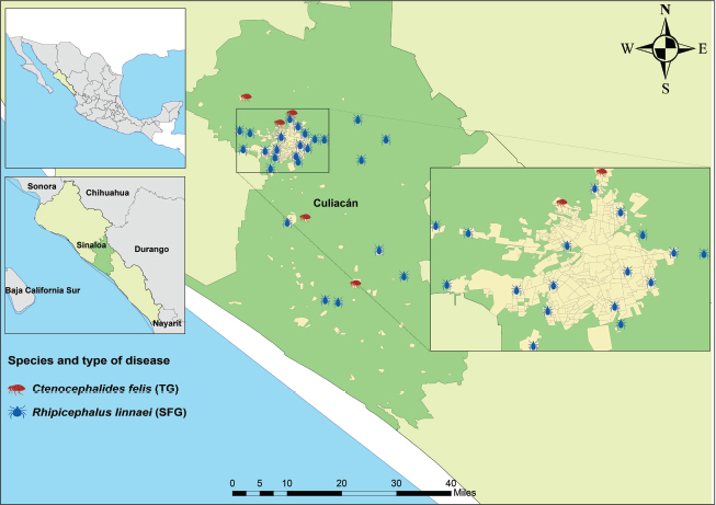
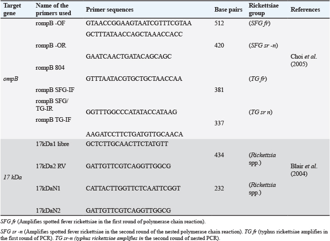
AbstractBackground: Rickettsioses are zoonotic diseases transmitted by ectoparasites, such as ticks and fleas, with increasing public health relevance in Mexico. Dogs act as hosts and epidemiological sentinels, reflecting the environmental circulation of Rickettsia spp. However, despite the favorable environmental conditions, information on the occurrence of Rickettsia spp. in dogs and their ectoparasites in Culiacán, Sinaloa, remains limited. Aim: To determine the serological and molecular presence of Rickettsia spp. in blood samples and ectoparasites collected from dogs in Culiacán’s urban and marginalized rural areas. Methods: A cross-sectional descriptive study was conducted on 30 dogs selected through convenience sampling. Blood and ectoparasite (ticks and fleas) samples were collected. Serological detection of R. rickettsii and R. typhi was performed using an indirect immunofluorescence assay (IFA). Molecular detection was performed using polymerase chain reaction (PCR) targeting the ompB and 17-kDa genes. Results: A total of 356 ectoparasites were collected, with the tick Rhipicephalus linnaei (86.5%) and the flea Ctenocephalides felis (13.5%) being the only identified species. Seroreactivity to R. rickettsii was the most frequently observed (50.0%). Concurrent seroreactivity to both R. rickettsii and R. typhi was detected in 23.33% of the dogs, whereas exclusive reactivity to R. typhi and seronegative results each accounted for 13.33%. PCR results were positive in 40.0% of blood samples and 86.7% of ectoparasite samples. The spotted fever group rickettsiae were detected mainly in ticks, whereas the typhus group rickettsiae were detected in fleas. Conclusion: The findings suggest exposure to and circulation of Rickettsia spp. in dogs and their ectoparasites, with seroreactivity to R. rickettsii being the most frequently detected pattern. Dogs may serve as valuable epidemiological sentinels, underscoring the need for integrated surveillance, ectoparasite control, and timely diagnosis within the One Health framework. Keywords: Dogs, Ectoparasites, Mexico, Rickettsia, Spotted fever, Typhus fever. IntroductionRickettsial diseases are zoonotic infections transmitted by ectoparasites, representing a growing threat to public health worldwide, including Mexico (Álvarez-Hernández et al., 2017). These infections are caused by bacteria of the genus Rickettsia and are primarily transmitted by ticks and fleas parasitizing domestic and wild animals (Abdad et al., 2018; Rymaszewska and Piotrowski, 2024). Based on genomic information, the genus Rickettsia is currently classified into five groups: Group I BG, Belli (the oldest initial Rickettsia group); Group II CG, canadensis; Group III typhus group (TG), typhus; Group IV, spotted fever group (SFG) II (formerly TGR); and Group V, SFG I (El Karkouri et al., 2022). Approximately 40 Rickettsia species have been described, of which approximately 15 are recognized as pathogens of public health and veterinary importance worldwide (Murray et al., 2016; Sánchez-Montes et al., 2021). Dogs play an important role as hosts for ectoparasites and as epidemiological sentinels because they may reflect the circulation of Rickettsia spp. in a specific area (Mesquita et al., 2023). Transmission of rickettsial agents usually occurs through tick bites within the first 24 hours of attachment, and infections in dogs or humans may cause a wide spectrum of clinical manifestations, from subclinical and inapparent infections to severe and potentially life-threatening diseases (Nicholson et al., 2010; Allison and Little, 2013). In Mexico, Rocky Mountain spotted fever (RMSF) is caused by Rickettsia rickettsii and other phylogenetically related rickettsiae, mainly transmitted by Rhipicephalus sanguineus sensu lato, currently recognized in many regions as Rhipicephalus linnaei, the latter being the most frequently reported species in dogs and distributed in tropical areas around the world (Álvarez -Hernández et al., 2024; Dantas-Torres et al., 2024). However, murine typhus (TM), caused by Rickettsia typhi and transmitted mainly by the cat flea Ctenocephalides felis, can also cause clinical symptoms in humans (Nogueras et al., 2013; Torres-Castro et al., 2022). The serological and molecular techniques, particularly indirect indirect immunofluorescence assay (IFA) and polymerase chain reaction (PCR), are the most commonly used methods for detecting Rickettsia spp. in humans and domestic animals, respectively (Oteo et al., 2014; Fomda et al., 2025). According to data published by the Ministry of Health in northwestern Mexico, specifically in Sinaloa, 155 probable cases were reported between 2024 and 2025, including eight cases of R. rickettsii and three cases of R. typhi (Secretary of Health of the State of Sinaloa). However, information on the presence of these bacteria and the ectoparasites that parasitize them in specific regions of Mexico remains scarce. The municipality of Culiacán is considered a high-risk area due to environmental factors, such as high temperatures and humidity, and sociodemographic factors, such as marginalized neighborhoods with poor hygienic conditions that favor the proliferation of vectors. Furthermore, the symptoms of rickettsiosis can be mistaken for other diseases such as dengue, leptospirosis, or typhoid fever, potentially leading to misdiagnosis and an increase in rickettsiosis cases in the population of Culiacán (Lokida et al., 2020; Gurrola et al., 2026). Therefore, improving diagnostic approaches is essential for the accurate detection of rickettsial agents. The present study aimed to determine the serological and molecular presence of Rickettsia spp. in blood samples and ectoparasites collected from dogs in Culiacán’s urban and marginalized rural areas to provide evidence of the circulation of these bacteria and their potential impact on public and veterinary health. Materials and MethodsStudy design and sampling methodsA cross-sectional descriptive study was conducted to detect Rickettsia spp. in blood samples and ectoparasites of dogs in the municipality of Culiacán, Sinaloa, which is located in northwestern Mexico, with an average altitude of 139 m above sea level and a warm sub-humid to dry climate, with an average annual temperature of 25°C and rains concentrated in summer (annual average: 790 mm) (INEGI, 2023). Its geographic coordinates are between latitudes 24°48’ and 27°02’ N and longitudes 105°23’ and 109°26’ W. From May to July 2023, convenience sampling was conducted in neighborhoods and rural communities near the city of Culiacán, with the area and sampling points shown in Figure 1. The inclusion criteria for selecting dogs were as follows: they lived in marginalized areas with unpaved floors, spent most of their time outdoors, and had ectoparasites. Sample collectionBefore sample collection, each dog’s owner provided informed consent for handling and sampling procedures. As a safety measure, the field staff muzzled the dogs prior to sample collection. Approximately 1–5 ml of whole blood was collected and transferred into sterile blood collection tubes without additives (dry tubes) and ethylenediaminetetraacetic acid (EDTA) anticoagulant tubes (BD Vacutainer®; Franklin Lakes, USA). All blood samples were transported to the laboratory under refrigeration (4°C). Serum was recovered from the dry tubes for subsequent IFA testing, while the tubes with EDTA were centrifuged at 3500 rpm for 10 minutes to extract the buffy coat for future PCR testing and stored at −70°C until molecular analysis. After blood sampling, all animals were physically inspected with forceps for the direct collection of ectoparasites, paying particular attention to body regions such as the ears, neck, interdigital spaces, and groin. To identify ectoparasites, we followed the taxonomic criteria of (Acosta and Morrone, 2003; Salceda-Sánchez, 2004; Guglielmone et al., 2006; Wall et al., 2008; Linardi and Santos, 2012) and compared the specimens with a local reference collection. Before the ectoparasites were processed using molecular procedures, morphological observations were made using a Labomed® Luxeo 4D stereomicroscope. Indirect immunofluorescence assay IFAAntigen preparationCanine serum samples were analyzed using an indirect IFA with crude antigens of R. rickettsii (spotted fever group) (Dzul-Rosado et al., 2013) and R. typhi (TG) (Zavala-Castro et al., 2014). For research, antigen slides were prepared in-house for research purposes following previously described protocols (Dzul Rosado et al., 2022; Villarreal-Jiménez et al., 2026). In brief, R. rickettsii and R. typhi were cultured in VERO 76 cells (ATCC CRL-1587), and infection progression was monitored through cytopathic effects and macroscopic changes in the culture medium (Tello-Martin et al., 2018). Infected cells were harvested, suspended in phosphate-buffered saline containing 10% poly-L-lysine, and adjusted to a final concentration of 5,000–10,000 cells/µl. Aliquots of 10 µl were placed onto 12-well slides containing 80%–100% confluence infected cells (La Scola and Raoult, 1997). Slides were dried at 37°C for 15 minutes, fixed according to standard protocols (Ammerman et al., 2008), and stored at −20°C until use.
Fig. 1. Culiacán Municipality and sampling sites. The central rectangle indicates the urban area, and the outer points indicate the rural areas. The blue symbols represent ticks on dogs with Rhipicephalus linnaei, and the red symbols represent fleas (Ctenocephalides felis). TG and SFG indicate the rickettsiae of the typhus and spotted fever groups, respectively. IFA procedureIFA was performed to detect anti-rickettsial antibodies in canine serum. For each assay batch, a hyperimmune serum (from serum of naturally infected dogs collected after the acute phase) was used as a positive control, and phosphate-buffered saline (PBS) containing 1% bovine serum albumin (BSA) was used as a negative control to ensure specificity and validate results. Serum samples were serially diluted in PBS with 1% BSA and added to antigen-coated wells (10 µl). Slides were incubated in a humidified chamber at 37°C for 30 minutes, then washed with PBS containing 0.1% Tween-20 to remove unbound antibodies. fluorescein isothiocyanate immunoglobulin G (IgG) conjugated antibody (Anti dog– Jackson® InmunoR 304-095-003), diluted 1:100 in PBS containing 1% BSA and 0.01% Tween 20, was used as the secondary antibody. After incubation under the same conditions, slides were washed again, including a final wash with PBS-Tween containing 0.01% Evans blue as a counterstain. Slides were examined under a fluorescence microscope at 400× magnification. Samples exhibiting specific fluorescence compatible with Rickettsia spp. at a dilution ≥ 1:64 were considered positive (Stewart and Stewart, 2021). Positive samples were further titrated by serial dilution to determine endpoint titers. deoxyribonucleic acid (DNA) extraction and polymerase chain reaction testingEach dog represented one sample, and DNA extraction was performed based on the ectoparasites of each dog. The ectoparasites were pooled, with up to five ticks or four fleas included per sample (30 samples total). Specimens were ground in a tissue homogenizer using 1.5 ml tubes with beads and 200 μl of AL buffer from the Qiagen kit. DNA was extracted from blood directly from the buffy coat after centrifugation. Both buffy coat and ectoparasite extractions were performed using the QIAamp® DNA Blood and Tissue Kit 69506 (QIAGEN, CA, USA). According to the manufacturer’s instructions, the extracted DNA was stored at −20°C until analysis. Molecular detection of Rickettsia spp. was performed by PCR using the GoTaq® PCR Core System (Promega, EE. UU.). The volumes of each reaction were as follows: 5 μl of buffer solution, 1.5 μl of MgCl2, 2.5 μl of dNTPs, 0.5 μl of each primer, 0.3 μl of DNA Taq polymerase, 11.7 μl of sterile nanopure water, and 3 μl of DNA from each sample. A nested PCR was performed to identify Rickettsia spp. using primers for the ompB gene of outer membrane protein B, where the expected amplicons were 512 base pairs (bp) in the first primer pair and 420 bp in the second for spotted fever rickettsiae (SFG), and 381 bp in the first amplification and 337 bp in the second round of the nested PCR for typhus rickettsiae. Therefore, PCR results were interpreted at the rickettsial group level (SFG or TG) and not at the species level (Choi et al., 2005). Similarly, a second nested PCR was performed to amplify a 17 kDa region, a highly conserved gene across all Rickettsia species. The amplicon from the first round was 434 bp in size, and that from the second round was 232 bp (Blair et al., 2004). The primer sequences used are shown in Table 1. Both PCRs were performed in a thermocycler under the following conditions: denaturation at 95°C for 5 minutes, followed by 40 cycles of 95°C for 15 seconds, 54°C for 15 seconds, and 72°C for 30 seconds, and a final extension at 72°C for 7 minutes. Each PCR run included positive and negative controls to ensure assay reliability and monitor contamination. The amplified products were analyzed by electrophoresis on vertical 8% polyacrylamide gels (acrylamide/bis-acrylamide, 29:1). The gel was stained with ethidium bromide (LifeScience USB) at a final concentration of 0.5 mg/ml and visualized under ultraviolet light using a photodocumentation device (Gel Doc XR, BioRad). Statistical analysisData obtained from the serological detection of antibodies against Rickettsia spp., determined by indirect immunofluorescence (IFA), as well as the frequency of molecular detection by PCR, were organized in an electronic database and analyzed using descriptive statistics. The categorical variables were summarized using absolute and relative frequencies (percentages). IFA and PCR results for canine blood samples and ectoparasites were described separately. All statistical analyses were performed using R version 4.3.1, and the results are presented descriptively without establishing inferential associations between the diagnostic tests. Table 1. Primers used for nested PCR amplification of the ompB and 17 kDa genes of Rickettsia spp.
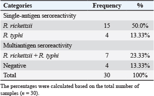
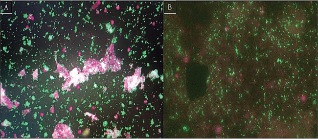
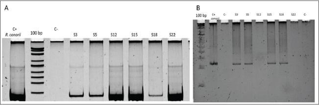
Ethical approvalThe study protocol was reviewed and approved by the Internal Committee for the Care and Use of Animals (CICUA), Faculty of Veterinary Medicine and Animal Science, Autonomous University of Sinaloa, Mexico. All procedures complied with institutional and national regulations for animal welfare in scientific research (approval number 26/FMVZ-UAS/CBI-CEBISFC-13-20080078). ResultsCharacteristics of the collected samples and ectoparasitesA total of 30 dogs were sampled from neighborhoods in the urban area and marginalized rural communities of the municipality of Culiacán. In total, 356 ectoparasites were collected from 25 dogs (176 females and 132 males), of which 308 corresponded to adult ticks identified as Rhipicephalus linnaei and 48 fleas identified as Ctenocephalides felis felis collected from 5 dogs. Indirect immunofluorescence assayThirty canine serum samples were analyzed. The most frequently observed category corresponded to positivity for R. rickettsii, with 15 (50.0%) samples. Concurrent seroreactivity to R. rickettsii and R. typhi was observed in seven samples (23.33%). Four samples showed exclusive seroreactivity to R. typhi (13.33%), and four samples were seronegative for both species (13.33%). The results of the IFA tests for the seroreactive samples for R. rickettsii and R. typhi are shown in Figure 2, and the serological reactivity values for these same species, as well as the cross-reactions between R. rickettsii and R. typhi and the negative samples, are shown in Table 2. PCR for detecting Rickettsia spp.Molecular detection targeting the ompB gene was positive in 12 of the 30 blood samples analyzed, corresponding to a positivity rate of 40.0%, in contrast to PCR performed on ectoparasites, which was positive in 26 of the 30 dogs sampled (86.7%). In the PCR analysis of ectoparasite samples, SFG rickettsiae were detected in ectoparasite pools obtained from 21 dogs infested with R. linnaei, whereas TG rickettsiae were detected in ectoparasite pools obtained from five dogs infested with C. felis. This finding should be considered preliminary and requires confirmation in larger sample-sized studies. Amplification of the ompB gene in blood samples allowed the identification of SFG rickettsiae in nine dogs and TG rickettsiae in three dogs, corresponding to the rickettsial group detected in ectoparasites collected from the same animals. PCR targeting the 17 kDa gene was positive in 12 dogs, seven of which were infested with R. linnaei ticks and five with C. felis fleas. The results of the positive samples on agarose gel imaging and amplification of the ompB and 17 kDa genes for blood and ectoparasites are shown in Figure 3, and the PCR values for both ompB and 17 kDa genes in blood and ectoparasite samples are shown in Figure 4. Table 2. Serological reactivity against R. rickettsii and R. typhi antigens determined by indirect immunofluorescence assay
Fig. 2. Indirect immunofluorescence images A. Indirect immunofluorescence test with titers of 1/512 for R. rickettsii. B. Indirect immunofluorescence IFA image with titers of 1/512 showing R. typhi fluorescent antibodies. DiscussionThis study provides evidence of the presence of Rickettsia spp. in dogs and their ectoparasites in the municipality of Culiacán. Previous studies have identified and associated R. linnaei and C. felis with Rickettsia spp., recognizing them as ectoparasites commonly associated with the transmission of rickettsial agents in canines and humans (Nogueras et al., 2013; Sosa-Gutiérrez et al., 2016; Ortega-Morales et al., 2019). Despite the small number of dogs included in this study and the fact that the dogs were selected based on clinical suspicion and ectoparasite infestation, the results are not representative of the general canine population in Culiacán and should not be interpreted as population-level prevalence estimates. Nevertheless, the high frequency of seroreactivity to rickettsial antigens suggests previous exposure of dogs to these pathogens, and similar results have been reported in endemic regions of Central America, such as Costa Rica, and other countries in Asia, Africa, and Europe, such as Sri Lanka, Angola, and Italy, where dogs act as hosts for ectoparasites and reflect the environmental circulation of Rickettsia spp. (Nanayakkara et al., 2013; Moreira-Soto et al., 2016; Barradas et al., 2017; Di Bella et al., 2024).
Fig. 3. Polyacrylamide gel images and expected amplicons of the ompB and 17 kDa genes for the detection of Rickettsia spp. in DNA samples extracted from buffy coat and ticks (R. linnaei). A. Nested PCR results (first round) for a canine buffy coat sample and ompB gene amplification, where the ~510 bp amplicons corresponding to SFG group rickettsiae align with the positive control of R. conorii. B. Electrophoresis analysis on an 8% polyacrylamide gel, where polymerase chain reaction (PCR) for R. linnaei tick samples and amplification of the 17 kDa protein gene result in an ~232 bp amplicon, which aligns with the positive control of R. conorii. Seroreactivity to R. rickettsii antigen (SFG) was the most frequently detected serological pattern among R. linnaei-infested dogs. In contrast, the lower frequency of seroreactivity to R. typhi antigen may reflect lower detection sensitivity or methodological limitations associated with factors such as the type of vector, particularly C. felis fleas, and is consistent with previous research in regions of Mexico and Latin America (Álvarez-Hernández et al., 2017; Sánchez-Montes et al., 2021; Gual-González et al., 2024). The association of the SFG rickettsiae with R. linnaei and the TG with C. felis is similar to previously reported associations, as documented in various epidemiological contexts in other states and regions of Mexico, including Baja California, Sonora, Coahuila, Puebla, and the Yucatán Peninsula (Peniche-Lara et al., 2015; Pieracci et al., 2019). The high frequency of molecular detection of Rickettsia spp. in ectoparasites, even in seronegative animals, suggests the possible environmental presence of rickettsial agents in the study area. These results are consistent with studies that have documented the presence of Rickettsia spp. in vectors, regardless of host serological status, which could reflect previous exposures or past infections with decreased antibody titers (Pacheco-Solano et al., 2019; Salceda-Sánchez et al., 2023). The discrepancy observed between the high seropositivity detected by indirect IFA and the lower proportion of PCR-positive samples in canine blood can be explained by the persistence of IgG antibodies for long periods after exposure, while rickettsemia is typically transient, thus limiting molecular detection in blood (Biggs et al., 2016; Stewart and Stewart, 2021). This pattern has been described in both dogs and humans, supporting the use of complementary diagnostic methods for a more comprehensive epidemiological assessment (Parola et al., 2003; Marques et al., 2024). In this context, dogs remain important epidemiological sentinels for rickettsial transmission in urban and peri-urban areas, as has been reported in other regions of the world (Shao et al., 2021; Tian et al., 2024). Therefore, strengthening integrated strategies for epidemiological surveillance, ectoparasite control, and timely diagnosis of rickettsial diseases in endemic regions of northwestern Mexico is essential. Such measures may include entomological surveillance, identification of vectors in domestic animals, and evaluation of infestation rates before and after interventions in accordance with the Mexican Official Standard (NOM-032-SSA2-2010).
Fig. 4. Percentage of positive and negative samples obtained using the PCR method. Results of amplification of the 17 kDa gene for ectoparasite samples and ompB for blood and ectoparasite samples. (TG) Typhus Group, Spot Fever Group. The close interaction among dogs, humans, and vectors, especially linked to socio-economic factors, highlights the need for a One Health approach that integrates animal, human, and environmental health. Joint surveillance of dogs and their ectoparasites represents an effective tool for monitoring the circulation of Rickettsia and other vector-borne pathogens, thus supporting public health decision-making (Vada et al., 2025). These findings suggest that environmental conditions are compatible with the circulation of rickettsial agents and highlight the need for surveillance of potential human exposure. This study has some limitations, including the small sample size, convenience sampling, and lack of molecular sequencing. However, it provides relevant evidence on Rickettsia spp. circulation in a region with limited epidemiological data. ConclusionThis study provides serological and molecular evidence of the presence of Rickettsia spp. in dogs and their ectoparasites from Culiacán and surrounding areas. The high seroreactivity detected by IFA and the molecular detection of rickettsial DNA in ectoparasites suggest the presence of these agents in the study area. These findings support the role of dogs as epidemiological sentinels and highlight the need for integrated surveillance and control strategies under the One Health approach. AcknowledgmentsThe authors express their gratitude to CONAHCYT, Mexico, for the fellowship support (CVU 861703), to veterinarians: Claudia Leticia de Jesús Gámez Payan and Brenda Cristina González León for their support in conducting the study and reviewing the dogs, and to Dr. Juan Alfredo Hernández García of the National Polytechnic Institute for providing technical support, training, and guidance during the experimental phase. Conflict of interestThe authors have no conflicts of interest to declare. FundingThis study was funded by a doctoral fellowship from the National Council of Science, Humanities and Technology (CONAHCYT), awarded to Aramis Gurrola Ramírez (CVU 861703). This work was also supported by CONAHCYT and FOSISS grant number 261885. Authors’ contributionsAGR performed the sampling and experimentation, data analysis, and writing of the manuscript. KRDR assisted with reviews, study design, reagent supply, and manuscript writing. NCD wrote the manuscript. JJPL designed the study, wrote the report, and analyzed the statistical data. HNNP conducted indirect IFA experiments. The OLC wrote and managed the information. IGL assisted with figure creation and manuscript writing. The DRC designed, wrote, and refined the manuscript. SMGC provided funding, supervised the study, contributed to manuscript preparation, and reviewed the final version of the manuscript. Data availabilityThe data generated in this study are included in the revised manuscript. ReferencesAbdad, M.Y., Abou Abdallah, R., Fournier, P.E., Stenos, J. and Vasoo, S. 2018. A concise review of the epidemiology and diagnostics of Rickettsioses: Rickettsia and Orientia spp. J. Clin. Microbiol. 56(8), e01728–17; doi:10.1128/JCM.01728-17 Acosta, R. and Morrone, J.J. 2003. Illustrated key for the identification of the supraspecific taxa of Siphonaptera from Mexico. Acta Zool. Mex. 89, 39–53. Allison, R.W. and Little, S.E. 2013. Diagnosis of rickettsial diseases in dogs and cats. Vet. Clin. Pathol. 42(2), 127–144; doi:10.1111/vcp.12040 Álvarez-Hernández, G., Roldán, J.F.G., Milan, N.S.H., Lash, R.R., Behravesh, C.B. and Paddock, C.D. 2017. Rocky Mountain spotted fever in Mexico: past, present, and future. Lancet Infect. Dis. 17(6), e189–e196; doi:10.1016/S1473-3099(17)30173-1. Álvarez-Hernández, G., López-Ridaura, R., Cortés-Alcalá, R., García Rodríguez, G., Calleja-López, J.R.T., Rivera-Rosas, C.N., Alomía-Zegarra, J.L., Brophy, M., Brito-Lorán, C.B., Del Carmen Candia-Plata, M., Ceballos-Liceaga, S.E., Correa-Morales, F., Dzul-Rosado, K.R., Foley, J., Galván-Moroyoqui, J.M., Ganta, R., Gutiérrez-Cedillo, V., Hernández-Milán, N.S., López-Pérez, A.M., López-Soto, L.F., Martínez-Soto, J.M., Mata-Pineda, A.L., Paddock, C.D., Ruiz-González, I.L.J., Salinas-Aguirre, J.E., Salzer, J.S., Sánchez-Montes, S., Soto-Guzmán, A., Tamez-Rivera, Ó., Wagner, D.M. and Walker, D.H. 2024. Rocky Mountain spotted fever in Mexico: A call to action. Am. J. Trop. Med. Hyg. 111(5), 1070–1077; doi:10.4269/ajtmh.24-0265 Ammerman, N.C., Beier-Sexton, M. and Azad, A.F. 2008. Laboratory maintenance of Rickettsia rickettsii. Curr. Protoc. Microbiol. 11:3A.5.1–3A.5.21; doi:10.1002/9780471729259.MC03A05S11 Barradas, P.F., Vilhena, H., Oliveira, A.C., Granada, S., Amorim, I., Ferreira, P., Cardoso, L., Gärtner, F. and de Sousa, R. 2017. Serological and molecular detection of spotted fever group Rickettsia in a group of pet dogs from Luanda, Angola. Parasit Vectors. 10(1), 271; doi:10.1186/s13071-017-2216-3 Biggs, H.M., Behravesh, C.B. and Bradley, K.K. et al. 2016. Diagnosis and management of tickborne rickettsial diseases: Rocky Mountain spotted fever and other spotted fever group rickettsioses, ehrlichioses, and anaplasmosis—United States. MMWR Recommendations Rep. 65(2), 1–44; doi:10.15585/mmwr.rr6502a1 Blair, P.J., Jiang, J., Schoeler, G.B., Moron, C., Anaya, E., Cespedes, M., Cruz, C., Felices, V., Guevara, C., Mendoza, L., Villaseca, P., Sumner, J.W., Richards, A.L. and Olson J.G., 2004. Characterization of spotted fever group rickettsiae in flea and tick specimens from Northern Peru. J. Clin. Microbiol. 42, 4961–4967; doi:10.1128/JCM.42.11.4961-4967.2004 Choi, Y.J., Jang, W.J., Kim, J.H., Ryu, J.S., Lee, S.H., Park, K.H., Paik, H.S., Koh, Y.S., Choi, M.S. and Kim, I.S. 2005. Spotted fever group and typhus group Rickettsioses in humans. South Korea. Emerg. Infect. Dis. J. 11, 237; doi:10.3201/eid1102.040603.16 Dantas-Torres, F., de Sousa-Paula, L.C. and Otranto, D., 2024. The Rhipicephalus sanguineus group: updated list of species, geographical distribution, and vector competence. Parasit. Vectors 17(1), 540; doi:10.1186/S13071-024-06572-3 Di Bella, S., Gambino, D., Pepe, D., Gentile, A., Blanda, V., Valenti, A., Santangelo, F., Ballatore, A., Spina, G., Barbaccia, G., Cannella, V., Cassata, G. and Guercio, A. 2024. Serosurvey of Rickettsia spp. and Ehrlichia canis in dogs from shelters in Sicily (Southern Italy). Pathogens 13(12), 1119; doi:10.3390/pathogens13121119 Dzul-Rosado, K., Peniche-Lara, G., Tello-Martín, R., Zavala-Velázquez, J., Pacheco Rde, C., Labruna, M.B., Sánchez, E.C. and Zavala-Castro, J. 2013. Rickettsia rickettsii isolation from naturally infected Amblyomma parvum ticks by centrifugation in a 24-well culture plate technique. Open Vet. J. 3(2), 101–105. Dzul-Rosado, K., Cámara Herrera, R., Miranda-Schaeubinger, M., Arias-León, J., Peniche-Lara, G., Gilman Robert, H., Mercado-Saavedra Brandon, N., Lugo-Caballero, C., López Ávila, K., Tello Martín, R. and Omodior, O. 2022. Socio-ecological determinants of rickettsial seroprevalence in a rural community of Yucatán, Mexico. Infect. Genet. Evol. 102, 105291; doi:10.1016/j.meegid.2022.105291 El Karkouri, K., Ghigo, E., Raoult, D. and Fournier, P.E. 2022. Genomic evolution and adaptation of arthropod-associated Rickettsia. Sci. Rep. 12, 3807; doi:10.1038/s41598-022-07725-z Fomda, B., Qadri, U., Rashid, R.U., Baqal, S. and Nisar, Q. 2025. Seroprevalence of spotted fever group rickettsiosis in Kashmir: a community-based study using indirect immunofluorescence assay. Indian J. Med. Microbiol. 56, 100902; doi:10.1016/j.ijmmb.2025.100902 Gual-González, L., Torres, M.E., Self, S.C.W., Cantillo-Barraza, O. and Nolan, M.S. 2024. Spotted fever group Rickettsia spp. Molecular and serological evidence among colombian vectors and animal hosts: a historical review. Insects. 15(3), 170; doi:10.3390/insects15030170 Guglielmone, A.A., Beati, L., Barros-Battesti, D.M., Labruna, M.B., Nava, S., Venzal, J.M., Mangold, A.J., Szabó, M.P., Martins, J.R., González-Acuña, D. and Estrada-Peña, A. 2006. Ticks (Ixodidae) on humans in South America. Exp. Appl. Acarol. 40(2), 83–100; doi:10.1007/s10493-006-9027-0 Gurrola Ramírez, A., Rossanet Dzul, K.R., Castro del Campo, N., Portillo Loera, J.J., Ricardo- Caldera, D., González-Gómez, J.P, López-Cuevas, O., Brango, H. and Gaxiola Camacho, S.M. 2026. Molecular detection and genetic analysis of Rickettsia, Ehrlichia, and Bartonella in ectoparasites from domestic dogs in Sinaloa, Mexico, Vet. Parasitol.: Reg. Stud. Reports 67, 101392; doi:10.1016/j.vprsr.2025.101392 La Scola, B. and Raoult, D. 1997. Laboratory diagnosis of rickettsioses: current approaches to diagnosis of old and new rickettsial diseases. J. Clin. Microbiol. 35(11), 2715–2727; doi:10.1128/jcm.35.11.2715-2727.1997 Linardi, P.M. and Santos, J.L.C. 2012. Ctenocephalides felis felis vs. Ctenocephalides canis (Siphonaptera: Pulicidae): some issues in correctly identify these species. Rev. Bras. Parasitol. Vet. 21, 345–354; doi:10.1590/S1984-29612012000400002 Lokida, D., Hadi, U., Lau, C.Y., Kosasih, H., Liang, C.J., Rusli, M., Sudarmono, P., Lukman, N., Laras, K., Asdie, R.H., Murniati, D., Utama, I.M.S., Mubin, R.H., Karyana, M., Gasem, M.H. and Alisjahbana, B. 2020. Underdiagnoses of Rickettsia in patients hospitalized with acute fever in Indonesia: observational study results. BMC Infect. Dis. 20(1), 364; doi:10.1186/s12879-020-05057-9 Marques, H.G., Ribeiro, A.J., Gadelha, A.K.O.A., Resende, C.A.A., Silva, D.R.D., Deus, D.P.M., Barcelos, I.C.D.S., Pereira, I.M., Paula, I.T.S., Lopes, L.D.S., Silva, L.S., Lopes, M.C.D.P., Chávez-Fumagalli, M.A., Coelho, E.A.F., Giunchetti, R.C., Gonçalves, A.A.M. and Galdino, A.S. 2024. Spotted fever diagnosis using molecular methods. Rev. Soc. Bras. Med. Trop. 57, e00204–2024; doi:10.1590/0037-8682-0226-2024 Mesquita, C.A.M., Guimarães, A.M., Guedes, E., Silveira, J.A.G., Rocha, G.C., Nogueira, C.I., Varaschin, M.S., de Souza Santos, M.A. and da Rocha, C.M.B.M. 2023. Dogs (Canis familiaris) as sentinels for determining the risk of occurrence of Rickettsia spp. and Anaplasma phagocytophilum in previously undiagnosed areas. Vet. Parasitol. Reg. Stud. Reports. 46, 100930; doi:10.1016/j.vprsr.2023.100930 Ministry of Health (SSA). 2010. NOM-032-SSA2-2010: for the epidemiological surveillance, prevention and control of vector-borne diseases. Moreira-Soto, A., Carranza, M.V., Taylor, L., Calderón-Arguedas, O., Hun, L. and Troyo, A. 2016. Exposure of dogs to spotted fever group rickettsiae in urban sites associated with human rickettsioses in Costa Rica. Ticks Tick Borne Dis. 7(5), 748–753; doi:10.1016/j.ttbdis.2016.03.007 Murray, G.G., Weinert, L.A., Rhule, E.L. and Welch, J.J. 2016. The phylogeny of Rickettsia using different evolutionary signatures: how tree-like is bacterial evolution? Syst. Biol. 65(2), 265–279; doi:10.1093/sysbio/syv084 Nanayakkara, D.M., Rajapakse, R.P., Wickramasinghe, S. and Kularatne, S.A. 2013. Serological evidence for exposure of dogs to Rickettsia conorii, Rickettsia typhi, and Orientia tsutsugamushi in Sri Lanka. Vector Borne Zoonotic Dis. 13(8), 545–549; doi:10.1089/vbz.2012.1049 National Institute of Statistics and Geography, INEGI., 2023, Available via https://www.inegi.org.mx/contenidos/app/areasgeograficas/resumen/resumen_25.pdf Nicholson, W.L., Allen, K.E., McQuiston, J.H., Breitschwerdt, E.B. and Little, S.E. 2010. The increasing recognition of rickettsial pathogens in dogs and people. Trends Parasitol. 26(4), 205–212; doi:10.1016/j.pt.2010.01.007 Nogueras, M.M., Pons, I., Ortuño, A., Miret, J., Pla, J., Castellà, J. and Segura, F. 2013. Molecular detection of Rickettsia typhi in cats and fleas. PLoS One 8(8), e71386; doi:10.1371/journal.pone.0071386 Ortega-Morales, A.I., Nava-Reyna, E., Avila-Rodríguez, V., Gonzalez-Alvarez, V.H., Castillo-Martínez, A., Siller-Rodríguez, Q.K., Cabezas-Cruz, A., Dantas-Torres, F. and Almazan, C., 2019. Detection of Rickettsia spp. in Rhipicephalus sanguineus (sensu lato) collected from free-roaming dogs in Coahuila state, northern Mexico. Parasit. Vectors 12(1), 130; doi:10.1186/S13071-019-3377-Z Oteo, J.A, Nava, S, Sousa, R.D, Mattar, S, Venzal, J.M, Abarca, K, Labruna, M.B and Zavala-Castro, J. 2014. Guías Latinoamericanas de la RIICER para el diagnóstico de las rickettsiosis transmitidas por garrapatas. Revista chilena de infectología 31(1), 54–65; doi:10.4067/S0716-10182014000100009 Pacheco-Solano, K., Barrantes-González, A., Dolz, G., Troyo, A., Jiménez-Rocha, A.E., Romero- Zuñiga, J.J and Taylor, L. 2019. Exposure of dogs to Rickettsia spp. in Costa Rica: risk factors for PCR-positive ectoparasites and seropositivity. Parasite. Epidemiol. Control 7, e00118; doi:10.1016/j.parepi.2019.e00118 Parola, P., et al. 2003. Update on tick-borne rickettsioses around the world: a geographic approach. Clin. Microbiol. Rev. 26(4), 657–702; doi:10.1128/CMR.00032-13. Peniche-Lara, G., Jimenez-Delgadillo, B. and Dzul-Rosado, K. 2015. Rickettsia rickettsii and Rickettsia felis infection in Rhipicephalus sanguineus ticks and Ctenocephalides felis fleas co-existing 18 in a small city in Yucatan, Mexico. J. Vector Ecol. 40(2), 422–424; doi:10.1111/jvec.12185 Pieracci, E.G., De La Rosa, J.D.P., Rubio, D.L., Perales, M.E.S., Contreras, M.V., Drexler, N.A., Nicholson, W.L., De La Rosa, J.J.P., Chung, I.H., Kato, C., Barton Behravesh, C., Enríquez, M.A.G., Roldan, J.F.G. and Villarino, M.E. 2019. Seroprevalence of spotted fever group rickettsiae in canines along the United States-Mexico border. Zoonoses Public Health. 66(8), 918–926; doi:10.1111/zph.12642 Rymaszewska, A. and Piotrowski, M. 2024. Rickettsia species: genetic variability, vectors, and rickettsiosis-a review. Pathogens. 13(8), 661; doi:10.3390/pathogens13080661 Salceda Sánchez, B. 2004. Key for the identification of adults of the common and most medically important flea species (Insecta: Siphonaptera) in Mexico. Salceda-Sanchez, B., Gasca-Zarate, C.M., Jimenez-Soto, K., Grostieta, E., Lopez- Sanchez, C.G., Soto-Gutierrez, J.J., Lammoglia-Villagomez, M., Huerta-Peña, J., Hernandez- Carbajal, G.R., Chagoya-Fuentes, J.L., Jacome-Sosa, E., Perez-Brígido, C. D., Ballados- Gonzalez, G.G., Becker, I. and Sanchez-Montes, S. 2023. Molecular detection of Rickettsia felis in fleas and ticks collected from dogs and cats of Puebla, Mexico. Zoonoses Public Health 70, 176–183; doi:10.1111/ZPH.13011 Sánchez-Montes, S., Colunga-Salas, P., Lozano-Sardaneta, Y.N., Zazueta-Islas, H.M., Ballados-González, G.G., Salceda-Sánchez, B., Huerta-Jiménez, H., Torres-Castro, M., Panti-May, J.A., Peniche-Lara, G., Muñoz-García, C.I., Rendón-Franco, E., Ojeda-Chi, M.M., Rodríguez-Vivas, R.I., Zavala-Castro, J., Dzul-Rosado, K., Lugo-Caballero, C., Alcántara-Rodríguez, V.E., Delgado-de la Mora, J., Licona-Enríquez, J.D., Delgado-de la Mora, D., López-Pérez, A.M., Álvarez-Hernández, G., Tinoco-Gracia, L., Rodríguez-Lomelí, M., Ulloa-García, A., Blum-Domínguez, S., Tamay-Segovia, P., Aguilar-Tipacamú, G., Cruz-Romero, A., Romero-Salas, D., Martínez-Medina, M.A. and Becker, I. 2021. The genus Rickettsia in Mexico: current knowledge and perspectives. Ticks Tick Borne Dis. 12(2), 101633; doi:10.1016/j.ttbdis.2020.101633 Shao, J.W., Yao, X.Y., Song, X.D., Li, W.J., Huang, H.L., Huang, S.J. and Zhang, X.L. 2021. Molecular detection and genetic diversity of Rickettsia spp. in pet dogs and their infesting ticks in Harbin, northeastern China. BMC Vet. Res. 17(1), 113; doi:10.1186/s12917-021-02823-y Sosa-Gutiérrez, C.G., Vargas-Sandoval, M., Torres, J. and Gordillo-Pérez, G. 2016. Tick- borne rickettsial pathogens in questing ticks, removed from humans and animals in Mexico. J. Vet. Sci. 17, 353–360 Secretary of Health of the State of Sinaloa. 2025. Epidemiological bulletin: epidemiological week ́ N◦ 22. Sinaloa. Stewart, A.G. and Stewart, A.G.A. 2021. An update on the laboratory diagnosis of Rickettsia spp. Infection Pathogens 10(10), 1319; doi: 10.3390/pathogens10101319 Tello-Martin, R., Dzul-Rosado, K., Zavala-Castro, J. and Lugo-Caballero, C. 2018. Approaches for the successful isolation and cell culture of American Rickettsia species. J. Vector Borne Dis. 55(4), 258–264; doi:10.4103/0972-9062.256560 Tian, Y., Juarez, J.G., Moller-Vasquez, A.M., Granados-Presa, M., Ferreira, F.C., Pennington, P.M., Padilla, N., Hamer, G.L. and Hamer, S.A. 2024. Dog ectoparasites as sentinels for pathogenic Rickettsia and Bartonella in rural Guatemala. Res Sq [Preprint]. rs.3.rs-4656611. doi:10.21203/rs.3.rs-4656611/v1 Torres-Castro, M., Reyes-Novelo, E., Noh-Pech, H., Sánchez-Montes, S., Colunga-Salas, P., Lugo-Caballero, C. and Rodríguez-Vivas, R.I. 2022. Rickettsia rickettsii and Rickettsia typhi in habitants from a rural community of southeast Mexico. Rev. Perú Med. Exp. Salud Publica. 39(1), 124–125; doi:10.17843/rpmesp.2022.391.10519 Vada, R., Zanet, S., Trisciuoglio, A. et al. 2025. A one-health approach to surveillance of tick- borne pathogens across different host groups. BMC Vet. Res. 21, 553; doi: 10.1186/s12917-025-04983-7 Villarreal-Jimenez, E., Dzul-Rosado, K., Puerto-Manzano, F., Guillermo-Herrera, J.C., Pech-Noh, H. and Mendez-Dominguez, N. 2026. Ecologic and sociodemographic factors associated with Seroprevalence of Rickettsia in Yucatan, Mexico. Epidemiologic (Basel). 7(2), 30; doi:10.3390/epidemiologia7020030. Wall, R. and Shearer, D. 2008. Veterinary ectoparasites: biology, pathology & control. John Wiley & Sons. Zavala-Castro, J.E., Dzul-Rosado, K.R., Peniche-Lara, G., Tello-Martín, R. and Zavala-Velázquez, J.E. 2014. Isolation of Rickettsia typhi from human, Mexico. Emerg. Infect. Dis. 20(8), 1411–1412; doi:10.3201/eid2008.130095 | ||

| How to Cite this Article |
| Pubmed Style Ramírez AG, Rosado KRD, Campo NCD, Loera JJP, Pech HNN, Cuevas OL, López IG, Ricardo-caldera D, Camacho SMG. Dogs as rickettsiosis sentinels: Serological and molecular detection of Rickettsia spp. in blood and ectoparasites of dogs from Culiacán, Mexico. Open Vet. J.. 2026; 16(8): 5158-5168. doi:10.5455/OVJ.2026.v16.i8.12 Web Style Ramírez AG, Rosado KRD, Campo NCD, Loera JJP, Pech HNN, Cuevas OL, López IG, Ricardo-caldera D, Camacho SMG. Dogs as rickettsiosis sentinels: Serological and molecular detection of Rickettsia spp. in blood and ectoparasites of dogs from Culiacán, Mexico. https://www.openveterinaryjournal.com/?mno=316971 [Access: August 08, 2026]. doi:10.5455/OVJ.2026.v16.i8.12 AMA (American Medical Association) Style Ramírez AG, Rosado KRD, Campo NCD, Loera JJP, Pech HNN, Cuevas OL, López IG, Ricardo-caldera D, Camacho SMG. Dogs as rickettsiosis sentinels: Serological and molecular detection of Rickettsia spp. in blood and ectoparasites of dogs from Culiacán, Mexico. Open Vet. J.. 2026; 16(8): 5158-5168. doi:10.5455/OVJ.2026.v16.i8.12 Vancouver/ICMJE Style Ramírez AG, Rosado KRD, Campo NCD, Loera JJP, Pech HNN, Cuevas OL, López IG, Ricardo-caldera D, Camacho SMG. Dogs as rickettsiosis sentinels: Serological and molecular detection of Rickettsia spp. in blood and ectoparasites of dogs from Culiacán, Mexico. Open Vet. J.. (2026), [cited August 08, 2026]; 16(8): 5158-5168. doi:10.5455/OVJ.2026.v16.i8.12 Harvard Style Ramírez, A. G., Rosado, . K. R. D., Campo, . N. C. D., Loera, . J. J. P., Pech, . H. N. N., Cuevas, . O. L., López, . I. G., Ricardo-caldera, . D. & Camacho, . S. M. G. (2026) Dogs as rickettsiosis sentinels: Serological and molecular detection of Rickettsia spp. in blood and ectoparasites of dogs from Culiacán, Mexico. Open Vet. J., 16 (8), 5158-5168. doi:10.5455/OVJ.2026.v16.i8.12 Turabian Style Ramírez, Aramis Gurrola, Karla Rossanet Dzul Rosado, Nohemí Castro Del Campo, Jesús José Portillo Loera, Henry Noé Noh Pech, Osvaldo López Cuevas, Irvin González López, Dina Ricardo-caldera, and Soila Maribel Gaxiola Camacho. 2026. Dogs as rickettsiosis sentinels: Serological and molecular detection of Rickettsia spp. in blood and ectoparasites of dogs from Culiacán, Mexico. Open Veterinary Journal, 16 (8), 5158-5168. doi:10.5455/OVJ.2026.v16.i8.12 Chicago Style Ramírez, Aramis Gurrola, Karla Rossanet Dzul Rosado, Nohemí Castro Del Campo, Jesús José Portillo Loera, Henry Noé Noh Pech, Osvaldo López Cuevas, Irvin González López, Dina Ricardo-caldera, and Soila Maribel Gaxiola Camacho. "Dogs as rickettsiosis sentinels: Serological and molecular detection of Rickettsia spp. in blood and ectoparasites of dogs from Culiacán, Mexico." Open Veterinary Journal 16 (2026), 5158-5168. doi:10.5455/OVJ.2026.v16.i8.12 MLA (The Modern Language Association) Style Ramírez, Aramis Gurrola, Karla Rossanet Dzul Rosado, Nohemí Castro Del Campo, Jesús José Portillo Loera, Henry Noé Noh Pech, Osvaldo López Cuevas, Irvin González López, Dina Ricardo-caldera, and Soila Maribel Gaxiola Camacho. "Dogs as rickettsiosis sentinels: Serological and molecular detection of Rickettsia spp. in blood and ectoparasites of dogs from Culiacán, Mexico." Open Veterinary Journal 16.8 (2026), 5158-5168. Print. doi:10.5455/OVJ.2026.v16.i8.12 APA (American Psychological Association) Style Ramírez, A. G., Rosado, . K. R. D., Campo, . N. C. D., Loera, . J. J. P., Pech, . H. N. N., Cuevas, . O. L., López, . I. G., Ricardo-caldera, . D. & Camacho, . S. M. G. (2026) Dogs as rickettsiosis sentinels: Serological and molecular detection of Rickettsia spp. in blood and ectoparasites of dogs from Culiacán, Mexico. Open Veterinary Journal, 16 (8), 5158-5168. doi:10.5455/OVJ.2026.v16.i8.12 |


