





| Research Article | ||
Open Vet. J.. 2026; 16(8): 5150-5157 !
Open Veterinary Journal, (2026), Vol. 16(8): 5150–5157 Research Article Enhancing venipuncture training: Evaluation of press-assisted shallow-angle insertion technique using a simulation modelTakuya Maruo1, Hideki Kayanuma1, Yuta Nishiyama2, Yusuke Banno2 and Kotaro Nishi3*1Laboratory of Veterinary Radiology, Azabu University, Sagamihara, Japan 2Veterinary Teaching Hospital, Azabu University, Sagamihara, Japan 3Laboratory of Small Animal Internal Medicine, Azabu University, Sagamihara, Japan *Corresponding Author: Kotaro Nishi. Laboratory of Small Animal Internal Medicine, Azabu University, Sagamihara, Japan. Email: k-nishi [at] azabu-u.ac.jp Submitted: 05/04/2026 Revised: 22/06/2026 Accepted: 30/06/2026 Published: 08/08/2026 © 2025 Open Veterinary Journal
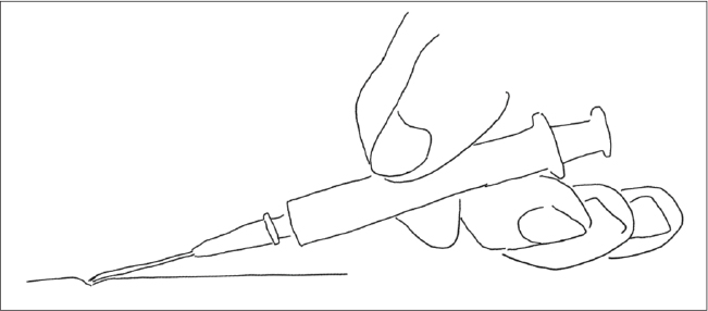
AbstractBackground: Venipuncture is a fundamental but technically demanding skill in veterinary medicine, particularly in small animals with superficial veins. Conventional guidance recommends inserting the needle at a steep angle and then adjusting it after skin penetration; however, this maneuver may increase the risk of posterior vessel-wall injury and needle instability. Aim: This study aimed to evaluate the mechanical characteristics and educational suitability of the press-assisted shallow-angle insertion (PASAI) technique compared with the conventional angle adjustment insertion (CAAI) technique. Methods: This study consisted of two phases. In Study 1, we qualitatively assessed skin–needle interactions during PASAI in 3 healthy beagle dogs using blunted needles to examine skin indentation and functional insertion angle. In Study 2, a crossover simulation-based evaluation was performed with 36 participants, including veterinary students, veterinarians, and veterinary nurses. After instruction and repeated practice with both PASAI and CAAI, performance was assessed using a venipuncture simulator by examining simulated posterior vessel wall injury, qualitative video analysis of needle and finger movement, and structured questionnaire responses. Results: Compared with CAAI, PASAI resulted in significantly fewer injuries to the opposite wall of the simulated vessel (5 vs. 72, p < 0.001). Qualitative analysis in beagle models showed that gentle skin indentation of approximately 1 mm created a localized skin slope and increased the functional insertion angle to an estimated 15°–30°, despite a shallow needle-shaft orientation. The video assessment demonstrated that PASAI was associated with less vertical needle-angle adjustment and reduced index-finger displacement during insertion. Experienced participants rated PASAI more favorably than CAAI in several categories, particularly regarding reduced need for post-entry angle adjustment and lower perceived risk of over-penetration, whereas novices initially found PASAI more difficult. Conclusion: PASAI appears to be a mechanically stable venipuncture technique associated with reduced simulated posterior vessel wall injury and unnecessary needle movement. Although this exploratory study warrants further validation, focused training may help beginners acquire the PASAI technique. The findings suggest that formal incorporation of PASAI into simulation-based veterinary training may improve procedural safety and help standardize instruction for SVA. Keywords: Procedural skills, Simulation-based training, Venipuncture, Veterinary education. IntroductionVenipuncture success depends heavily on the needle insertion angle. Blood collection guidelines recommend inserting the needle at an initial angle of 30° and subsequently adjusting it along the vein at the easiest angle of entry to the vein (Ho et al., 2002; WHO, 2010; Chang et al., 2025). However, beginners may find it difficult to follow the guideline-recommended needle-angle adjustment because the cephalic vein in dogs and cats runs immediately beneath the skin within the subcutaneous tissue (Evans and de Lahunta, 2013; Dyce et al., 2017; Plumb, 2020). Practitioners sometimes adopt a shallow, nearly parallel needle orientation and gently press the skin with the needle tip before insertion (Fig. 1). This “Press-Assisted” component is not a deviation from safety standards but rather a mechanical strategy to stabilize the superficial vein and optimize the entry angle. Although this approach appears to facilitate venipuncture in superficial veins, its mechanical characteristics and educational suitability have not been evaluated in detail.
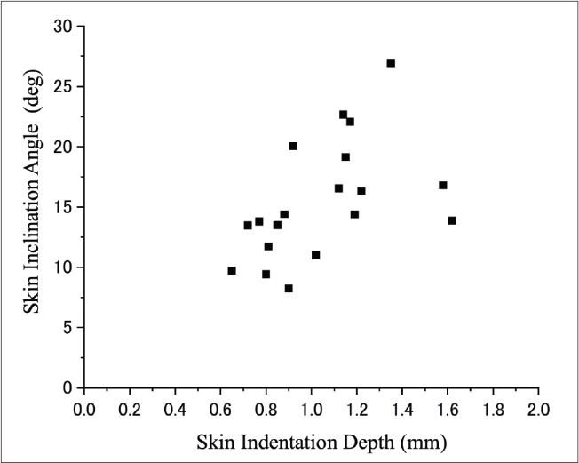
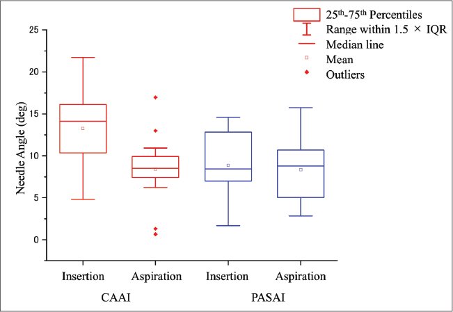
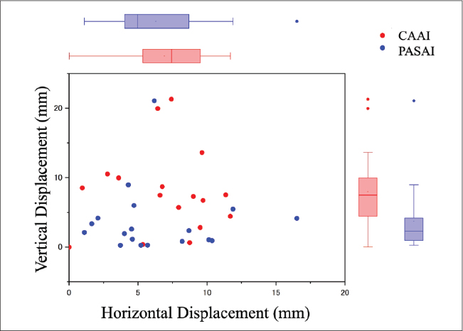
Fig. 1. Schematic of the PASAI technique. The needle is held at a shallow angle, and the needle tip gently presses and indents the skin surface. We hypothesized that this press-assisted shallow-angle insertion (PASAI) technique—defined as a shallow (≈10°) needle orientation combined with gentle, tip-induced skin indentation—would allow venipuncture to be performed without the need for substantial angle adjustment after skin puncture. We further hypothesized that PASAI would be intuitive for learners and advantageous when accessing superficial veins in small animals. This study aimed to characterize venipuncture mechanics using the PASAI technique and compare it with the CAAI technique. In particular, we (1) analyzed skin and needle-tip movement in beagle models and (2) assessed venipuncture performance and perceptions of participants after performing venipuncture with both techniques through a crossover practice session, technique assessment, and questionnaire. Materials and MethodsDefinitionPASAI: The needle was held at a shallow angle as it was inserted into the vessel, and the needle tip was used to gently press the skin surface before insertion (Fig. 1). CAAI: The needle was inserted at a steeper (~30°) angle, and the angle was adjusted toward a more parallel alignment after entry. Study 1: Skin and needle movement in BeaglesThree healthy beagle dogs (8–12 kg) were enrolled, and the forearm hair was clipped. A straight guideline was drawn over the cephalic vein using a skin marker to visualize the skin indentation region. The PASAI technique was evaluated. All procedures were performed following the guidelines of the Laboratory Animal Committee of Azabu University (No. 251211-1). To prevent skin penetration, a blunted 23-G, 1-inch needle (Terumo Co., Tokyo) was attached to a 2.5-ml syringe (Nipro Co., Osaka). The needle was held at a shallow angle, nearly parallel to the skin’s surface. The needle tip was then used to gently press and indent the skin by approximately 1–2 mm, creating a localized skin indentation. Image acquisition and qualitative evaluationLateral-view images were recorded using an iPhone (Apple Inc., USA), and each dog underwent five trials per technique. ImageJ (version 1.54g; National Institutes of Health, Bethesda, MD) was used to visually assess needle tip and skin movement. Study 2: Participant performance, preferred technique, and questionnaire responsesVeterinary students, veterinarians, and veterinary nurses were recruited as participants. Study 2-1: Crossover instruction and practiceThe PASAI and CAAI techniques were explained. Each participant then practiced both methods five times using a venipuncture simulator. The order in which the techniques were explained was counterbalanced among the participants to minimize sequence bias. After practice, the rubber tubing was collected to evaluate the simulated vessel wall injury. Study 2-2: QuestionnaireParticipants rated both techniques with respect to ease of insertion, needle-tip visibility, perceived risk of misalignment or over-penetration, stability during insertion, need for post-entry angle adjustment, and feasibility in case studies. Clinically experienced participants were also asked whether they used a shallow-angle insertion approach in practice. Study 2-3: Assessment of the preferred venipuncture techniqueThe participants then performed venipuncture using their preferred technique on the same simulation model. Lateral-view video recordings were obtained, and needle behavior was assessed qualitatively. In particular, we evaluated whether the needle tip pressed downward on the rubber tubing before or during insertion, the needle angle at simulated skin puncture, the needle-tip angle during aspiration, and the magnitude and direction of index-finger movement. All observations were descriptively interpreted to characterize general behavioral patterns rather than to derive precise numerical measurements. Each participant’s preferred technique was classified into one of the following two categories: PASAI and CAAI. Analysis of the puncture modelThe simulated vessel (latex rubber tubing) was incised longitudinally, and needle-induced damage was inspected on the opposite wall. Simulation model setupA 20-ml syringe barrel (Nipro Co., Osaka, Japan) was used to approximate the size of the forelimbs of small dogs and cats. To simulate a small superficial vein, natural latex rubber tubing (“Amegomu,” inner diameter 2 mm, outer diameter 3.5 mm; Hagitec Co., Ltd., Tokyo) was filled with water and taped to the 20-ml syringe. A 23-G, 1-inch needle attached to a 2.5-ml syringe was used for all the procedures. The tubing was advanced by 1 cm after each puncture to provide a new segment, and the needle was replaced. StatisticsFor Study 2-1, the frequency of rubber-tube damage between PASAI and CAAI was compared using the paired Wilcoxon signed-rank test. For Study 2-2, the Wilcoxon signed-rank test was used to analyze the questionnaire responses to compare paired ratings between PASAI and CAAI. The Mann–Whitney U test was used to compare experienced and novice (student) participants. Bonferroni correction was applied to the 19 exploratory comparisons in the questionnaire to account for multiple testing, and adjusted p-values were used for significance testing. A significance level of p < 0.05 was considered significant for all inferential analyses. Nurses with minimal venipuncture experience were classified as novices, whereas students with frequent clinical experience during their clerkships as senior veterinary students were classified as experienced. All statistical analyses were conducted using the Statistical Package for the Social Sciences, version 28 (IBM Corp., Armonk, NY). ResultsStudy 1The skin indentation was approximately 1 mm, and the resulting skin inclination angle was estimated to be approximately 10°–15° (Fig. 2). Even if the needle-shaft angle was shallower than the recommended angle, the functional insertion angle increased due to local skin depression. Study 2-1: Damage modelThe median number of posterior wall injuries per participant was 2 [interquartile range (IQR), 1–3] for CAAI and 0 (IQR, 0–0) for PASAI, corresponding to aggregated counts of 72 and 5, respectively (p < 0.001). This difference indicates that the PASAI technique may reduce the likelihood of puncturing or traumatizing the simulated vessel’s posterior wall. Study 2-2: QuestionnaireA total of 36 participants, including veterinarians (n=11), veterinary nurses (n=5), and students (n=20), participated in the practice and questionnaire components. Table 1 summarizes the aggregated questionnaire responses. Participants rated PASAI significantly higher than CAAI in perceived risk of misalignment or over-penetration (4.0 ± 1.0 vs. 1.9 ± 0.9, p < 0.05) and no need for post-entry angle adjustment (4.4 ± 0.7 vs. 1.6 ± 0.8, p < 0.05). In addition, experienced participants tended to rate PASAI higher than novices. Of the 18 people (11 veterinarians, 4 senior veterinary students, and 3 veterinary nurses), 13 responded that they were implementing PASAI. However, novices reported that performing PASAI was more difficult than performing CAAI. Study 2-3: Analysis of needle and finger movementVideo analysis revealed two distinct venipuncture patterns corresponding to PASAI (n=19) and CAAI (n=17). Participants classified as using PASAI held the needle at a shallow angle and lightly pressed the tip against the tubing, causing the needle to bend slightly. After entry, the vertical adjustment of the needle angle was minimal (Fig. 3), and the index-finger vertical displacement remained small, allowing the shallow angle to be maintained throughout the procedure (Fig. 4). On the contrary, users with CAAI inserted the needle at a steeper angle and then adjusted the angle after entry (Fig. 3). This adjustment resulted in greater vertical movement of the needle and larger index-finger displacement (Fig. 4). “Knocking,” in which the needle contacted or pierced the posterior wall during redirection, was also observed. DiscussionThis study yielded four major findings regarding the suitability of the PASAI technique for educational purposes. The survey results in Study 2-2 indicated that a shallow puncture angle is commonly and intuitively used in clinical practice. Human studies have reported that experienced nurses often insert needles at approximately 15° (Fujii, 2013). Many clinicians, who adopt a shallow-angle insertion in practice, may be applying the PASAI mechanism. The fact that many participants reproduced PASAI on a simulation model and that experienced participants rated the PASAI as superior to the CAAI in all categories in Study 2-2 suggests that PASAI is a clinically important venipuncture technique that should be mastered.
Fig. 2. Relationship between the skin indentation depth and skin inclination angle during PASAI. Pressing the skin with the needle tip creates a localized inclination of the skin surface, which increases the functional insertion angle. Second, Study 1 revealed that a light skin indentation of approximately one needle diameter (≈1 mm) naturally creates an effective functional insertion angle. This intentional indentation serves two critical purposes: (1) it increases local skin tension to reduce penetration resistance, and (2) it creates a temporary “micro-slope” that guides the needle at the ideal 15°–30° angle while the practitioner maintains a stable, shallow hand position. Even when the needle shaft is held at a shallow angle (≈10°), gentle pressure from the needle tip can depress the skin by about 1 mm and generate a functional angle of approximately 15°–30°, which is within the range recommended by blood collection guidelines (Plumb, 2020). Previous reports have also noted that increasing skin tension stabilizes the surface, reduces resistance during penetration, and facilitates needle insertion (Irwin, 2021). The skin tension generated by the needle tip during PASAI may, therefore, provide an additional stabilizing mechanism that contributes to smoother insertion. However, the force required to produce this skin indentation was not quantified in the present study. Because excessive pressure may distort tissue geometry or alter the position of superficial veins, whereas insufficient pressure may fail to generate the intended functional insertion angle, quantification of the applied force represents an important area for future investigation. Future studies should incorporate force-sensing devices, pressure-sensitive films, or instrumented simulation models to determine the range of forces associated with successful PASAI performance. Establishing an objective target force may facilitate standardization of the technique, improve training reproducibility, and support the development of competency-based educational criteria for novice learners. Third, Study 2-2 found that changing the needle angle within the tissue may cause tissue injury by the needle tip. In the conventional CAAI technique, adjusting the needle angle after skin entry may cause the needle tip to momentarily pass deeper than the intended vascular axis, increasing the risk of unintended tissue damage (Morris, 2021; Saudi Society for Clinical Chemistry, 2025). In superficial veins, nerve paralysis may occur if the angle is not shallow (Sheu and Yuan, 2001). Previous reports also suggest that even needle gauge influences complications, with thinner needles reducing the incidence of nerve injury (Umemura et al., 2024). On the contrary, the shallow insertion angle characteristic of PASAI appeared to limit unnecessary penetration depth and reduce simulated vascular trauma. In addition, PASAI required less index-finger movement—especially in the vertical direction—indicating greater control and stability during insertion, according to Study 2-3. Collectively, these findings suggest that the PASAI technique may mitigate tissue injury by maintaining a stable needle trajectory and reducing excessive hand motion. Table 1. Perceptions and Ratings of CAAI and PASAI Techniques
Fourth, although Study 2-2 supported both techniques, participants consistently rated PASAI more favorably regarding the need for post-entry angle adjustment and lower in perceived risk of misalignment or over-penetration compared to CAAI. PASAI simplifies the procedure by reducing the need for angle modification after skin puncture—a critical yet error-prone step in CAAI. Simulation models also demonstrated less up-and-down movement of the index finger with PASAI, suggesting that finger movement and needle trajectory are more easily aligned. Simplified and standardized procedural steps, which minimize unnecessary variability in hand movements and decision-making, have been shown to improve learning efficiency and procedural performance reproducibility in novice learners (Haji et al., 2016; Burgess et al., 2020). However, while novices rated the PASAI technique as more difficult to perform, experienced participants rated it more highly. This finding indicates that PASAI belongs to a category of “expert-level motor skills” that may feel counterintuitive to beginners who are taught to “aim and thrust” at steep angles. Therefore, this study’s educational value lies in highlighting the need to transition students from the error-prone CAAI to the safer, more stable PASAI through focused simulation training. Taken together, these results suggest the need for PASAI skill acquisition for novices in venipuncture training. In this context, PASAI may be considered compatible with the principles of simulation-based procedural training, as techniques that minimize unnecessary hand movements and procedural complexity have been shown to facilitate skill acquisition and learner confidence in venipuncture education (Lyon et al., 2013). This study has several limitations. First, the simulation model did not replicate the anatomical or biomechanical properties of canine skin and veins, which may limit direct extrapolation to clinical cases. Second, needle–skin interactions were assessed using only two-dimensional lateral images, and precise insertion angles could not be measured; therefore, angle–depth relationships should be interpreted qualitatively. Third, although a crossover design with counterbalanced instructional order was used to reduce inter-individual variability and minimize potential order effects, the final dataset did not retain individual sequence information. Consequently, the potential influence of order effects, carryover effects, and learning effects could not be formally evaluated. These factors may have influenced the observed differences between techniques and the estimated effect sizes. Therefore, the present findings should be considered exploratory and interpreted with caution. Fourth, the clinical experience of the participants varied, and differences in baseline skill levels may have influenced performance and subjective ratings. Fifth, the study focused on mechanical characteristics and simulated vessel injury rather than clinical outcomes such as first-attempt success, hematoma formation, or patient discomfort. Finally, the press-assisted component of PASAI was not quantified in terms of applied force, and excessive pressure could theoretically increase the risk of contamination or distort tissue anatomy. To address these limitations, future studies using anatomically realistic models, three-dimensional motion analysis, retained sequence data, force measurement, and clinical outcome evaluation are needed.
Fig. 3. Box plot of the needle angle between PASAI and CAAI. The needle angle during insertion and aspiration when the tube is held horizontal is shown. The CAAI (red) showed a large change in angle during insertion and aspiration, whereas the PASAI (blue) showed little change. Boxes indicate the interquartile range (25th–75th percentiles), and the median is shown as a horizontal line. Whiskers represent values within 1.5 × IQR. The mean is shown as an open square, and outliers are plotted as diamonds. Gently depressing the skin by approximately one needle diameter passively creates a functional 15°–30° insertion angle, enabling smooth penetration with minimal need for angle adjustment. Although similar behaviors are often performed intuitively, formalizing the PASAI technique could standardize training and reduce procedural variability—particularly when accessing superficial veins. Stable needle orientation associated with PASAI may also aid learning by improving depth control and reducing the risk of PWI. Although further studies incorporating retained sequence data, objective force measurements, and clinical outcome assessments are needed, these exploratory findings suggest that PASAI can support training in simulation-based procedural skills.
Fig. 4. Scatter and box plots of finger movements for CAAI and PASAI. Finger movement was plotted using a tube held horizontally during aspiration. The index finger position at the start of the insertion was used as the reference point (0, 0), and the index finger position during aspiration was expressed as the absolute coordinate. Compared with PASAI (blue), CAAI (red) showed greater vertical movement. Boxes indicate the interquartile range (25th–75th percentiles), and the median is shown as a horizontal line. Whiskers represent values within 1.5 × IQR. The mean is shown as an open square, and outliers are plotted as diamonds. AcknowledgmentsThe authors would like to thank the participants of this study for their time and cooperation. Gemini (Google LLC, Mountain View, CA) provided editing assistance to improve grammar and clarity. All scientific content was developed and verified by the authors. Authors' contributionsConceptualization: Takuya Maruo and Yuta Nishiyama. Animal study: Yusuke Banno. Investigation (Questionnaire and analysis): Takuya Maruo, Yuta Nishiyama, and Hideki Kayanuma. Writing–Original Draft: Takuya Maruo and Kotaro Nishi. Supervision: Hideki Kayanuma Review & Editing: All authors have read and agreed to the manuscript’s publication. Conflict of interestThe authors declare no conflict of interest. FundingThis study received no specific grant. Ethical approvalThis study involved voluntary, anonymous responses from students and clinicians as part of routine educational activities. The Laboratory Animal Committee of Azabu University approved this study (Approval Number 260205-1). Data availabilityAll data supporting this study’s findings are available within the manuscript. ReferencesBurgess, A., Van Diggele, C., Roberts, C. and Mellis, C. 2020. Tips for teaching procedural skills. BMC Med. Educ. 20(2), 458; doi: 10.1186/s12909-020-02284-1 Chang, J., Choi, S., Cho, H., Kim, S., Chung, J.W., Yoo, S.J., Song, E.Y. and Chun, S. 2025. Standards and practice guidelines for venous blood collection: consensus recommendations from the Korean Society for Laboratory Medicine. Ann. Lab. Med. 45(4), 343–357; doi: 10.3343/alm.2025.0022 Dyce, K.M., Sack, W.O., Wensing, C.J.G. 2017. Veins of the thoracic limb. In Textbook of veterinary anatomy, 5th ed. Eds., Dyce, K.M., Sack, W.O., and Wensing, C.J.G. St. Louis, MO: Elsevier, pp. 423–425. Evans, H.E., de Lahunta, A. 2013. Veins of the thoracic limb. In Miller’s anatomy of the dog, 4th ed. Eds., Evans, H.E., de Lahunta, A. St. Louis, MO: Elsevier Saunders, pp. 434–436. Fujii, C. 2013. Clarification of the characteristics of needle-tip movement during vacuum venipuncture to improve safety. Vasc. Health Risk Manag. 9, 381–390. Haji, F.A., Cheung, J.J.H., Woods, N., Regehr, G., De Ribaupierre, S. and Dubrowski, A. 2016. Thrive or overload? The effect of task complexity on novices' simulation-based learning. Med. Educ. 50(9), 955–968; doi: 10.1111/medu.13086 Ho, A. 2002. Venepuncture and cannulation. In: The Royal Marsden Hospital Manual of Clinical Nursing Procedures. 6th ed. Eds. Dougherty, L. and Lister, S. Oxford, UK: Blackwell Science, pp: 267–276. Irwin, T., Speirs, A. and Merrett, C. 2021. The effect of skin tension, needle diameter and insertion velocity on the fracture properties of porcine tissue. J. Mech. Behav. Biomed. Mater. 123, 104660; doi: 10.1016/j.jmbbm.2021.104660 Lyon, C., Schultz, J. and et al. 2013. Simulation-based peripheral intravenous catheter insertion training: improving performance and procedural confidence. J. Infus. Nurs. 36(5), 346–351. Morris, W. 2011. Complications. In Venepuncture and cannulation. 2nd ed. Eds., Phillips, S., Collins, M., Dougherty, L. Oxford: Wiley-Blackwell, pp. 118–120. Plumb, D.C. 2020. Venous access and injection sites. In Plumb’s veterinary drug handbook. 9th ed. Eds., Plumb, D.C. Hoboken, NJ: Wiley-Blackwell, pp. 1248–1249. Saudi Society for Clinical Chemistry. 2025. Guidelines for venous blood collection. 2nd ed. Saudi Society for Clinical Chemistry. Sheu, J.J. and Yuan, R.Y. 2001. Superficial radial neuropathy following venepuncture. Int. J. Clin. Pract. 55(6), 422–423; doi: 10.1016/j.intjcp.2001.01.03 Umemura, H., Takahashi, H., Fukuda, Y., Soma, H., Aoki, R., Takei, N. and Nakayama, T. 2024. Use of finer needles for venipuncture increases in vitro hemolysis despite reducing persistent pain and nerve injury: a retrospective study. Ann. Clin. Biochem. 61(2), 107–114; doi: 10.1177/00045632231196045 WHO. 2010. WHO Guidelines on blood drawing best practices in phlebotomy. Geneva, Switzerland: World Health Organization. | ||

| How to Cite this Article |
| Pubmed Style Maruo T, Kayanuma H, Nishiyama Y, Banno Y, Nishi K. Enhancing venipuncture training: Evaluation of press-assisted shallow-angle insertion technique using a simulation model. Open Vet. J.. 2026; 16(8): 5150-5157. doi:10.5455/OVJ.2026.v16.i8.11 Web Style Maruo T, Kayanuma H, Nishiyama Y, Banno Y, Nishi K. Enhancing venipuncture training: Evaluation of press-assisted shallow-angle insertion technique using a simulation model. https://www.openveterinaryjournal.com/?mno=316420 [Access: August 08, 2026]. doi:10.5455/OVJ.2026.v16.i8.11 AMA (American Medical Association) Style Maruo T, Kayanuma H, Nishiyama Y, Banno Y, Nishi K. Enhancing venipuncture training: Evaluation of press-assisted shallow-angle insertion technique using a simulation model. Open Vet. J.. 2026; 16(8): 5150-5157. doi:10.5455/OVJ.2026.v16.i8.11 Vancouver/ICMJE Style Maruo T, Kayanuma H, Nishiyama Y, Banno Y, Nishi K. Enhancing venipuncture training: Evaluation of press-assisted shallow-angle insertion technique using a simulation model. Open Vet. J.. (2026), [cited August 08, 2026]; 16(8): 5150-5157. doi:10.5455/OVJ.2026.v16.i8.11 Harvard Style Maruo, T., Kayanuma, . H., Nishiyama, . Y., Banno, . Y. & Nishi, . K. (2026) Enhancing venipuncture training: Evaluation of press-assisted shallow-angle insertion technique using a simulation model. Open Vet. J., 16 (8), 5150-5157. doi:10.5455/OVJ.2026.v16.i8.11 Turabian Style Maruo, Takuya, Hideki Kayanuma, Yuta Nishiyama, Yusuke Banno, and Kotaro Nishi. 2026. Enhancing venipuncture training: Evaluation of press-assisted shallow-angle insertion technique using a simulation model. Open Veterinary Journal, 16 (8), 5150-5157. doi:10.5455/OVJ.2026.v16.i8.11 Chicago Style Maruo, Takuya, Hideki Kayanuma, Yuta Nishiyama, Yusuke Banno, and Kotaro Nishi. "Enhancing venipuncture training: Evaluation of press-assisted shallow-angle insertion technique using a simulation model." Open Veterinary Journal 16 (2026), 5150-5157. doi:10.5455/OVJ.2026.v16.i8.11 MLA (The Modern Language Association) Style Maruo, Takuya, Hideki Kayanuma, Yuta Nishiyama, Yusuke Banno, and Kotaro Nishi. "Enhancing venipuncture training: Evaluation of press-assisted shallow-angle insertion technique using a simulation model." Open Veterinary Journal 16.8 (2026), 5150-5157. Print. doi:10.5455/OVJ.2026.v16.i8.11 APA (American Psychological Association) Style Maruo, T., Kayanuma, . H., Nishiyama, . Y., Banno, . Y. & Nishi, . K. (2026) Enhancing venipuncture training: Evaluation of press-assisted shallow-angle insertion technique using a simulation model. Open Veterinary Journal, 16 (8), 5150-5157. doi:10.5455/OVJ.2026.v16.i8.11 |


