





| Research Article | ||
Open Vet. J.. 2026; 16(8): 5202-5214 !
Open Veterinary Journal, (2026), Vol. 16(8): 5202–5214 Research Article Proteomic mapping of seminal plasma and spermatozoa in Indonesia-bred Simmental bulls housed at an artificial insemination facilityNi Wayan Kurniani Karja1, Brilla Widya Witri2, Iis Arifiantini1, Wahono Esthi Prasetyaningtyas3, Sri Suharti4, Masayasu Taniguchi5 and Mokhamad Fahrudin3*1Division of Reproduction and Obstetrics, School of Veterinary Medicine and Biomedical Sciences, IPB University, Bogor, Indonesia 2Veterinary Biomedical Science Study Program, School of Veterinary Medicine and Biomedical Science, IPB University, Bogor, Indonesia 3Division of Anatomy, Histology and Embryology, School of Veterinary Medicine and Biomedical Sciences, IPB University, Bogor, Indonesia 4Department of Animal Nutrition and Feed Technology, Faculty of Animal Science, IPB University, Bogor, Indonesia 5Laboratory of Theriogenology, Joint Faculty of Veterinary Medicine, Yamaguchi University, Yamaguchi, Japan *Corresponding Author: Mokhamad Fahrudin. Division of Anatomy, Histology, and Embryology, School of Veterinary Medicine and Biomedical Science, IPB University, Bogor, Indonesia. Email: mfahrudin [at] apps.ipb.ac.id Submitted: 28/03/2026 Revised: 22/06/2026 Accepted: 03/07/2026 Published: 08/08/2026 © 2025 Open Veterinary Journal
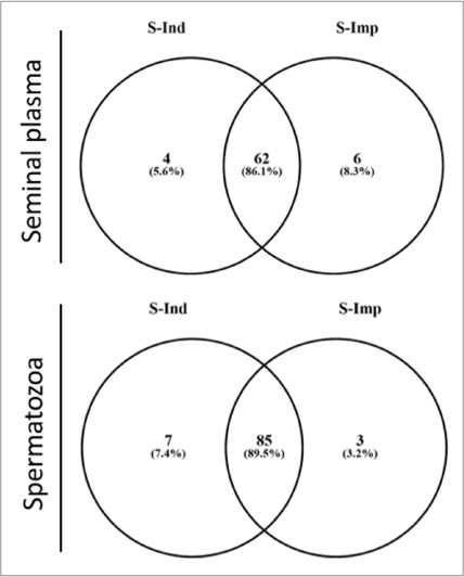
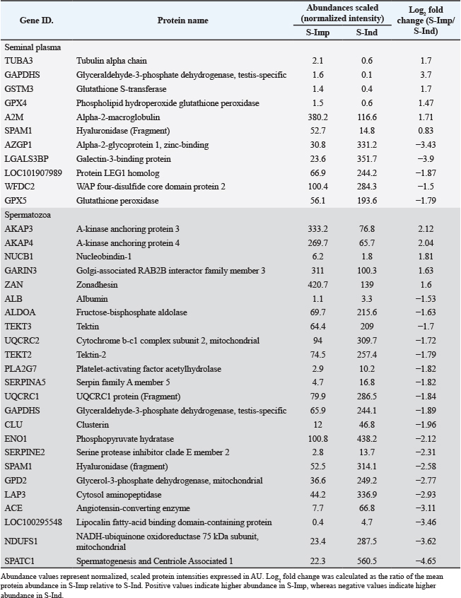
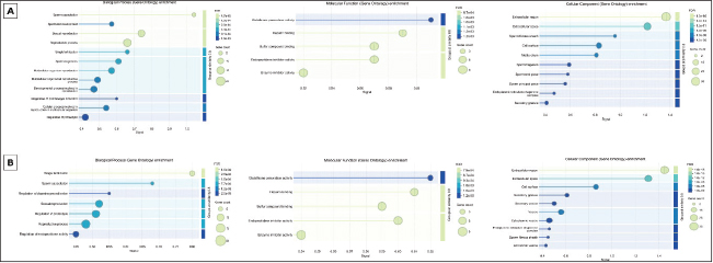
AbstractBackground: Bull fertility is a critical determinant of success in an artificial insemination (AI) center. Fertility is strongly influenced by proteins present in seminal plasma and spermatozoa. Proteomics is an advanced analytical approach for identifying and characterizing these proteins and their associations with sperm fertility. Aim: This study aimed to identify and characterize the proteomic profiles of seminal plasma and spermatozoa derived from Indonesian-bred Simmental bulls and compare them with imported Simmental bulls housed at the same AI facility. Methods: Semen samples were collected from three Indonesian-bred (S-Ind) and three imported (S-Imp) Simmental bulls using an artificial vagina. Seminal plasma and sperm pellets were separated and analyzed using liquid chromatography-tandem mass spectrometry (LC-MS/MS) analysis following protein extraction and 1D SDS-PAGE separation. Protein identification was performed using Proteome Discoverer 2.2 software with Uniprot protein database. Gene Ontology and STRING analyses were conducted to assess protein functions and interactions. Results: LC-MS/MS analysis identified 68 and 92 proteins in seminal plasma and spermatozoa, respectively. Four proteins were unique to the S-Ind group in seminal plasma, while six proteins were unique to the S-Imp group. In spermatozoa, seven proteins were identified exclusively in the S-Ind group, whereas three proteins were detected only in the S-Imp group. The identified proteins were involved in spermatogenesis, sperm motility, capacitation, the acrosome reaction, fertilization, oxidative stress balance, and energy metabolism. Conclusion: The proteomic landscape and key biological functions of proteins in seminal plasma and spermatozoa from bulls bred in Indonesia were comparable to those observed in imported bulls. These proteins are involved in spermatogenesis, sperm capacitation, the acrosome reaction, fertilization, energy metabolism, and sperm protection against oxidative stress. Despite this overall similarity, distinct differences in protein composition and abundance were observed. S-Ind exhibited a higher abundance of proteins involved in metabolism and cellular protection, whereas S-Imp exhibited a greater abundance of structural proteins associated with sperm function. Keywords: Fertility, Proteomics, Seminal plasma, Simmental bulls, Spermatozoa. IntroductionArtificial insemination (AI) programs using frozen semen place significant importance on the productivity of the bull, as it serves as an indicator of efficiency and overall performance in the production of frozen semen. Therefore, male fertility is a particularly important determinant of AI center success and overall cattle herd productivity (DeJarnette et al., 2004). Specific proteins in seminal plasma and spermatozoa strongly influence bull fertility and regulate sperm quality. Spermatozoa undergo a series of biochemical and functional modifications as they travel from the testes to the distal region of the epididymis, ultimately acquiring the capacity to fertilize (Gervasi and Visconti, 2017). During ejaculation and subsequent deposition within the female reproductive tract, spermatozoa are transported within a complex biological fluid known as seminal plasma (Maxwell et al., 2007). Seminal plasma is a dynamic and complex mixture of fluids originating from multiple male reproductive organs, including the epididymis, prostate gland, seminal vesicles, and bulbourethral glands (Rodríguez‐Martínez et al., 2011), containing molecules that influence sperm motility and capacitation, cell protection, acrosome reaction, fertilization, and embryonic development (Moura et al., 2018). Proteins in seminal plasma and spermatozoa play crucial roles in regulating sperm function throughout various stages of spermatogenesis (Ruprecht and Kunji, 2020). They are involved in sperm capacitation, the acrosome reaction, and fertilization (Gómez-Torres et al., 2022; Ramírez-López et al., 2023). In addition, these proteins are associated with sperm motility (Shibata et al., 2024) and have been reported to protect spermatozoa from oxidative stress within the epididymis (Kraus et al., 2005), as well as after ejaculation and within the female reproductive tract (Pournasir et al., 2021). They are also implicated in energy metabolism processes (Gitlits et al., 2000). Proteins are expressed in different ways and play a crucial role in sperm; this may be a key factor in explaining reproductive success. Therefore, the identification of proteins involved in cellular functions within cells has become a critically important area of research, particularly for the discovery of fertility-associated biomarkers (Panner Selvam et al., 2019). Currently, omics-based technologies, particularly proteomics, are employed as advanced analytical tools to identify and characterize proteins in seminal plasma and spermatozoa. Although the biological role of the seminal plasma proteome in sperm physiology remains complex, it is now possible to accurately characterize both the spermatozoa and seminal plasma proteomes using high-resolution mass spectrometry (Druart and de Graaf, 2018). These approaches facilitate the examination of protein functions and interactions, thereby enabling the establishment of associations between molecular profiles and male fertility (Bustamante-Filho et al., 2022). Frozen semen for AI in Indonesia is produced by both national and regional AI centers. High-quality bulls are acquired either through domestic procurement or by importation from abroad. To date, no studies have analyzed and compared the proteomic profiles of these two groups of bulls. Therefore, this study aimed to identify and characterize seminal plasma and spermatozoa derived from Indonesian bulls and compare them with those obtained from imported cattle. Materials and MethodsAnimals and the collection of semenSimmental bulls raised at the AI station were used in this study. Three of the bulls were born at one of the government's breeding centers (S-Ind), while the other three were imported (S-Imp). All bulls were approximately 3 years old and were raised under the same husbandry system in the same AI station for approximately 1 year before semen collection. The bulls used in this study were routinely subjected to semen collection twice weekly and consistently exhibited 60%–70% sperm motility. Semen was collected using an artificial vagina. After collection, semen from three bulls was pooled and centrifuged at 3,000 g for 30 minutes to separate the seminal plasma from the sperm pellet. The seminal plasma and sperm pellet samples were then stored at −20°C until further analysis. Protein extraction and 1D SDS-PAGESperm pellet protein was extracted using PRO-PREP™ protein extraction solution (iNtRON Biotechnology, Korea) according to the manufacturer’s instructions. In brief, 400 µl of PRO-PREP solution was added to the sperm pellet. The mixture was incubated at −20°C for 20 minutes, centrifuged at 13,200 g (4°C) for 5 minutes, and the supernatant was transferred to a sterile tube. The total protein concentration in seminal plasma and sperm pellets was measured in the same volume from each sample using the bicinchoninic acid (BCA) protein assay (Thermo Scientific™, United States). SDS-PAGE was performed to separate proteins into gel bands by molecular weight. Protein separation was performed using SurePAGE™, Bis-Tris, 10 cm × 8 cm, 12-well, 4%–20% gradient gel (M00656; GenScript) (SurePAGE, GenScript Biotech Corp., Hong Kong) with Tris-MOPS-SDS running buffer (M00138; GenScript). Electrophoresis was performed at 200 V and 100–120 mA for 50 minutes. The gel was stained with Coomassie Brilliant Blue R-250 (Bio-Rad, USA). The marker used was the Broad Multi-Color Pre-stained Protein Standard (M00624; GenScript) with a molecular weight range of ~5–120 kDa. In-gel tryptic digestionThe protein bands from SDS-PAGE were cut from the acrylamide gel and placed in sterile microcentrifuge tubes. Stains were removed by adding 200 µl destaining solution (80 mg ammonium bicarbonate in 20 ml ACN and ultrapure water) and incubating at 37°C for 30 minutes with gentle agitation. This was repeated twice. Cysteine groups were reduced by adding 30 µl reducing solution (3.3 µl TCEP in 30 µl digestion buffer) and incubating at 60°C for 10 minutes. After discarding the reducing solution, 30 µl of alkylation solution (iodoacetamide in digestion buffer) was added, and the mixture was incubated in the dark at room temperature for 1 hour. The gel fragments were washed twice with 200 µl decolorization solution at 37°C for 15 minutes. For in-gel digestion, gel fragments were dehydrated by adding 50 µl acetonitrile (ACN) and incubating for 15 minutes, followed by air-drying for 5–10 minutes. Subsequently, 10 µl of active trypsin solution (10 ng/µl) was added, and the samples were incubated for 15 minutes to allow enzyme absorption. Thereafter, 25 µl of digestion buffer was added, and digestion was performed for 4 hours at 37°C or overnight at 30°C with continuous stirring. The digestion reaction was terminated by adding 10 µl of 1% trifluoroacetic acid (TFA), followed by incubation for 5 minutes. Peptides were purified using a Pierce C18 spin column according to the manufacturer’s protocol. In brief, the resin was activated with 200 µl of 50% ACN and equilibrated with 200 µl of 0.5% TFA in 5% ACN. A 150 µl peptide sample was loaded onto the column and centrifuged at 1,500 × g for 5 minutes. The column was then washed with 200 µl of equilibration buffer and centrifuged again. The peptides were eluted with 20 µl of 70% ACN and subsequently dried under vacuum centrifugation (SpeedVac). The resulting peptide samples were stored until LC–MS/MS analysis. LC-MS/MS analysisThe dried peptide sample was dissolved in 50 μl of a solvent mixture consisting of 2% ACN, 98% ultrapure water, and 0.1% formic acid. The peptide sample was centrifuged at 11,200 g for 10 minutes. Next, 2.5 μl of the peptide was fractionated using the Nano LC Ultimate 3000 Series System Tandem Q Exactive Plus Orbitrap HRMS (Thermo Fisher Scientific). The trap column used had a diameter of 30 μm and a length of 5 mm (Thermo Scientific™ 164649), combined with a PepMap RSLC C18 capillary column (inner diameter 75 μm × 15 cm, particle size 3 μm, pore size 100, part number ES 800, Thermo Scientific) and a flow rate of 300 nl/minutes. The eluents used were H₂O + 0.1% formic acid (A) and acetonitrile + 0.1% formic acid (B). The elution process involved a gradient from 2% to 35% solvent B over 27 minutes, followed by a gradient from 35% to 99% solvent B over 10 minutes, then 99% solvent B for 15 minutes, and finally 2% solvent B for 30 minutes. Signal peptides were detected using an LTQ-Orbitrap mass spectrometer (Thermo Scientific, Bremen, Germany) with a mass range of 200–2,000 m/z. Data analysis, protein classification, and bioinformatics analysisThe data collected from the LC-MS/MS instrument were analyzed using Proteome Discoverer 2.2 software (Thermo Fisher Scientific) with the Sequest HT search engine and the Uniprot (https://www.uniprot.org/) database of bovine (Bos taurus) proteins. For protein identification, only those with an HT sequence score > 0 and at least two unique peptides, with a mass tolerance of 10 ppm, were considered. Following identification, the protein functions were analyzed using the online PANTHER classification system (https://pantherdb.org/). Venn diagrams illustrating group intersections were generated using Venny 2.1.0 (https://bioinfogp.cnb.csic.es/tools/venny/). Finally, protein interactions were examined using STRING version 12.0 (https://string-db.org/). Ethical approvalThe use of animals in this study was approved by the Animal Ethics Committee of the School of Veterinary Medicine and Biomedical Sciences, IPB University, under number 344/KEH/SKE/VII/2025. ResultsLC-MS/MS analysis identified 68 and 92 proteins in seminal plasma and sperm samples, respectively (Fig. 1). Among the seminal plasma proteins, four proteins (5.6%) (CTSD, LAP3, ACR, and IZUMO4) were uniquely identified in the S-Ind group, whereas six proteins (8.3%) (SLC25A31, RAB2A, CCIN, COX4I1, C10H15orf48, and CABYR) were uniquely identified in the S-Imp group. In spermatozoa, seven proteins (7.4%) (GPX5, WFDC2, GPX3, CES5A, TEKT4, MELTF, and PLET1) were identified exclusively in the S-Ind group, whereas three proteins (3.2%) (HEXB, NGF, and BPIFB) were detected only in the S-Imp group. The abundance of each protein was determined based on fold change values (>1.5), revealing differences between the two groups of bulls (Table 1). In addition to proteins that are exclusively found in either seminal plasma or spermatozoa within each group, the same proteins identified in both groups of bulls show differences in their abundance levels. TUBA3, GAPDHS, GSTM3, GPX4, A2M, SPAM1, and KRT1 were more abundant in the S-Imp group’s seminal plasma. In contrast, AZGP1, LGALS3BP, LOC101907989, WFDC2, and GPX5 were more abundant in the S-Ind group. In spermatozoa, AKAP4, AKAP3, NUCB1, GARIN3, and ZAN were more abundant in the S-Imp group, whereas ALB, ALDOA, TEKT3, UQRC2, TEKT2, PLA2G7, SERPINA5, UQCRC1, GAPDHS, CLU, ENO1, SERPINE2, SPAM1, GPD2, LAP3, ACE, LOC100295548, NDUFS1, and SPATC1 were more abundant in the S-Ind group. Gene Ontology (GO) analysis groups proteins in seminal plasma into molecular function, biological process, and cellular component categories (Fig. 2). Enrichment of proteins involved in biological processes, such as sperm capacitation (GO:0048240), single fertilization (GO:0007338), sexual reproduction (GO:0019953), reproductive process (GO:0022414), and regulation of plasminogen activation (GO:0010755), was found in the seminal plasma of both groups of bulls. Proteins involved in the biological processes of spermatogenesis (GO:0007283), spermatid development (GO:0007286), multicellular organism reproduction (GO:0032504), and cellular processes involved in reproduction in multicellular organisms (GO:0022412) were enriched (FDR < 0.05) in the S-Imp group (Fig. 2B), whereas the biological process of endopeptidase activity regulation (GO:0052548) was enriched only in the S-Ind group. Molecular function enrichment analysis revealed the same enrichment patterns in both seminal plasma and spermatozoa, such as enzyme inhibitor activity (GO:0004857), endopeptidase inhibitor activity (GO:0004866), heparin binding (GO:0008201), sulfur compound binding (GO:1901681), and glutathione peroxidase activity (GO:0004602). Cellular component analysis of seminal plasma indicated that the proteins identified in both groups of bulls were predominantly located in the extracellular region (GO:0005576) and extracellular space (GO:0005615) (Fig. 2).
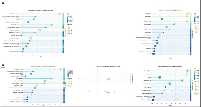
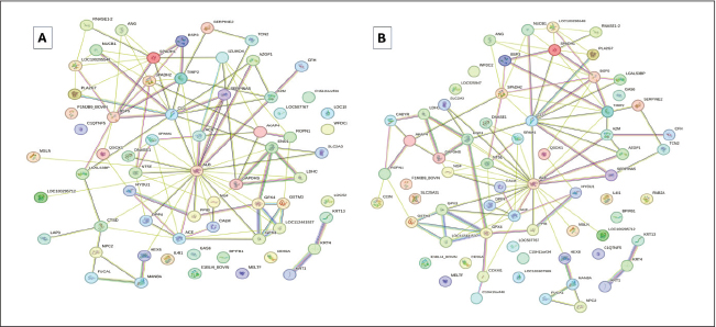
Fig. 1. Venn diagram of seminal plasma (upper) and sperm (lower) of Indonesian-bred (S-Ind) and imported (S-Imp) Simmental bulls. The GO analysis of proteins identified in the spermatozoa of both groups of bulls showed the same enrichment related biological processes, such as the ATP metabolic process (GO: 0046034), the pyruvate metabolic process (GO: 0006090), the generation of precursor metabolites and energy (GO: 0006091), and the purine-containing compound metabolic process (GO:0072521). No molecular functions were enriched in the S-Imp group, whereas oxidoreductase activity (GO:0016491) was enriched in the S-Ind group. Proteins detected in the spermatozoa of both bull groups were predominantly associated with specific cellular components, including the sperm flagellum (GO:0036126), midpiece (GO:0097225), sperm fibrous sheath (GO:0035685), mitochondrion (GO:0005739), acrosome vesicle (GO:0001669), and cytoplasm (GO:0005737) (Fig. 3). STRING analysis revealed complex protein interactions in both seminal plasma and spermatozoa. The resulting interaction network showed that ALB and CLU had the highest number of interactions in both bull groups. These two proteins directly interact with BSP3, BSP5, SPADH1, SPADH2, SPAM1, and SERPINA5, all of which are involved in reproductive processes. Proteins found exclusively in the S-Imp group, such as CCIN, COX4I1, and CABYR, were located in the peripheral regions of the network. In contrast, proteins unique to the S-Ind group, including ACR and IZUMO4, were directly connected to the network core and showed a stronger association with fertilization functions (Fig. 4). Table 1. Summary of abundant proteins in the seminal plasma and spermatozoa of Indonesian-bred (S-Ind) and imported (S-Imp) Simmental bulls (foldchange > 1.5).
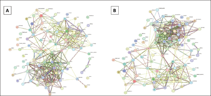
The STRING network of sperm proteins exhibits more complex and stronger interactions than those found in seminal plasma (Fig. 5). The most densely connected cluster is primarily associated with mitochondrial energy production, involving ATP5F1A, ATP5F1B, UQCRC2, VDAC2, VDAC3, COX2, and ALDOA. Additional clusters include proteins related to sperm structure and fertilization processes, such as TEKT2, TEKT3, TEKT5, AKAP3, AKAP4, SPACA1, ODF2, and CABYR. Although similar interaction clusters were observed between the bull groups, certain proteins were uniquely present in each group. Specifically, HEXB, NGF, and BPIFB were identified in the S-Imp group, whereas GPX5, WFDC2, GPX3, CES5A, TEKT4, MELTF, and PLET1 were found in the S-Ind group. DiscussionIn this study, 86.1% and 89.5% of the same proteins were identified in seminal plasma and spermatozoa, respectively, in both groups of bulls. Although certain proteins were detected exclusively in seminal plasma or spermatozoa, many of these proteins were also identified in the corresponding counterparts across the two bull groups. These findings indicate that the proteins identified in seminal plasma and spermatozoa contribute to similar biological processes, molecular functions, and cellular components across the groups. Furthermore, the protein–protein interaction networks showed a high degree of similarity between the two bull groups.
Fig. 2. Enrichment gene ontology seminal plasma of Imported (S-Imp: A) and Indonesian-bred (S-Ind: B) Simmental bulls. The classification is based on biological processes, molecular function, and cellular components. FDR=False Discovery Rate.
Fig. 3. Enrichment gene ontology of the sperm of Imported (S-Imp: A) and Indonesian-Bred (S-Ind: B) Simmental bulls. The classification is based on biological processes, molecular functions, and cellular components. FDR=False Discovery Rate.
Fig. 4. STRING protein–protein interaction network showing the interactions of seminal plasma proteins identified in Indonesian-bred (A) and imported (B) Simmental bulls. Proteins involved in spermatogenesis, motility, capacitation, acrosome reaction, and fertilizationComplex protein networks drive spermatogenesis and sperm function. Protein clusters involved in spermatogenesis were identified in this study, particularly CCIN, SPATC1, C7orf61, and the SPACA1 complex. CCIN plays a role in remodeling the sperm head structure during spermiogenesis (Fan et al., 2022), whereas SPATC1 participates in spermatogenesis (Kim et al., 2018). C7orf61 (Chromosome 7 open reading frame 61) is a testis-specific gene that plays a critical role in acrosome formation and nucleus shaping during spermatogenesis and the sperm-egg fusion process (Gómez-Torres et al., 2022). SPACA1, a membrane protein localized in the equatorial segment of spermatozoa, is essential for these processes. Disruption of Spaca1 leads to the disappearance of the nuclear plate and failure of acrosomal expansion during spermiogenesis, resulting in the degeneration and disappearance of the acrosome in mature spermatozoa (Fujihara et al., 2012). In addition, protein clusters associated with sperm motility were identified in the spermatozoa flagella. Tektins (TEKT2, TEKT3, TEKT4, and TEKT5) are microtubule inner proteins (MIPs) that localize at the inside lumen of doublet microtubules (DMTs) of cilia/flagella are critical for sperm motility (Matsuyama et al., 2005; Geng et al., 2024). ROPN1 (ropporin-1, rhophilin-associated tail protein 1) is a critical protein found in the fibrous sheath of the sperm flagellum principal piece, plays a crucial role in regulating sperm motility (Fujita et al., 2000). In addition, Pelloni et al. (2018) found a positive correlation between ROPN1/CABYR gene expression and motility of the sperm (Pelloni et al., 2018). Proteins found in seminal plasma and spermatozoa of both groups regulated sperm capacitation and acrosome reaction. BSP family (BSP3, BSP5) and spermadhesin (SPADH1, SPADH2, RNASE1-2, and F1MJB9_Bovin). BSP and SPADH proteins bind to the cell membranes of mammalian spermatozoa, influencing their functionality, and were identified in seminal plasma and sperm in this study. The BSP protein is characterized by two tandemly repeated fibronectin type 2 (Fn2) domains. These proteins are secreted to the seminal plasma by the seminal vesicle and bind to choline phospholipids in the sperm membrane during ejaculation (Ardon and Suarez, 2013). BSP proteins specifically interact with high-density lipoproteins and glycosaminoglycans to promote sperm capacitation (Manjunath and Thérien, 2002). Similarly, SPADH is a major protein in seminal plasma that is primarily associated with sperm capacitation (Ramírez-López et al., 2023) and plays several vital roles in reproductive processes (Özbek et al., 2021). Capacitation-related proteins (ALB, CLU, and GPD) also support sperm function through distinct mechanisms. ALB is closely related to the quality and function of spermatozoa and regulates intracellular redox homeostasis (Kasimanickam et al., 2019). Clusterin (CLU) is an extracellular chaperone for proteins under stress, preventing aggregation and precipitation. The chaperone role of CLU may be important for sperm maturation and capacitation (Saewu et al., 2017). Glycerol-3-phosphate dehydrogenase 2 (GPD2) enables tyrosine phosphorylation during sperm capacitation, a critical biochemical modification for sperm preparation (Kota et al., 2010). The A-kinase anchoring protein (AKAP) family, including AKAP3 and AKAP4, regulates sperm function, including motility, capacitation, and the acrosome (Vizel et al., 2015). CABYR protein is a fibrous sheath calcium-binding tyrosine phosphorylation-regulated protein (Ficarro et al., 2003), and it is involved in calcium binding when phosphorylated during capacitation (Young et al., 2016). IZUMO4, a member of the immunoglobulin group and Izumo protein family, plays a role in the pre-fertilization stages, such as capacitation and acrosome reaction (Guasti et al., 2020).
Fig. 5. STRING protein–protein interaction network showing the interactions of the spermatozoa proteins identified in Indonesian-bred (A) and imported (B) Simmental bulls. Sperm binding to the zona pellucida (ZP) and subsequent fertilization were supported by some proteins. One of them is SPACA1, a membrane protein that localizes to the equatorial segment of spermatozoa in mammals and functions in sperm-egg fusion (Jones et al., 2008). Sperm adhesion molecule 1 (SPAM1) is a hyaluronidase that facilitates sperm-zona pellucida binding by dispersing the cumulus-oocyte matrix (Gómez-Torres et al., 2022). In addition, proteins involved in sperm ZP binding, including GARIN3, ZAN, and ACE, are also found in seminal plasma. All GARINs, including GARIN 3, are indispensable for sperm head morphogenesis; GARIN loss impairs sperm head morphology and ZP binding, resulting in ZP penetration failure (Wang et al., 2024). ZAN confers species specificity to sperm-ZP adhesion (Tardif et al., 2010). Angiotensin-converting enzyme (ACE), a zinc-dependent dipeptidyl carboxypeptidase, plays an important role in binding to the zonae pellucidae (Shibata et al., 2024). Proteins involved in oxidative stress balanceThe protein cluster of enzymes identified in the seminal plasma and spermatozoa of both groups of bulls was related to the oxidative balance. Low levels of reactive oxygen species (ROS) are essential for normal physiological processes, such as sperm capacitation, hyperactivation, and the acrosome reaction (Lee et al., 2017). Excessive ROS production can be harmful to sperm cells, causing oxidative stress. The identified proteins, including GPX3, GPX4, GPX5, GSTM3, GSTO2, and LAP3, play critical roles in glutathione metabolism and reactive oxygen species detoxification. The presence of these proteins in both seminal plasma and spermatozoa neutralizes highly reactive oxygen molecules such as superoxide, hydrogen peroxide, and hydroxyl radicals, converting them into harmless compounds such as water and oxygen (Wang et al., 2025). GPX enzymes are some of the most important antioxidant proteins identified in this study. GPX (GPX5, GPX3) is one of the most important antioxidant enzymes, which converts hydrogen peroxide (H₂O₂) into H₂O and O₂ (Pournasir et al., 2021). Selenoprotein glutathione peroxidase 4 (GPX4) is an essential antioxidant enzyme that plays a critical role in oxidative stress protection by eliminating ROS, such as lipid peroxides, and converting them to their benign alcohol analogs using glutathione as a cofactor (Margis et al., 2008). LAP3 is involved in glutathione metabolism and thus plays a role in strengthening the antioxidant defenses of spermatozoa (Guo et al., 2025). Glutathione S-transferase Mu 3 (GSTM3), which is detected in both seminal plasma and spermatozoa, plays a vital role in cellular protection against oxidative stress, thereby preserving sperm function and fertilizing ability (Llavanera et al., 2019). Similarly, GSTO2's primary function in the seminal plasma and on the spermatozoa surface is to protect against oxidative stress, thereby facilitating sperm capacitation (Hamilton et al., 2019). Proteins involved in glycolysis and transketolase or transaldolase activityA group of proteins (ENO1, PKM, TPI1, ALDOA, and GAPDHS) has been detected to play a role in glycolysis and transketolase or transaldolase activity. ENO1 is one of three enolase isoenzymes found in mammals that are most abundant in the tail of mature spermatozoa (Gitlits et al., 2000). ENO1 acts as a glycolytic enzyme that generates energy in microtubules (Harrison and White, 1972). PKM (pyruvate kinase M), particularly the PKM2 isoform, is a pivotal glycolytic enzyme found in seminal plasma and spermatozoa. PKM catalyzes the final step of glycolysis, converting phosphoenolpyruvate to pyruvate. This is essential for ATP production, which drives sperm motility (Qian et al., 2024). This terminal enzymatic reaction represents a critical control point in the glycolytic pathway, directly linking metabolic flux to the energy demands of sperm flagellar movement and reproductive function. TPI1 is essential for providing energy (ATP) to maintain the motility of spermatozoa. Triosephosphate isomerase 1 (TPI1) is an enzyme involved in glycolysis and gluconeogenesis, primarily responsible for the reversible conversion of dihydroxyacetone phosphate (DHAP) to glyceraldehyde 3-phosphate (GAP) (Vilagran et al., 2016). Aldolase (ALDOA) is a glycolytic enzyme that catalyzes the conversion of fructose 1-6-diphosphate to glyceraldehyde 3-phosphate and dihydroxyacetone phosphate via the glycolysis metabolic pathway, directly supporting sperm energy metabolism and regulating the motility required for fertilization (Gaitskell-Phillips et al., 2022). GAPDHS (glyceraldehyde-3-phosphate dehydrogenase) is a glycolytic enzyme essential for ATP production and plays critical roles in sperm motility and male fertility. GAPDHS binds to the sperm fibrous sheath, strategically positioning it within the flagellar apparatus where ATP demand is highest. This localization ensures that the enzyme-catalyzed oxidation of glyceraldehyde 3-phosphate directly supports the energetic requirements of the contractile machinery that drives sperm movement and enables successful fertilization (Gomes et al., 2020). Mitochondrial proteinsNADH-ubiquinone oxidoreductase 75 kDa subunit (NDUFS1) and cytochrome b-c1 complex subunit 2 (UQCRC2) are mitochondrial complex subunits I and III, respectively. NDUFS1 and UQCRC2 expression is associated with ATP production (Dias et al., 2020). In spermatozoa, mitochondria are crucial for cellular energy production, survival, and function (Ferramosca and Zara, 2017). Reduced NDUFS1 and UQCRC2 expression indicates mitochondrial dysfunction and abnormal ROS production, which can lead to oxidative stress and consequently affect sperm function, particularly the ability of spermatozoa to undergo capacitation and bind to the egg (de Lamirande and O’Flaherty, 2008). SLC25A31 is a mitochondrial carrier protein (SLC25) involved in the exchange of cytoplasmic ADP with mitochondrial ATP, which supports high-energy requirements, particularly during spermatogenesis and many cellular processes (Ruprecht and Kunji, 2020). The exclusive expression of mitochondrial energy proteins, specifically COX4I1 and SLC25A31 in the S-Imp group, represents a compensatory metabolic response to thermal acclimatization demands. When introduced to tropical environments, cattle must allocate additional metabolic resources to maintain thermoregulation, creating a metabolic trade-off that diverts energy from reproductive functions (Brito et al., 2004). Heat stress significantly impacts mitochondrial function in spermatozoa, leading to reduced mitochondrial membrane potential, decreased ATP content, and impaired mitochondrial respiratory complex activities (Abdelnour et al., 2022). Studies on bovine sperm have demonstrated that elevated temperatures disrupt the electron transport chain, resulting in excessive ROS production and oxidative damage to sperm cells (Boe-Hansen et al., 2018). The presence of COX4I1 exclusively in S-Imp seminal plasma may represent a compensatory upregulation to counteract heat-induced mitochondrial dysfunction. Previous research has shown that heat stress decreases mitochondrial protein import and activates glycogen synthase kinase-3α, further compromising mitochondrial integrity (Gong et al., 2017). COX4I1, also known as the critical subunit of the mitochondrial respiratory chain complex, plays a pivotal role in oxidative phosphorylation and ATP production. The upregulation of this protein in the S-Imp group suggests an adaptive response to meet the elevated energy demands required for maintaining homeostasis under heat stress conditions (Abdelnour et al., 2022). Similarly, SLC25A31 functions as an adenine nucleotide translocator that facilitates the exchange of cytoplasmic ADP with mitochondrial ATP, supporting high-energy requirements during spermatogenesis and cellular processes (Ruprecht and Kunji, 2020). The exclusive detection of SLC25A31 in S-Imp seminal plasma indicates a compensatory mechanism to ensure adequate ATP supply to spermatozoa despite the metabolic burden imposed by acclimatization to tropical climates. The unique expression of structural proteins CCIN and CABYR in the S-Imp group further supports the hypothesis of adaptive compensation to environmental stress. CCIN, a perinuclear theca protein essential for sperm head morphogenesis, has been reported to increase in abundance as a protective mechanism against thermal stress-induced chromatin instability (Fan et al., 2022). CABYR plays a critical role during capacitation and is essential for sperm-oocyte interaction (Ficarro et al., 2003). The elevated expression of these structural proteins in imported bulls may serve to maintain sperm head integrity and functional competence when exposed to tropical heat conditions that differ markedly from their temperate origin habitats. This finding aligns with previous observations that heat stress causes chromatin remodeling and structural instability, necessitating compensatory increases in stabilizing proteins (Boe-Hansen et al., 2018). In contrast, the S-Ind group demonstrated a more efficient adaptive strategy rooted in evolutionary adaptation to tropical conditions. In Indonesian tropical environments, local Simmental bulls have undergone generations of natural selection, resulting in optimized metabolic pathways that do not require the same degree of compensatory protein upregulation. The absence of COX4I1 and SLC25A31 in the S-Ind suggests that these animals maintain adequate mitochondrial function through baseline metabolic efficiency rather than compensatory overexpression. These proteomic differences have significant implications for AI programs using imported Simmental bulls in Indonesia. The compensatory protein expression patterns observed in the S-Imp bulls suggest that they may benefit from targeted nutritional supplementation with mitochondrial support compounds, antioxidants, and energy substrates. ConclusionThe proteomic landscape and key biological functions of proteins in seminal plasma and spermatozoa from bulls bred in Indonesia were comparable to those observed in imported bulls. These proteins are involved in spermatogenesis, sperm capacitation, the acrosome reaction, fertilization, energy metabolism, and sperm protection against oxidative stress. Differentially expressed proteins with critical roles in seminal plasma and spermatozoa were identified and characterized. Despite this overall similarity, distinct differences in protein composition and abundance were observed. S-Ind exhibited a higher abundance of proteins involved in metabolism and cellular protection, whereas S-Imp exhibited a greater abundance of structural proteins associated with sperm function. AcknowledgmentsThe authors would like to thank the Central Java Artificial Insemination Station for providing semen samples and the necessary facilities. FundingThis research was funded by the Directorate General of Research and Development of the Ministry of Higher Education, Science, and Technology in accordance with the contract for the implementation of the 2025 fiscal year Research Program Number 59547/lT3.D10/PT.01.03/P/B/2025. Authors' contributionsNWKK: conception, study design, sample collection, data analysis, and manuscript drafting. BWW: collected the sample and performed data analysis, improving the manuscript. IA: conception, design of the study, sample collection, and improvement of the manuscript. WEP: design of the study, data analysis, and manuscript improvement. SS conception and manuscript improvement. MT: improvement of the manuscript. MF: conception, study design, data analysis, and manuscript supervision. Conflict of interestThe authors declare no conflicts of interest. Data availabilityAll data supporting this study’s findings are available within the manuscript. ReferencesAbdelnour, S.A., Swelum, A.A., Sindi, R.A., Barkat, R.A., Khalifa, N.E., Amin, A.A., El‐Raghi, A.A., Tufarelli, V., Losacco, C. and Abd El‐hack, M.E. 2022. Responses of sperm mitochondria functionality in animals to thermal stress: the mitigating effects of dietary natural antioxidants. Reprod. Domest. Anim. 57(10), 1101–1112; doi:10.1111/rda.14193 Ardon, F. and Suarez, S.S. 2013. Cryopreservation increases coating of bull sperm by seminal plasma binder of sperm proteins BSP1, BSP3, and BSP5. Reproduction 146(2), 111–117; doi:10.1530/REP-12-0468 Boe‐Hansen, G.B., Rêgo, J.P.A., Satake, N., Venus, B., Sadowski, P., Nouwens, A., Li, Y. and Mcgowan, M. 2018. Effects of increased scrotal temperature on semen quality and seminal plasma proteins in Brahman bulls. Mol. Reprod. Dev. 87(5), 574–597; doi:10.1002/mrd.23328 Brito, L.F.C., Silva, A.E.D.F., Barbosa, R.T. and Kastelic, J.P. 2004. Testicular thermoregulation in Bos indicus, crossbred and Bos taurus bulls: relationship with scrotal, testicular vascular cone and testicular morphology, and effects on semen quality and sperm production. Theriogenology 61(2- 3), 511–528; doi:10.1016/s0093-691x(03)00231-0 Bustamante-Filho, I.C., Pasini, M. and Moura, A.A. 2022. Animal reproduction spermatozoa and seminal plasma proteomics: too many molecules, too few markers. The case of bovine and porcine semen. Anim. Reprod. Sci. 247, 107075; doi:10.1016/j.anireprosci.2022.107075 Dejarnette, J.M., Marshall, C.E., Lenz, R.W., Monke, D.R., Ayars, W.H. and Sattler, C.G. 2004. Sustaining the fertility of artificially inseminated dairy cattle: the role of the artificial insemination industry. J. Dairy. Sci. 87(E. Suppl.), E93–E104. Dias, T.R., Agarwal, A., Pushparaj, P.N., Ahmad, G. and Sharma, R. 2020. New insights on the mechanisms affecting fertility in men with non-seminoma testicular cancer before cancer therapy. World J. Mens. Health 38(2), 198–207; doi:10.5534/wjmh.180099 Druart, X. and De Graaf, S. 2018. Seminal plasma proteomes and sperm fertility. Anim. Reprod. Sci. 194, 33–40; doi:10.1016/j.anireprosci.2018.04.061 Fan, Y., Huang, C., Chen, J., Chen, Y., Wang, Y., Yan, Z., Yu, W., Wu, H., Yang, Y., Nie, L., Huang, S., Wang, F., Wang, H., Hua, Y., Lyu, Q., Kuang, Y. and Lei, M. 2022. Mutations in CCIN cause teratozoospermia and male infertility. Sci. Bull. 67(20), 2112–2123; doi:10.1016/j.scib.2022.09.026 Ferramosca, A. and Zara, V. 2017. Mitochondria and fertility: the mitochondria critical role on spermatozoa function. JDReAM 1(1), 21–26; doi:10.1285/i25327518v1i1p21 Ficarro, S., Chertihin, O., Westbrook, V.A., White, F., Jayes, F., Kalab, P., Marto, J.A., Shabanowitz, J., Herr, J.C., Hunt, D.F. and Visconti, P.E. 2003. Phosphoproteome analysis of capacitated human sperm: evidence of tyrosine phosphorylation of a kinase-anchoring protein 3 and valosin-containing protein/p97 during capacitation. J. Biol. Chem. 278(13), 11579–11589; doi:10.1074/jbc. M202325200 Fujihara, Y., Satouh, Y., Inoue, N., Isotani, A., Ikawa, M. and Okabe, M. 2012. SPACA1-deficient male mice are infertile with abnormally shaped sperm heads reminiscent of globozoospermia. Development 139, 3583–3589; doi:10.1242/dev.081778 Fujita, A., Nakamura, K.I., Kato, T., Watanabe, N., Ishizaki, T., Kimura, K., Mizoguchi, A. and Narumiya, S. 2000. Ropporin, a sperm-specific binding protein of rhophilin, that is localized in the fibrous sheath of sperm flagella. J. Cell Sci. 113(1), 103–112; doi:10.1242/jcs.113.1.103 Gaitskell-Phillips, G., Martín-Cano, F.E., Ortiz-Rodríguez, J.M., Da Silva-álvarez, E., Masot, J., Redondo, E., Gil, M.C., Ortega-Ferrusola, C. and Peña, F.J. 2022. Seminal plasma proteins as potential biomarkers for sperm motility and velocities. Theriogenology 177, 34–41; doi:10.1016/j.theriogenology.2021.10.007 Geng, X.Y., Jin, H.J., Xia, L., Wang, B.B. and Chen, S.R. 2024. Tektin bundle interacting protein, TEKTIP1, functions to stabilize the tektin bundle and axoneme in mouse sperm flagella. Cell. Mol. Life Sci. 81(1), 118; doi:10.1007/s00018-023-05081-3 Gervasi, M.G. and Visconti, P.E. 2017. Molecular changes and signaling events occurring in sperm during epididymal maturation. Andrology 5(2), 204–218; doi:10.1111/andr.12320 Gitlits, V.M., Toh, B.H., Loveland, K.L. and Sentry, J.W. 2000. The glycolytic enzyme enolase is present in sperm tail and displays nucleotide-dependent association with microtubules. Eur. J. Cell Biol. 79(2), 104–111; doi:10.1078/S0171-9335(04)70012-6 Gomes, F.P., Park, R., Viana, A.G., Fernandez-Costa, C., Topper, E., Kaya, A., Memili, E., Yates, J.R. and Moura, A.A. 2020. Protein signatures of seminal plasma from bulls with contrasting frozen-thawed sperm viability. Sci. Rep. 10, 14661; doi:10.1038/s41598-020-71015-9 Gómez-Torres, M.J., Sáez-Espinosa, P., Manzano-Santiago, P., Robles-Gómez, L., Huerta-Retamal, N. and Aizpurua, J. 2022. Sperm adhesion molecule 1 (SPAM1) distribution in selected human sperm by hyaluronic acid test. Biomedicines 10, 2553. Gong, Y., Guo, H., Zhang, Z., Zhou, H., Zhao, R. and He, B. 2017. Heat stress reduces sperm motility via activation of glycogen synthase kinase-3α and inhibition of Mitochondrial Protein Import. Front. Physiol. 8, 718; doi:10.3389/fphys.2017.00718 Guasti, P.N., Souza, F.F., Scott, C., Papa, P.M., Camargo, L.S., Schmith, R.A., Monteiro, G.A., Hartwig, F.P. and Papa, F.O. 2020. Equine seminal plasma and sperm membrane: functional proteomic assessment. Theriogenology 156, 70–81; doi:10.1016/j.theriogenology.2020.06.014 Guo, L., Gu, Z., Wang, B., Wang, Y., Chen, J., Li, Y., Zheng, Q., Zhao, J., Ding, H., Liu, H., Fang, Y., Wang, J. and Lyu, W. 2025. Integrated proteomics and metabolomics reveal spermine enhances sperm freezability via antioxidant pathways. Antioxidants 14(7), 861; doi:10.3390/antiox14070861 Hamilton, L.E., Zigo, M., Mao, J., Xu, W., Sutovsky, P., O’Flaherty, C. and Oko, R. 2019. GSTO2 isoforms participate in the oxidative regulation of the plasmalemma in eutherian spermatozoa during capacitation. Antioxidants 8(12), 601; doi:10.3390/antiox8120601 Harrison, R.A. and White, I.G. 1972. Glycolytic enzymes in the spermatozoa and cytoplasmic droplets of bull, boar and ram, and their leakage after shock. J. Reprod. Fertil. 30(1), 105–115. Jones, R., James, P.S., Oxley, D., Coadwell, J., Suzuki-Toyota, F. and Howes, E.A. 2008. The equatorial subsegment in mammalian spermatozoa is enriched in tyrosine phosphorylated proteins. Biol. Reprod. 79, 421–431. Kasimanickam, R.K., Kasimanickam, V.R., Arangasamy, A. and Kastelic, J.P. 2019. Sperm and seminal plasma proteomics of high-versus low-fertility Holstein bulls. Theriogenology 126, 41–48; doi:10.1016/j.theriogenology.2018.11.032 Kim, J., Kwon, J.T., Jeong, J., Kim, J., Hong, S.H., Kim, J., Park, Z.Y., Chung, K.H., Eddy, E.M. and Cho, C. 2018. SPATC1L maintains the integrity of the sperm head-tail junction. EMBO. Rep. 19(9), e45991; doi:10.15252/embr.201845991 Kota, V., Rai, P., Weitzel, J., Middendorff, R., Bhande, S.S. and Shivaji, S. 2010. Role of glycerol-3-phosphate dehydrogenase 2 in mouse sperm capacitation. Mol. Reprod. &. Develop. 77(9), 773–783; doi:10.1002/mrd.21218 Kraus, M., Tichá, M., Železná, B., Pěknicová, J. and Jonáková, V. 2005. Characterization of human seminal plasma proteins homologous to boar AQN spermadhesins. J. Reprod. Immunol. 65(1), 33–46; doi:10.1016/j.jri.2004.10.001 Lamirande, E. and O’Flaherty, C. 2008. Sperm activation: role of reactive oxygen species and kinases. Biochim. Biophys. Acta. 1784(1), 106–115; doi:10.1016/j.bbapap.2007.08.024 Lee, D., Moawad, A.R., Morielli, T., Fernandez, M.C. and O'Flaherty, C. 2017. Peroxiredoxins prevent oxidative stress during human sperm capacitation. Mol. Hum. Reprod. 23, 106–115; doi:10.1093/molehr/gaw081 Llavanera, M., Mateo-Otero, Y., Bonet, S., Barranco, I., Fernández-Fuertes, B. and Yeste, M. 2019. The triple role of glutathione S-transferases in mammalian male fertility. Cell. Mol. Life. Sci. 77(12), 1–12; doi:10.1007/s00018-019-03405-w Manjunath, P. and Thérien, I. 2002. Role of seminal plasma phospholipid-binding proteins in sperm membrane lipid modification that occurs during capacitation. J. Reprod. Immunol. 53(1-2), 109–119; doi:10.1016/S0165-0378(01)00098-5 Margis, R., Dunand, C., Teixeira, F.K. and Margis-Pinheiro, M. 2008. Glutathione peroxidase family—An evolutionary overview. FEBS J. 275(15), 3959–3970; doi:10.1111/j.1742-4658.2008.06542.x Matsuyama, T., Honda, Y., Doiguchi, M. and Iida, H. 2005. Molecular cloning of a new member of TEKTIN Family, Tektin4, located to the flagella of rat spermatozoa. Mol. Reprod. Develop. 72, 120–128; doi:10.1002/mrd.20331 Maxwell, W.M.C., De Graaf, S.P., Ghaoui, R.E. and Evans, G. 2007. Seminal plasma effects on sperm handling and female fertility. Soc. Reprod. Fertil. 64, 13–38; doi:10.5661/rdr-vi-13 Moura, A.A., Memili, E., Portela, A.M.R., Viana, A.G., Velho, A.L.C., Bezerra, M.J.B. and Vasconselos, F.R. 2018. Seminal plasma proteins and metabolites: effects on sperm function and potential as fertility markers. Anim. Reprod. 15(1), 691–702; doi:10.21451/1984-3143-AR2018-0029 Özbek, M., Hitit, M., Kaya, A., Jousan, F.D. and Memili, E. 2021. Sperm functional genome associated with bull fertility. Front. Vet. Sci. 8, 610888; doi:10.3389/fvets.2021.610888 Panner Selvam, M.K., Agarwal, A., Pushparaj, P.N., Baskaran, S. and Bendou, H. 2019. Sperm proteome analysis and identification of fertility-associated biomarkers in unexplained male infertility. Genes 10(7), 522; doi:10.3390/genes10070522 Pelloni, M., Paoli, D., Majoli, M., Pallotti, F., Carlini, T., Lenzi, A. and Lombardo, F. 2018. Molecular study of human sperm RNA: ropporin and CABYR in asthenozoospermia. J. Endocrinol. Invest. 41(7), 781–787; doi:10.1007/s40618-017-0804-x Pournasir, M., Ghorbian, S., Ghasemnejad, T., Fattahi, A. and Nouri, M. 2021. Glutathione peroxidase 3 (extracellular isoform) levels and functional polymorphisms in fertile and infertile men. Middle East Fertil. Soc. J. 26(1), 13; doi:10.1186/s43043-021-00057-4 Qian, G., Wang, X., Zhang, X. and Shen, B. 2024. Pyruvate kinase M in germ cells is essential for sperm motility and male fertility but not spermatogenesis. Asian J. Androl. 26(2), 212–219; doi:10.4103/aja202350 Ramírez-López, C.J., Barros, E., Vidigal, P.M.P., Silva Okano, D., Duarte Rodrigues, J.N., Lopes Gomes, L., Montes-Vergara, J.C., Petro Hernandez, V.G., Baracat-Pereira, M.C., Guimarães, S.E.F. and Guimarães, J.D. 2023. Relative abundance of spermadhesin-1 in the seminal plasma of young Nellore bulls is in agreement with reproductive parameters. Vet. Sci. 10(10), 610; doi:10.3390/vetsci10100610 Rodríguez‐Martínez, H., Kvist, U., Ernerudh, J., Sanz, L. and Calvete, J.J. 2011. Seminal plasma proteins: what role do they play?. Am. J. Reprod. Immunol. 66(1), 11–22; doi:10.1111/j.1600-0897.2011.01033.x Ruprecht, J.J. and Kunji, E.R.S. 2020. The SLC25 mitochondrial carrier family: structure and mechanism. Trends. Biochem. Sci. 45(3), 244–258; doi:10.1016/j.tibs.2019.11.001 Saewu, A., Kadunganattil, S., Raghupathy, R., Kongmanas, K., Diaz-Astudillo, P., Hermo, L. and Tanphaichitr, N. 2017. Clusterin in the mouse epididymis: possible roles in sperm maturation and capacitation. Reproduction 154(6), 867–880; doi:10.1530/REP-17-0518 Shibata, T., Bhat, S.A., Cao, D., Saito, S., Bernstein, E.A., Nishi, E., Medenilla, J.D., Wang, E.T., Chan, J.L., Pisarska, M.D., Tourtellotte, W.G., Giani, J.F., Bernstein, K.E. and Khan, Z. 2024. Testicular ACE regulates sperm metabolism and fertilization through the transcription factor PPARγ. J. Biol. Chem. 300(1), 105486; doi:10.1016/j.jbc.2023.105486 Tardif, S., Wilson, M.D., Wagner, R., Hunt, P., Gertsenstein, M., Nagy, A., Lobe, C., Koop, B.F. and Hardy, D.M. 2010. Zonadhesin is essential for species specificity of sperm adhesion to the egg zona pellucida. J. Biol. Chem. 285(32), 24863–24870; doi:10.1074/jbc.M110.123125 Vilagran, I., Castillo-Martín, M., Prieto-Martínez, N., Bonet, S. and Yeste, M. 2016. Triosephosphate isomerase (TPI) and epididymal secretory glutathione peroxidase (GPX5) are markers for boar sperm quality. Anim. Reprod. Sci. 165, 22–30; doi:10.1016/j.anireprosci.2015.12.001 Vizel, R., Hillman, P., Ickowicz, D. and Breitbart, H. 2015. AKAP3 degradation in sperm capacitation is regulated by its tyrosine phosphorylation. Biochimica. Et. Biophysica. Acta. 1850(9), 1912–1920; doi:10.1016/j.bbagen.2015.06.005 Wang, H., Iida-Norita, R., Mashiko, D., Pham, A.H., Miyata, H. and Ikawa, M. 2024. Golgi associated RAB2 interactor protein family contributes to murine male fertility to various extents by assuring correct morphogenesis of sperm heads. PLos Genet. 20(6), e1011337; doi:10.1371/journal.pgen.1011337 Wang, Y., Fu, X. and Li, H. 2025. Mechanisms of oxidative stress-induced sperm dysfunction. Front. Endocrinol. 16, 1520835; doi:10.3389/fendo.2025.1520835 Young, S.A.M., Miyata, H., Satouh, Y., Aitken, R.J., Baker, M.A. and Ikawa, M. 2016. CABYR is essential for fibrous sheath integrity and progressive motility in mouse spermatozoa. J. Cell Sci. 129(23), 4379–4387; doi:10.1242/jcs.193151 | ||
| How to Cite this Article |
| Pubmed Style Karja NWK, Witri BW, Arifiantini I, Prasetyaningtyas WE, Suharti S, Taniguchi M, Fahrudin M. Proteomic mapping of seminal plasma and spermatozoa in Indonesia-bred Simmental bulls housed at an artificial insemination facility. Open Vet. J.. 2026; 16(8): 5202-5214. doi:10.5455/OVJ.2026.v16.i8.16 Web Style Karja NWK, Witri BW, Arifiantini I, Prasetyaningtyas WE, Suharti S, Taniguchi M, Fahrudin M. Proteomic mapping of seminal plasma and spermatozoa in Indonesia-bred Simmental bulls housed at an artificial insemination facility. https://www.openveterinaryjournal.com/?mno=315542 [Access: August 08, 2026]. doi:10.5455/OVJ.2026.v16.i8.16 AMA (American Medical Association) Style Karja NWK, Witri BW, Arifiantini I, Prasetyaningtyas WE, Suharti S, Taniguchi M, Fahrudin M. Proteomic mapping of seminal plasma and spermatozoa in Indonesia-bred Simmental bulls housed at an artificial insemination facility. Open Vet. J.. 2026; 16(8): 5202-5214. doi:10.5455/OVJ.2026.v16.i8.16 Vancouver/ICMJE Style Karja NWK, Witri BW, Arifiantini I, Prasetyaningtyas WE, Suharti S, Taniguchi M, Fahrudin M. Proteomic mapping of seminal plasma and spermatozoa in Indonesia-bred Simmental bulls housed at an artificial insemination facility. Open Vet. J.. (2026), [cited August 08, 2026]; 16(8): 5202-5214. doi:10.5455/OVJ.2026.v16.i8.16 Harvard Style Karja, N. W. K., Witri, . B. W., Arifiantini, . I., Prasetyaningtyas, . W. E., Suharti, . S., Taniguchi, . M. & Fahrudin, . M. (2026) Proteomic mapping of seminal plasma and spermatozoa in Indonesia-bred Simmental bulls housed at an artificial insemination facility. Open Vet. J., 16 (8), 5202-5214. doi:10.5455/OVJ.2026.v16.i8.16 Turabian Style Karja, Ni Wayan Kurniani, Brilla Widya Witri, Iis Arifiantini, Wahono Esthi Prasetyaningtyas, Sri Suharti, Masayasu Taniguchi, and Mokhamad Fahrudin. 2026. Proteomic mapping of seminal plasma and spermatozoa in Indonesia-bred Simmental bulls housed at an artificial insemination facility. Open Veterinary Journal, 16 (8), 5202-5214. doi:10.5455/OVJ.2026.v16.i8.16 Chicago Style Karja, Ni Wayan Kurniani, Brilla Widya Witri, Iis Arifiantini, Wahono Esthi Prasetyaningtyas, Sri Suharti, Masayasu Taniguchi, and Mokhamad Fahrudin. "Proteomic mapping of seminal plasma and spermatozoa in Indonesia-bred Simmental bulls housed at an artificial insemination facility." Open Veterinary Journal 16 (2026), 5202-5214. doi:10.5455/OVJ.2026.v16.i8.16 MLA (The Modern Language Association) Style Karja, Ni Wayan Kurniani, Brilla Widya Witri, Iis Arifiantini, Wahono Esthi Prasetyaningtyas, Sri Suharti, Masayasu Taniguchi, and Mokhamad Fahrudin. "Proteomic mapping of seminal plasma and spermatozoa in Indonesia-bred Simmental bulls housed at an artificial insemination facility." Open Veterinary Journal 16.8 (2026), 5202-5214. Print. doi:10.5455/OVJ.2026.v16.i8.16 APA (American Psychological Association) Style Karja, N. W. K., Witri, . B. W., Arifiantini, . I., Prasetyaningtyas, . W. E., Suharti, . S., Taniguchi, . M. & Fahrudin, . M. (2026) Proteomic mapping of seminal plasma and spermatozoa in Indonesia-bred Simmental bulls housed at an artificial insemination facility. Open Veterinary Journal, 16 (8), 5202-5214. doi:10.5455/OVJ.2026.v16.i8.16 |


