





| Research Article | ||
Open Vet. J.. 2026; 16(8): 5193-5201 !
Open Veterinary Journal, (2026), Vol. 16(8): 5193–5201 Research Article Zoonotic transmission of Fasciola hepatica in Northern Iraq: Combined coprological and molecular evidence from livestock and humansOthman Jamal Nassrullah1, Brwa M. Ali Essamaddin2, Khalid Jabar Aziz3*, Basim Abdulwahid Ali1, Hardi F. Marif1, Nivar Othamn Jamal1, Kazhin Azad Muhammad1, Sima Sdiq Hassan1 and Yadgar Salah Rasul11Department of Clinic and Internal Medicine, College of Veterinary Medicine, University of Sulaimani, Sulaymaniyah, Iraq 2Department of Surgery and Theriogenology, College of Veterinary Medicine, University of Sulaimani, Sulaymaniyah, Iraq 3Department of Basic Sciences, College of Veterinary Medicine, Salahaddin University-Erbil, Erbil, Kurdistan Region, Iraq *Corresponding Author: Khalid Jabar Aziz. Department of Basic Sciences, College of Veterinary Medicine, Salahaddin University-Erbil, Erbil, Kurdistan Region, Iraq. Email:khalid.aziz1 [at] su.edu.krd Submitted: 28/03/2026 Revised: 29/06/2026 Accepted: 13/07/2026 Published: 08/08/2026 © 2025 Open Veterinary Journal
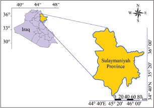
AbstractBackground: Fascioliasis remains a significant neglected zoonotic infection affecting both livestock and humans worldwide, leading to considerable economic losses and public health concerns, particularly in endemic regions. Aim: This study aimed to determine the prevalence and molecular characterization of Fasciola spp. in livestock and humans and to identify the circulating species using molecular and phylogenetic analyses. Methods: Between 2024 and 2025, 364 fecal samples were collected from sheep (n=120), goats (n=56), cattle (n=76), and humans (n=112). Samples were initially screened for Fasciola eggs using microscopic examination, followed by molecular confirmation using 28S rRNA polymerase chain reaction and species differentiation using cox1-based primers. Positive isolates were sequenced and phylogenetically analyzed. Statistical associations between infection and host factors (species, age, and sex) were evaluated using Fisher’s exact test. Results: Microscopic examination revealed prevalence rates of 9.1%, 8.9%, 11.8%, and 11.6% in sheep, goats, cattle, and humans, respectively, with no significant difference among host species (p=0.88). Infection was not significantly associated with age or sex among animals (n=252). The age-specific prevalence in animals was 9.3% in those <1 year, 10.8% in those aged 1–3 years, and 9.0% in those aged >3 years (p=0.897), whereas the sex-related prevalence was 5.7% in males and 13.1% in females (p=0.061). Molecular analysis confirmed that 38 positive samples produced the expected 618-bp amplicon, including 11 sheep, 5 goats, 9 cattle, and 13 human samples. Species identification using cox1 primers detected five Fasciola hepatica isolates, while no amplification was obtained for Fasciola gigantica. Sequence analysis (GenBank accession numbers: PX529836 and PX530408–PX530411) revealed 99%–99.5% similarity with global F. hepatica 28S rRNA sequences. Phylogenetic analysis clustered the isolates within F. hepatica lineage I and showed close relationships with isolates from Iran and Chile. Conclusion: The findings confirm that F. hepatica is the predominant species infecting both livestock and humans, with low genetic diversity suggesting common environmental transmission pathways between animals and humans. Keywords: Fasciola hepatica, Iraq, Molecular epidemiology, One Health. IntroductionFasciola hepatica, commonly referred to as the liver fluke, is a parasitic flatworm that represents a major concern for both veterinary and public health sectors worldwide (Angles et al., 2024; Ardo and Aliyara, 2014). Adult flukes inhabit the bile ducts of mammals, particularly grazing livestock such as cattle and sheep, where they cause fascioliasis, a chronic liver disease associated with reduced animal performance and notable morbidity in humans (Bargues et al., 2017). The parasite has a complex indirect life cycle that involves two obligatory hosts (Chand et al., 2009). Sexual reproduction occurs in the mammalian host, whereas asexual multiplication occurs within freshwater snails belonging to the family Lymnaeidae (Charlier et al., 2008; Dalton, 2021). Eggs expelled in the feces of infected hosts develop in aquatic environments and release miracidia, which penetrate suitable snail hosts (Delgado et al., 2023). Following several developmental stages, cercariae emerge from the snails and encyst on aquatic vegetation or in the surrounding water as metacercariae, the stage responsible for initiating infection in mammals (Garedaghi et al., 2023). In farm animals, infection is mainly acquired during grazing or drinking when metacercariae attached to pasture plants or suspended in water are ingested (Hecker et al., 2024). Immature flukes migrate through the liver tissue before settling in the bile ducts, where they mature and persist after excystation in the intestine. This migratory process and chronic biliary infection lead to liver damage, anemia, poor weight gain, decreased milk yield, and reduced reproductive performance (Howell et al., 2015). Consequently, the livestock industry suffers considerable economic losses due to lower productivity, increased control costs, and condemnation of affected livers at slaughter (Howell and Williams, 2020). Humans become infected through similar environmental routes, most commonly by consuming raw or inadequately washed aquatic plants, such as watercress, or by drinking water contaminated with metacercariae (Angles et al., 2024; Howell et al., 2019). In certain settings, transmission may also occur indirectly through the use of contaminated water during food preparation. Human fascioliasis is a neglected foodborne parasitic disease that is predominantly reported in rural and agricultural communities where close interactions between people, livestock, and freshwater ecosystems favor ongoing transmission (Knubben-Schweizer & Torgerson, 2015). The shared pathways of infection between animals and humans highlight the need for integrated control approaches that simultaneously address animal reservoirs, environmental management, and food and water safety (Lalor et al., 2021). Despite the significant veterinary and public health importance of F. hepatica, molecular data on its genetic diversity and phylogenetic relationships remain limited in many endemic regions, including Iraq. Therefore, this study aimed to molecularly characterize F. hepatica isolates using the Cox1 and 28S rRNA genes and analyze their phylogenetic relationships with global isolates to improve the understanding of fascioliasis epidemiology. Materials and MethodsStudy region and sampling methodThe research was conducted in Sulaimanyah Province, Kurdistan Region, northern Iraq. The study area included selected districts such as Sulaimanyah Center, Chamchamal, and Halabja. The geographic coordinates of the region range approximately between 35°33’00”N and 36°37’00”N latitude and 44°45’00”E and 46°33’00”E longitude (Fig. 1). Sulaimanyah Province shares borders with both Iran to the east and Kirkuk Province to the southwest (Jalal et al., 2022). Sulaimanyah is characterized by diverse topography, including mountainous and semi-mountainous terrain of the Zagros Mountain range, with elevations ranging from approximately 500 m in the lowland plains to over 3,000 m in high mountain areas. The province falls within a Mediterranean climatic zone, experiencing cold, snowy winters in the highlands and moderately hot, dry summers in the plains. The mean annual temperature varies between 8°C and 20°C depending on altitude, while the annual rainfall ranges from 400 to 1,100 mm, mostly occurring between November and April. The environmental and climatic conditions across the districts are generally similar, with minor variations influenced by elevation and proximity to mountainous areas (Kurdistan Region: Directorate of Meteorology, 2024).
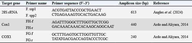
Fig. 1. Study area’s geographic location in Sulaimanyah Province, Kurdistan, Iraq (Jalal et al., 2022). Sample collection and laboratory examinationThis field-based investigation was conducted in various geographical regions of Sulaimaniyah Province, Iraq, from November 2024 to May 2025. A total of 252 fresh fecal samples were collected directly from the rectum of 76 cattle, 56 goats, and 120 sheep. Additionally, 112 fecal samples were obtained from human participants, including patients presenting with clinical signs suggestive of fascioliasis (e.g., abdominal pain, hepatomegaly, eosinophilia, or abnormal hepatobiliary ultrasonographic findings) or other liver disorders, as determined by specialist physicians. Fecal samples were transported under cooled conditions to the Parasitology Laboratory, Faculty of Veterinary Medicine, University of Sulaimani, and processed within 8 hours of collection. All samples were examined using the sedimentation technique described by Correa et al. (2016) for detecting eggs of Fasciola spp. Demographic and epidemiological data were collected from each participant using a structured questionnaire administered during sample collection. The recorded variables included age, sex, source of drinking water, consumption of raw vegetables, contact history with domestic animals, and residential environment. Direct interviews with the participants were used to obtain information regarding living conditions and personal hygiene practices. Clinical evaluations were performed, and relevant laboratory findings (microscopic detection of Fasciola spp. eggs) were documented for each participant. DNA extractionThe fecal samples positive for Fasciola eggs from both animals and humans were selected for molecular analysis. In addition, one adult Fasciola worm was recovered from the bile duct of an infected animal during postmortem examination. Eggs were concentrated from 3 g of fecal samples using a modified sedimentation technique. The recovered eggs were washed in phosphate-buffered saline (PBS) and pooled. The samples were treated with proteinase K, 1% SDS, and glass beads to disrupt the eggshells, followed by repeated freeze–thaw cycles. Genomic DNA was extracted from both fecal egg suspensions and the adult worm using the ADD-Bio Genomic DNA Extraction Kit (Add-Bio, South Korea), according to the manufacturer’s protocol. The DNA samples were stored at −20°C until polymerase chain reaction (PCR). Polymerase chain reactionPCR was performed to detect and differentiate Fasciola species using primers targeting the 28S rRNA gene and the mitochondrial cytochrome c oxidase subunit 1 (cox1) gene, as listed in Table 1. The genus-specific 28S rRNA primers amplified a fragment of approximately 613 bp for the detection of Fasciola spp. Additionally, species-specific primers targeting the cox1 gene were used to distinguish F. hepatica and Fasciola gigantica, generating 440 and 240 bp amplicons, respectively. Each PCR reaction was performed in a 20 μl reaction mixture containing 10 μl of 2× PCR Master Mix (Genet Bio, Korea), 1 μl of each primer (10 pmol/μl), 4 μl of DNA template, and 4 μl of nuclease-free water. Thermocycling conditions for amplification of the 28S rRNA gene consisted of an initial denaturation at 94°C for 5 minutes, followed by 35 cycles of denaturation at 94°C for 30 seconds, annealing at 58°C for 30 seconds, and extension at 72°C for 1 minute, with a final extension at 72°C for 5 minutes (Angles et al., 2024). For the amplification of the cox1 gene, PCR was performed under the following conditions: initial denaturation at 95°C for 5 minutes, followed by 35 cycles of denaturation at 95°C for 30 seconds, annealing at 55°C for 30 seconds, and extension at 72°C for 45–60 seconds, with a final extension at 72°C for 7 minutes, and a final hold at 4°C (Ardo and Aliyara, 2014). Agarose gel electrophoresis (AGGE)The PCR products were separated on 1% agarose gels containing SYBR Safe DNA Gel Stain (Invitrogen, USA). Electrophoresis was performed at 120 V for 40 minutes in 1× TBE buffer. DNA fragments were visualized under UV illumination and compared with a 100-bp DNA ladder (Thermo Fisher Scientific, USA) to determine product sizes. Bioinformatics and phylogenetic analysisSequence similarity analyses with previous sequences published in GenBank were performed using the BLAST program (http://www.ncbi.nlm.nih.gov/BLAST). The phylogenetic analysis included four samples that represent Sulaimanyah province. The MEGA7 version (http://www.megasoftware.net, July 2016) was employed for multiple sequence alignment. The Muscle software was used to align the sequences with the partial cox1 gene of F. hepatica sequences derived from GenBank (Edgar, 2004). Table 1. Primers used for the molecular identification of Fasciola spp.
Table 2. Prevalence of F. hepatica eggs by host species.
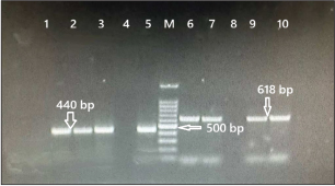
Statistical analysisThe relationship between categorical characteristics (e.g., species, sex, age group, grazing, and deworming) was assessed using the chi-square test or Fisher’s exact test, as applicable. A binomial logistic regression model was executed in GenStat (12th Edition, VSN International) to ascertain the intensity and direction of these correlations, estimating odds ratios (ORs) and their accompanying 95% confidence intervals (CIs). A two-tailed p value of 0.05 indicated statistical significance (Payne et al., 2009). Ethical approvalThis study was conducted in compliance with the ethical standards and under the supervision of the Ethics Committee of Sulaimani University/College of Veterinary Medicine (VMUS.EC.030510). ResultsOf the 364 collected samples, 38 were found to be positive for Fasciola spp. eggs by microscopic examination, including 11/121 sheep (9.1%), 5/56 goats (8.9%), 9/76 cattle (11.8%), and 13/112 human samples (11.6%) (Table 2). These microscopy-positive samples were subsequently subjected to PCR amplification using the 28S rRNA primer set, and all 38 samples yielded the expected amplicon size of 618 bp. To differentiate the Fasciola species, all 38 samples positive by 28S rRNA PCR were further analyzed using two sets of cox1 gene primers specific for F. hepatica and F. gigantica. All 38 samples showed amplification with the F. hepatica-specific primer set, producing the characteristic 440-bp amplicon, whereas no amplification was detected with the F. gigantica-specific primer set, confirming that all infections were caused by F. hepatica. Four representative PCR products were selected for sequencing analysis (Fig. 2). A total of 364 sheep (n=120), goats (n=56), cattle (n=76), and human samples (n=112) were examined. The prevalence rates were 9.1% in sheep, 8.9% in goats, 11.8% in cattle, and 11.6% in humans. The 95% CIs for these estimates overlapped, indicating similar infection levels among species. Statistical comparison using Fisher’s exact test (Monte Carlo simulation) showed no significant difference in prevalence among the four species (p=0.88). This finding was consistent with the results of the chi-square test for independence, which also indicated no association between species and infection status. Therefore, the infection appears to be evenly distributed across all examined hosts, with no species exhibiting significantly higher risk. In total, 252 animals were examined to assess the association between age and infection status. The overall prevalence was 9.9% (25/252). Animals aged <1 year showed a prevalence of 9.3% (5/54; 95% CI: 4.0%–19.9%), whereas those aged 1–3 years had a prevalence of 10.8% (13/120; 95% CI: 6.4%–17.7%). Animals older than 3 years demonstrated a prevalence of 9.0% (7/78; 95% CI: 4.4%–17.4%). Statistical analysis revealed no significant difference among the age categories (p=0.897), indicating that age did not have a measurable effect on the prevalence of infection in the study population.
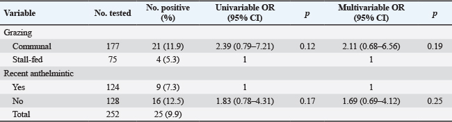
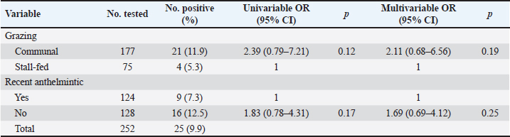
Fig. 2. AGGE of PCR products amplified from field samples of Fasciola spp. using cox1 and 28S rRNA gene primers. PCR amplification targeting the cox1 gene of F. hepatica produced the expected 440-bp band in lanes 1, 2, 3, and 5, confirming the positivity of these samples for F. hepatica. Amplification using 28S rRNA primers, designed for genus-level detection of Fasciola spp., showed the expected 618-bp band in lanes 6, 7, 9, and 10, indicating the presence of Fasciola spp. DNA. Lane M contains a standard DNA ladder, with the 500-bp marker indicated for reference. No amplification is visible in the remaining lanes, indicating negative results. A total of 252 animals were examined to evaluate the influence of sex on the prevalence of infection. The overall prevalence was 9.9% (25/252). Male animals showed a prevalence of 5.7% (8/140; 95% CI: 2.9%–10.9%), whereas females exhibited a higher prevalence of 13.1% (17/130; 95% CI: 8.3%–19.9%). Although the infection rate was greater in females than in males, the difference was not statistically significant (p=0.061). These results suggest a tendency toward increased susceptibility in females; however, sex was not confirmed as a significant risk factor in this study (Table 3). A total of 252 animals were included in the risk factor analysis, of which 25 (9.9%) were positive. Univariable logistic regression analysis showed that animals raised under a communal grazing system had higher odds of infection compared with stall-fed animals (OR=2.39; 95% CI: 0.79–7.21); however, this association was not statistically significant (p=0.12). Similarly, animals that had not received recent anthelmintic treatment exhibited increased odds of infection compared with treated animals (OR=1.83; 95% CI: 0.78–4.31), but this difference also failed to reach statistical significance (p=0.17). After adjustment for both variables in the multivariable logistic regression model, the association strength decreased slightly. Communal grazing remained associated with a higher likelihood of infection (adjusted OR=2.11; 95% CI: 0.68–6.56; p=0.19), and the absence of recent anthelmintic treatment continued to show elevated risk (adjusted OR=1.69; 95% CI: 0.69–4.12; p=0.25). Nevertheless, neither factor demonstrated a significant independent effect on infection status (Table 4). Table 3. Prevalence by sex within each animal species.
Table 4. Risk factors associated with F. hepatica infection identified by logistic regression analysis.
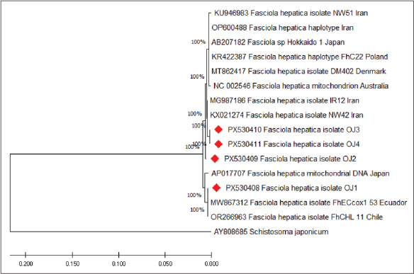
Only five positive PCR products were sequenced and submitted to GenBank for molecular confirmation. The sequence representing Fasciola spp. was assigned accession number PX529836, while the F. hepatica sequences were assigned accession numbers PX530408–PX530411. The last accession number (PX530411) corresponds to the human-derived sample. Phylogenetic analysisPhylogenetic analysis using partial 28S rRNA gene sequences demonstrated that all isolates from the current study (OJ1–OJ4) clustered within the F. hepatica clade (Fig. 3). The sequences exhibited high genetic similarity to previously documented F. hepatica isolates from various geographical regions deposited in GenBank. In the constructed phylogenetic tree, the Iraqi isolates formed a well-supported cluster with F. hepatica sequences from Iran (KU946983, OP600488, MG987186, and KX021274), Poland (KR422387), Denmark (MT862417), Japan (AB207182 and AP017707), Australia (NC002546), Ecuador (MW867312), and Chile (OR266963). The four isolates obtained in this study (OJ1–OJ4) were grouped within the same lineage, indicating a high degree of genetic similarity. The high bootstrap values (100%) strongly supported the clustering of these isolates with previously reported F. hepatica sequences, confirming their taxonomic identity as F. hepatica. The phylogenetic tree also clearly separated the F. hepatica clade from the outgroup species Schistosoma japonicum (AY808685), which formed a distinct branch. This separation confirms the reliability of the phylogenetic reconstruction and the accurate identification of the isolates. Overall, the phylogenetic analysis indicated that the F. hepatica isolates identified in this study share close genetic relationships with global isolates, particularly those reported from neighboring regions such as Iran, suggesting limited genetic divergence among these populations (Fig. 4). DiscussionThis study provides new epidemiological and molecular evidence on the zoonotic circulation of F. hepatica in Sulaymaniyah Province, Iraq, demonstrating concurrent infections in livestock and humans and confirming the exclusive presence of F. hepatica through molecular characterization. The identification of F. hepatica as the only circulating species is consistent with its ecological adaptation to temperate and semi-humid environments and its association with lymnaeid snail habitats (Mas-Coma et al., 2018). The overlapping prevalence observed among sheep, goats, cattle, and humans suggests that transmission occurs within a shared ecological system, as reported in endemic regions worldwide (Marcos et al., 2007; Mas-Coma et al., 2018). Environmental conditions, such as persistent moisture, moderate temperatures, and the presence of aquatic vegetation, promote the survival of Lymnaeidae snails, which are essential for the life cycle of F. hepatica (Mas-Coma et al., 2001). These ecological characteristics are compatible with the Sulaymaniyah geography and may explain the continuous circulation of the parasite among hosts. Although age was not significantly associated with infection in the present study, the sustained prevalence across all age groups indicates constant exposure to infective metacercariae, reflecting persistent environmental contamination. Similarly, females exhibited higher prevalence than males, which may be partially explained by physiological stress related to reproduction, lactation, and hormonal fluctuations that can suppress immune responses and increase susceptibility to parasitic infections (Moazeni and Ahmadi, 2016).
Fig. 3. Phylogenetic tree based on partial cytochrome c oxidase subunit 1 (cox1) gene sequences showing the genetic relationships between F. hepatica isolates and reference sequences retrieved from GenBank. The neighbor-joining method was used to construct the tree, and branch support values are indicated by bootstrap percentages (1,000 replicates). Isolates generated in this study are marked with red diamonds (OJ1–OJ4). Schistosoma japonicum (AY808685) was used as the outgroup to root the tree. Animals raised under communal grazing systems showed higher odds of infection, supporting previous reports that grazing practices strongly influence fasciolosis transmission (Novobilský and Höglund, 2015). Communal grazing increases contact with contaminated water sources and snail habitats, thereby facilitating the ingestion of metacercariae from pasture and aquatic plants (Pinilla et al., 2020). In addition, the lack of recent anthelmintic treatment contributes to the persistence of infection and environmental contamination, particularly in traditional livestock production systems (Ryan et al., 2020). The detection of F. hepatica in human samples confirms the presence of fasciolosis in the study area. Human infection primarily occurs through the ingestion of raw aquatic plants or contaminated water carrying metacercariae, rather than through the consumption of animal products (Marcos et al., 2007; Sabourin et al., 2018). Infection in humans and animals highlights the public health significance of the disease and the need for integrated control strategies. Comparable prevalence patterns have been reported in small ruminants in Nigeria, with infection rates of 0.32% in sheep and 0.23% in goats (Sanchez-Vazquez and Lewis, 2013). Differences in infection between animal species are often explained by grazing behavior, as sheep graze closer to the ground and in wetter areas where metacercariae accumulate, whereas goats browse higher vegetation, reducing their exposure risk (Sargison, 2008; Tanabe et al., 2024). Although no significant interspecies difference was observed in the present study, such behavioral factors likely influence local transmission dynamics. Overall, the ecological suitability for intermediate host snails, traditional husbandry practices, limited veterinary interventions, and possible emerging anthelmintic resistance create favorable conditions for sustained transmission of F. hepatica in Sulaymanyah (Tidman et al., 2023; Utrera-Quintana et al., 2022; Yilma & Malone, 1998). The sequencing analysis of the 28S rRNA gene further supported the molecular identification of F. hepatica, with the obtained isolates showing 99%–99.5% similarity to reference sequences deposited in GenBank. This high level of sequence conservation suggests low genetic divergence among F. hepatica populations and reflects this ribosomal marker’s relatively stable evolutionary nature. Phylogenetic analysis placed the Iraqi isolates within F. hepatica lineage I and demonstrated close clustering with isolates from Iran and Chile, indicating possible shared ancestral origins or conserved transmission patterns across geographically distant endemic regions. These findings emphasize the need for a coordinated health approach involving livestock management, snail control, water sanitation, and community education to effectively reduce the burden of fasciolosis.
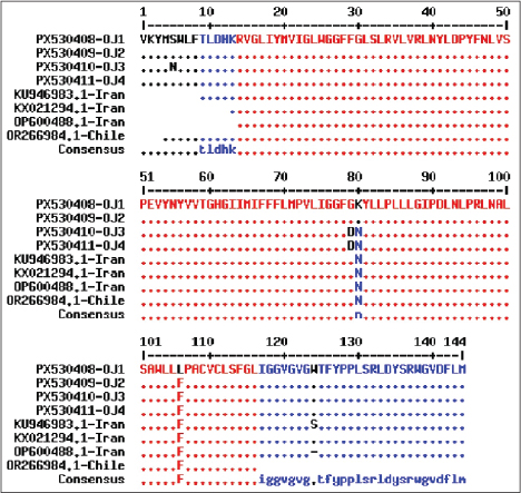
Fig. 4. Amino acid alignment of partial cytochrome oxidase subunit I (cox1) gene sequences from F. hepatica field isolates compared with reference sequences from Iran and Chile. Amino acid sequences of the four field isolates (PX530408–OJ1, PX530409–OJ2, PX530410–OJ3, and PX530411–OJ4 human) translated from the partial cox1 gene (approximately 144 amino acid region) were aligned with representative F. hepatica sequences from GenBank (KU946983, KX021294, OP600488, and OR266984). Alignment was performed in MEGA X using ClustalW. Residues identical to the consensus are shown as dots; variable sites are displayed as their amino acids. ConclusionThis study provides the first comprehensive molecular and epidemiological evidence of the zoonotic circulation of F. hepatica in Sulaimanyah Province, Iraq, demonstrating persistent transmission across different host species and age groups, which reflects continuous environmental contamination and ongoing exposure within the region. These findings emphasize the need to develop coordinated and sustainable control strategies based on a single health approach, integrating improved livestock management, control of snail habitats, enhanced water and food hygiene, routine veterinary interventions, and strengthened public health education. Implementing such measures is essential to reduce the economic burden of fasciolosis on livestock production, safeguard human health, and prevent further expansion of this neglected zoonotic disease. AcknowledgmentThe authors gratefully acknowledge the support and approval of the College of Veterinary Medicine, Salahaddin University-Erbil, and the College of Veterinary Medicine, University of Sulaimaniyah, for facilitating this research. Conflict of interestThe authors have no conflicts of interest to declare. FundingThis study received no external funding. Authors’ contributionsOthman Jamal Nassrullah was responsible for the following tasks: conceptualization, methodology, formal analysis, investigation, data curation, writing the original draft, and supervision. Nivar Othamn Jamal and Kazhin Azad Muhammad: Acquisition, resources, validation, and investigation of funding. Sima Sdiq Hassan and Yadgar Salah Rasul: Investigation, data curation, Writing—review & editing. Brwa M. Ali Essamaddin: Methodology, formal analysis, resources, and writing—review and editing. Hardi F. Marif: Supervision, project administration, data curation, investigation, and writing—review and editing. Khalid Jabar Aziz: Formal analysis and writing—review and editing. Data availabilityAll data are provided in the manuscript. Accession numbers: Px529836, Px530408, Px530409, Px530410, and Px530411. ReferencesAngles, R., Buchon, P., Valero, M.A., Bargues, M.D. and Mas-Coma, S. 2024. One Health action against human fascioliasis in the Bolivian Altiplano: food, water, housing, behavioural traditions, social aspects, and livestock management linked to disease transmission and infection sources. Int. J. Environ. Res. Public Health 19, 1120; doi:10.3390/ijerph19031120 Ardo, M.B. and Aliyara, Y.H. 2014. Prevalence of fasciolosis in small ruminants slaughtered at Yola Modern Abattoir, Adamawa State, Nigeria. Bayero J. Pure Appl. Sci. 7, 13–16; doi:10.4314/bajopas.v7i2.3 Bargues, M.D., Gayo, V., Sanchis, J., Artigas, P., Khoubbane, M., Birriel, S. and Mas-Coma, S. 2017. DNA multigene characterization of Fasciola hepatica and Lymnaea neotropica and its fascioliasis transmission capacity in Uruguay, with historical correlation, human report review and infection risk analysis. PLos Negl. Trop. Dis. 11, 5352; doi:10.1371/journal.pntd.0005352 Chand, M.A., Herman, J.S., Partridge, D.G., Hewitt, K. and Chiodini, P.L. 2009. Imported human fascioliasis, United Kingdom. Emerg. Infect. Dis. 15, 1876; doi:10.3201/eid1511.090779 Charlier, J., De Meulemeester, L., Claerebout, E., Williams, D. and Vercruysse, J. 2008. Qualitative and quantitative evaluation of coprological and serological techniques for the diagnosis of fasciolosis in cattle. Vet. Parasitol. 153, 44–51; doi:10.1016/j.vetpar.2008.01.035 Correa, S., Martínez, Y., López, J. and Velásquez, L. 2016. Evaluación de la técnica modificada de Dennis para el diagnóstico de fasciolosis bovina. Biomédica 36(Supl. 1), 64–68; doi:10.7705/biomedica.v36i2.2875 Dalton, J.P. 2021. Fasciolosis. Wallingford, UK: CABI. Delgado, N.U., Pereira, A.E., Martínez, R.A., Muñoz, A.A.F. and Pinilla, J.C. 2023. Seroprevalence and coprological prevalence of liver fluke Fasciola hepatica in cattle and sheep from Santander Department, Colombia. Rev. Bras. Parasitol. Vet. 32, 9923; doi:10.1590/S1984-29612023048 Edgar, R.C. 2004. MUSCLE: multiple sequence alignment with high accuracy and high throughput. Nucleic Acids Res. 32(5), 1792–1797; doi: 10.1093/nar/gkh340 Garedaghi, Y., Firouzivand, Y., Khanmiri, H.H. and Shabestari Asl, A. 2023. A review of the most important antiparasitic compounds effective on human fascioliasis from the past until now. Curr. Drug Ther. 18, 365–376; doi:10.2174/1574885518666230418094116 Hecker, A.S., Raulf, M.K., König, S., Knubben-Schweizer, G., Wenzel, C., May, K. and Strube, C. 2024. In-herd prevalence of Fasciola hepatica and Calicophoron/Paramphistomum spp. infections in German dairy cows with comparison of two coproscopical methods and establishment of real-time pyrosequencing for rumen fluke species differentiation. Vet. Parasitol. 327, 110142; doi:10.1016/j.vetpar.2024.110142 Howell, A., Baylis, M., Smith, R., Pinchbeck, G. and Williams, D. 2015. Epidemiology and impact of Fasciola hepatica exposure in high-yielding dairy herds. Prev. Vet. Med. 121, 41–48; doi:10.1016/j.prevetmed.2015.06.013 Howell, A.K. and Williams, D.J.L. 2020. The epidemiology and control of liver flukes in cattle and sheep. Vet. Clin. North Am. Food Anim. Pract. 36, 109–123; doi:10.1016/j.cvfa.2019.11.002 Howell, A.K., Mccann, C.M., Wickstead, F. and Williams, D.J.L. 2019. Co-infection of cattle with Fasciola hepatica or F. gigantica and Mycobacterium bovis: a systematic review. PLos One 14, 226300; doi:10.1371/journal.pone.0226300 Jalal, S.J., Musa, T.A., Din, A.H.M., Wan Aris, W.A. and Pa’suya, M.F. 2022. Improving the accuracy of local gravimetric geoid modelling using simulated terrestrial gravity data. Pure Appl. Geophys. 179(8), 2869–2887; doi: 10.1007/s00024-022-03104-4 Knubben-Schweizer, G. and Torgerson, P.R. 2015. Bovine fasciolosis: control strategies based on the location of Galba truncatula habitats on farms. Vet. Parasitol. 208, 77–83; doi:10.1016/j.vetpar.2014.12.019 Lalor, R., Cwiklinski, K., Calvani, N.E.D., Dorey, A., Hamon, S., Corrales, J.L., Dalton, J.P. and De Marco Verissimo, C. 2021. Pathogenicity and virulence of the liver flukes Fasciola hepatica and Fasciola gigantica that cause the zoonosis fasciolosis. Virulence 12, 2839–2867; doi:10.1080/21505594.2021.1996520 Marcos, L.A., Yi, P., Machicado, A., Andrade, R., Samalvides, F., Sanchez, J. and Mas-Coma, S. 2007. Hepatic fibrosis and Fasciola hepatica infection in cattle. J. Helminthol. 81, 381–386; doi:10.1017/S0022149X07850296 Mas-Coma, S., Bargues, M.D. and Valero, M.A. 2018. Human fascioliasis infection sources, their diversity, incidence factors, analytical methods and prevention measures. Parasitology 145, 1665–1699; doi:10.1017/S0031182018000914 Mas-Coma, S., Funatsu, I.R. and Bargues, M.D. 2001. Fasciola hepatica and lymnaeid snails occurring at very high altitude in South America. Parasitology 123, 115–127; doi:10.1017/S003118200100803X Moazeni, M. and Ahmadi, A. 2016. Controversial aspects of the life cycle of Fasciola hepatica. Exp. Parasitol. 169, 81–89; doi:10.1016/j.exppara.2016.08.005 Novobilský, A. and Höglund, J. 2015. First report of closantel treatment failure against Fasciola hepatica in cattle. Int. J. Parasitol. Drugs. Drug. Resist. 5, 172–177; doi:10.1016/j.ijpddr.2015.07.001 Pinilla, J.C., Florez, A.A., Orlandoni, G., Tobón, J.C. and Ortíz, D. 2020. Current status of prevalence and risk factors associated with liver fluke Fasciola hepatica in cattle raised in different altitudinal regions of Colombia. Vet. Parasitol. Reg. Stud. Rep. 22, 100487; doi:10.1016/j.vprsr.2020.100487 Payne, R., Murray, D., Harding, S., Baird, D. and Soutou, D. 2009. GenStat for Windows, 12th ed. UK: VSN International, Hemel Hempstead. Ryan, S., Shiels, J., Taggart, C.C., Dalton, J.P. and Weldon, S. 2020. Fasciola hepatica-derived molecules as regulators of the host immune response. Front. Immunol. 11, 2182; doi:10.3389/fimmu.2020.02182 Sabourin, E., Alda, P., Vázquez, A., Hurtrez-Boussès, S. and Vittecoq, M. 2018. Impact of human activities on fasciolosis transmission. Trends. Parasitol. 34, 891–903; doi:10.1016/j.pt.2018.08.001 Sanchez-Vazquez, M.J. and Lewis, F.I. 2013. Investigating the impact of fasciolosis on cattle carcase performance. Vet. Parasitol. 193, 307–311; doi:10.1016/j.vetpar.2012.11.030 Sargison, N. 2008. Fluke diseases of UK ruminant livestock part 1: life cycles, economic consequences and diagnosis. UK Vet. Livestock. 13, 59–67; doi:10.1111/j.2044-3870.2008.tb00175.x Tanabe, M.B., Caravedo, M.A., White AC Jr. and Cabada, M.M. 2024. An update on the pathogenesis of fascioliasis: what do we know?. Res. Rep. Trop. Med. 15, 13–24; doi:10.2147/RRTM.S397258 Tidman, R., Kanankege, K.S.T., Bangert, M. and Abela-Ridder, B. 2023. Global prevalence of four neglected foodborne trematodes targeted for control by WHO: a scoping review to highlight the gaps. PLos Negl. Trop. Dis. 17, 11073; doi:10.1371/journal.pntd.0011073 Utrera-Quintana, F., Covarrubias-Balderas, A., Pérez-Elizalde, R. and Durán-Díaz, A. 2022. Fasciolosis prevalence, risk factors and economic losses due to bovine liver condemnation in abattoirs. Microb. Pathog. 173, 105851; doi:10.1016/j.micpath.2022.105851 Yilma, J.M. and Malone, J.B. 1998. A geographic information system forecast model for strategic control of fasciolosis in Ethiopia. Vet. Parasitol. 78, 103–127; doi:10.1016/S0304-4017(98)00134-0 | ||

| How to Cite this Article |
| Pubmed Style Nassrullah OJ, Essamaddin BMA, Aziz KJ, Ali BA, Marif HF, Jamal NO, Muhammad KA, Hassan SS, Rasul YS. Zoonotic transmission of Fasciola hepatica in Northern Iraq: Combined coprological and molecular evidence from livestock and humans. Open Vet. J.. 2026; 16(8): 5193-5201. doi:10.5455/OVJ.2026.v16.i8.15 Web Style Nassrullah OJ, Essamaddin BMA, Aziz KJ, Ali BA, Marif HF, Jamal NO, Muhammad KA, Hassan SS, Rasul YS. Zoonotic transmission of Fasciola hepatica in Northern Iraq: Combined coprological and molecular evidence from livestock and humans. https://www.openveterinaryjournal.com/?mno=315533 [Access: August 08, 2026]. doi:10.5455/OVJ.2026.v16.i8.15 AMA (American Medical Association) Style Nassrullah OJ, Essamaddin BMA, Aziz KJ, Ali BA, Marif HF, Jamal NO, Muhammad KA, Hassan SS, Rasul YS. Zoonotic transmission of Fasciola hepatica in Northern Iraq: Combined coprological and molecular evidence from livestock and humans. Open Vet. J.. 2026; 16(8): 5193-5201. doi:10.5455/OVJ.2026.v16.i8.15 Vancouver/ICMJE Style Nassrullah OJ, Essamaddin BMA, Aziz KJ, Ali BA, Marif HF, Jamal NO, Muhammad KA, Hassan SS, Rasul YS. Zoonotic transmission of Fasciola hepatica in Northern Iraq: Combined coprological and molecular evidence from livestock and humans. Open Vet. J.. (2026), [cited August 08, 2026]; 16(8): 5193-5201. doi:10.5455/OVJ.2026.v16.i8.15 Harvard Style Nassrullah, O. J., Essamaddin, . B. M. A., Aziz, . K. J., Ali, . B. A., Marif, . H. F., Jamal, . N. O., Muhammad, . K. A., Hassan, . S. S. & Rasul, . Y. S. (2026) Zoonotic transmission of Fasciola hepatica in Northern Iraq: Combined coprological and molecular evidence from livestock and humans. Open Vet. J., 16 (8), 5193-5201. doi:10.5455/OVJ.2026.v16.i8.15 Turabian Style Nassrullah, Othman Jamal, Brwa M. Ali Essamaddin, Khalid Jabar Aziz, Basim Abdulwahid Ali, Hardi F. Marif, Nivar Othamn Jamal, Kazhin Azad Muhammad, Sima Sdiq Hassan, and Yadgar Salah Rasul. 2026. Zoonotic transmission of Fasciola hepatica in Northern Iraq: Combined coprological and molecular evidence from livestock and humans. Open Veterinary Journal, 16 (8), 5193-5201. doi:10.5455/OVJ.2026.v16.i8.15 Chicago Style Nassrullah, Othman Jamal, Brwa M. Ali Essamaddin, Khalid Jabar Aziz, Basim Abdulwahid Ali, Hardi F. Marif, Nivar Othamn Jamal, Kazhin Azad Muhammad, Sima Sdiq Hassan, and Yadgar Salah Rasul. "Zoonotic transmission of Fasciola hepatica in Northern Iraq: Combined coprological and molecular evidence from livestock and humans." Open Veterinary Journal 16 (2026), 5193-5201. doi:10.5455/OVJ.2026.v16.i8.15 MLA (The Modern Language Association) Style Nassrullah, Othman Jamal, Brwa M. Ali Essamaddin, Khalid Jabar Aziz, Basim Abdulwahid Ali, Hardi F. Marif, Nivar Othamn Jamal, Kazhin Azad Muhammad, Sima Sdiq Hassan, and Yadgar Salah Rasul. "Zoonotic transmission of Fasciola hepatica in Northern Iraq: Combined coprological and molecular evidence from livestock and humans." Open Veterinary Journal 16.8 (2026), 5193-5201. Print. doi:10.5455/OVJ.2026.v16.i8.15 APA (American Psychological Association) Style Nassrullah, O. J., Essamaddin, . B. M. A., Aziz, . K. J., Ali, . B. A., Marif, . H. F., Jamal, . N. O., Muhammad, . K. A., Hassan, . S. S. & Rasul, . Y. S. (2026) Zoonotic transmission of Fasciola hepatica in Northern Iraq: Combined coprological and molecular evidence from livestock and humans. Open Veterinary Journal, 16 (8), 5193-5201. doi:10.5455/OVJ.2026.v16.i8.15 |


