





| Research Article | ||
Open Vet. J.. 2026; 16(8): 5230-5241
Open Veterinary Journal, (2026), Vol. 16(8): 5230–5241 Research Article In vitro and in vivo evaluation of a topical marine collagen lotion derived from sea cucumber (Stichopus herrmanii)Rini Pramesti1, Wilis Ari Setyati1*, Subagiyo Subagiyo1, Belinda Aureliawati Wibowo1, Josua Gabriel Lumban Gaol2 and Hermawan Istiadi31Departement of Marine Science, Faculty of Fisheries and Marine Science, Universitas Diponegoro, Semarang, Central Java, Indonesia 2Department of Biology, Faculty of Mathematics and Natural Sciences, Universitas Sumatera Utara, Medan, North Sumatra, Indonesia 3Anatomical Pathology Department, Faculty of Medicine, Universitas Diponegoro, Semarang, Central Java, Indonesia *Corresponding Author: Wilis Ari Setyati. Department of Marine Science, Faculty of Fisheries and Marine Science, Diponegoro University, Semarang, Indonesia. Email: wilisarisetyati [at] yahoo.co.id Submitted: 23/03/2026 Revised: 21/06/2026 Accepted: 02/07/2026 Published: 08/08/2026 © 2025 Open Veterinary Journal
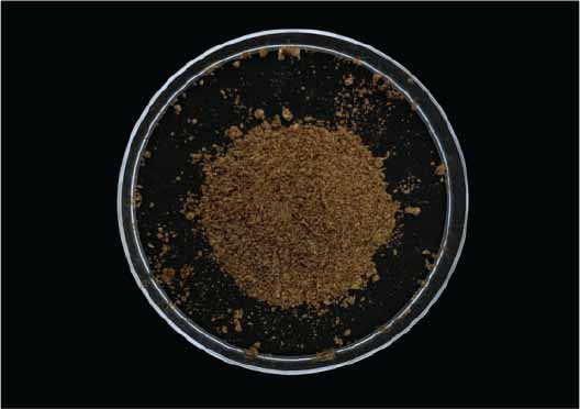
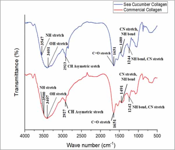
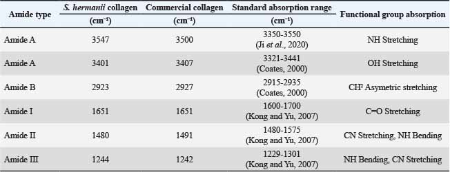
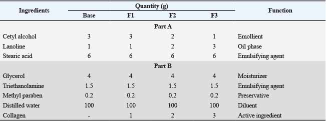
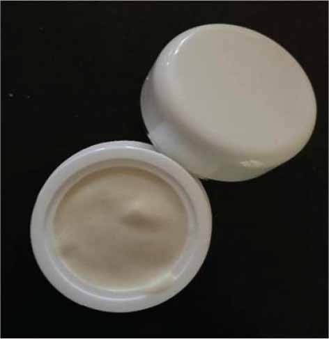
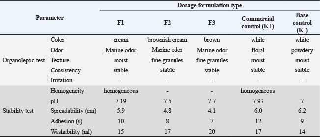
AbstractBackground: The sea cucumber Stichopus herrmanni is a marine-derived source of collagen with potential as a natural active ingredient in topical formulations. Collagen is integral to maintaining skin moisture, elasticity, and regeneration. Aim: This study aimed to assess the characteristics and biological activity of a sea cucumber collagen-based lotion through in vitro and in vivo testing. Methods: Collagen was characterized using the Hopkins–Cole test, ninhydrin test, pH measurement, water content analysis, and Fourier transform infrared (FTIR) spectroscopy. The lotion was formulated with varying collagen concentrations and evaluated through organoleptic testing, homogeneity, pH, spreadability, adhesion, and washability. In vivo testing was conducted on Mus musculus mice, with histological analysis of the skin, including measurements of epidermal thickness and fibroblast count, which were statistically analyzed using Analysis of Variance. Results: The collagen yield and water content were 1.26% and 3.1%, respectively. The Hopkins–Cole test indicated the absence of detectable tryptophan residues, while the ninhydrin test confirmed the presence of free amino acids. FTIR analysis identified the characteristic functional groups of collagen. The F1 formulation, which incorporated 1 g of collagen, exhibited the most favorable physical characteristics and stability compared to the other formulations. In vivo results showed that F1 produced the highest epidermal thickness of 91.14 μm, compared to the positive control, which measured 75.80 μm. However, the fibroblast count in F1 (12.50) was lower than that in the positive-control group (14.25). Conclusion: The F1 sea cucumber collagen lotion formulation exhibited favorable physical characteristics and a superior biological response compared to the untreated and negative control groups, particularly in terms of enhancing epidermal thickness. These findings suggest the potential of S. herrmanni collagen as a natural ingredient in topical lotion formulations, although further research is required to thoroughly evaluate its biological efficacy. Keywords: Marine collagen, Skin histological evaluation, Skin regeneration, Stichopus herrmanni, Topical lotion formulation. IntroductionIn 2022, global fisheries and aquaculture production reached 223.2 million tons, underscoring the increasingly significant role of marine resources, including marine biocomponents, in food security, biotechnology, and pharmaceutical sectors (FAO, 2024). Utilizing farmed sea cucumbers as a collagen source aligns with sustainable fisheries management principles, aiming to prevent the overexploitation of sea cucumber populations in their natural habitats (Dvoretsky and Dvoretsky, 2021). This commodity has been traditionally used as a food ingredient, in traditional medicine, and in cosmetics because of its high nutritional content, which includes proteins, amino acids, minerals, and bioactive compounds. The sea cucumber species S. herrmanni is particularly promising as a marine collagen source because its body walls are rich in collagen. Research indicates that approximately 70% of the total protein in the sea cucumber body wall is collagen, predominantly type I collagen fibers, which constitute the major structural components of the tissue (Barzkar et al., 2023). Collagen is crucial for tissue regeneration, wound healing, and maintaining skin elasticity (Geahchan et al., 2022). This positions golden sea cucumbers as a promising alternative collagen source for the pharmaceutical and cosmetic industries (Pramesti et al., 2025). Collagen, the primary structural protein in the extracellular matrix, comprises approximately 25%–35% of the total protein in vertebrates. Its characteristic triple helix structure, composed of the amino acids glycine, proline, and hydroxyproline, contributes to the strength and stability of the molecule (Senadheera et al., 2020). The body wall of sea cucumbers is predominantly composed of collagen, making it a potential source of bioactive ingredients for the bioindustry. Marine collagen is considered superior to terrestrial collagen because of its high biocompatibility, low molecular weight, and enhanced absorption by the body. In addition, sea cucumber collagen contains other bioactive components that support skin regeneration (Prajaputra et al., 2024). This suggests that sea cucumber collagen is beneficial not only for aesthetic purposes but also for overall skin health improvement. Human skin, the body's largest organ, serves protective and aesthetic functions that are closely linked to the quality of life. Conditions such as dry, dull, and wrinkled skin are often associated with premature aging, increasing the demand for skin care products. Among these, lotions are popular for their moisturizing properties and ability to strengthen skin barriers. However, the long-term use of synthetic lotions has been reported to potentially cause various skin issues, including irritation, contact dermatitis, and other disorders (Anitha et al., 2023). The high incidence of skin disorders caused by synthetic cosmetic ingredients underscores the need for safer, skin-friendly alternatives. Marine biota, particularly sea cucumbers, offer natural active ingredients that are rich in collagen and bioactive compounds with high efficacy. In the modern cosmetic industry, products aim not only to enhance beauty but also to promote skin health, a concept known as cosmeceuticals (Jadach et al., 2024). Consequently, lotion formulations can provide a glowing and moisturizing effect on the skin. (Aguirre-Cruz et al., 2020). This study proposes that collagen extracted from S. herrmanni can be formulated into a topical lotion, potentially facilitating skin regeneration by enhancing the epidermal structure and cellular responses within the dermis. The regenerative potential was assessed using in vivo histological parameters, specifically epidermal thickness and fibroblast counts. The novelty of this research lies in the formulation and comprehensive evaluation of a topical lotion containing marine collagen derived from the sea cucumber S. herrmanni, a source that has been infrequently explored in previous studies. In contrast to earlier research, which predominantly focused on fish-derived collagen, this study integrated in vitro and in vivo characterization to assess the physicochemical properties, stability, and biological potential of the formulated lotion. Although this study directly employed a biological model approach, all procedures adhered to the 3R principles (Replacement, Reduction, and Refinement) to ensure ethical, minimal, and welfare-conscious use of test organisms (Rinwa et al., 2024). Materials and MethodsPreparation and deproteinization of non-collagen proteinsStichopus herrmanni samples were procured from aquaculture harvests distributed by local collectors. The samples utilized were adult sea cucumbers, each weighing approximately ±200 g and measuring a body length of ±20 cm. The sea cucumber flesh was separated from the internal organs and washed with clean water until it was free of impurities. The samples were then sun-dried for ±6 h, cut into pieces of approximately 1 cm, and mechanically pulverized. Deproteinization was conducted to remove non-collagen proteins using a 0.1 M NaOH solution at a ratio of 1:10 (w/v) for 48 hours, with the solvent being replaced every 6 hours. During deproteinization, the samples were maintained at 4 °C (Rajagukguk et al., 2025). Subsequently, the samples were washed with running water until a neutral pH was achieved, before further testing. Extraction process of sea cucumber collagenCollagen extraction was performed using the acetic acid extraction method (CH₃COOH) at a concentration of 0.5 M and a 1:10 (w/v) ratio. The extraction process was conducted without solvent replacement at 4 °C for 48 h to maintain the stability of collagen (Afifah et al., 2023). Next, the residue was extracted using distilled water at a ratio of 1:2 (w/v) for 2 hours at 40°C–45°C on a hotplate with stirring. The extract was then filtered using a blancu cloth to separate the solid materials from the filtrate for easier centrifugation. Collagen was separated from the solvent by centrifugation at 6000 rpm for 30 min (Zhang et al., 2017). The resulting supernatant was dried in an oven at 40 °C for 5 hours. Low drying temperatures were employed to minimize alterations in protein structure due to excessive heat exposure. Collagen characterizationThe collagen yield percentage was calculated by comparing the dry weight of the extracted collagen with the initial weight of the material (Gaikwad and Kim, 2024). Qualitative identification of collagen was conducted using the Hopkins–Cole test, which involved the use of 1% oxalic acid, magnesium powder, and concentrated H₂SO₄. A positive result was indicated by the formation of purple rings. The ninhydrin test was performed by adding 1 M NaOH and 1% ninhydrin reagent to the sample, followed by heating, which resulted in a color change indicative of the presence of amino acids. The ninhydrin test was conducted to detect free amino acids, whereas the Hopkins–Cole test served as a supplementary GAGS test to confirm the absence of tryptophan residues, which are typically absent in collagen. The pH of the collagen was measured using a pH meter (Lutron pH-220S) after dissolving the sample in distilled water (Indriastuti et al., 2023). Moisture content was determined gravimetrically by drying a 0.5-g sample at 105 °C until a constant weight was achieved (Nirmaan et al., 2020). The functional group characterization of collagen was analyzed using FTIR (Shimadzu IRTracer-100) within the wavenumber range of 4000–400 cm⁻1 to identify the characteristic absorption bands of collagen (Saallah et al., 2021). Furthermore, the characterization conducted in this study focused on the preliminary evaluation of the physicochemical properties of collagen and its application in topical lotions. Preparation of lotion formulation from sea cucumber collagenThe lotion was formulated as an oil-based emulsion by separating oil phase A and aqueous phase B. Phase A comprised cetyl alcohol, lanolin, and stearic acid, while phase B included glycerol, triethanolamine, methyl paraben, and sea cucumber collagen dissolved in distilled water. Each ingredient was weighed according to the formula and placed in separate 50-ml beakers for mixing. Subsequently, phase A was heated until all the components melted and became homogeneous, while phase B was dissolved in 20-ml of distilled water and heated in a water bath until homogeneous. Both phases were then brought to the same temperature before emulsification. The oil and water phases were heated to the same temperature, and the oil phase was poured into a mortar to form a paste. The aqueous phase was then gradually added while homogenizing with a pestle until a stable, lotion-textured emulsion was formed. The remaining solution was added gradually while stirring continuously until an even mixture was achieved. Distilled water was added as required to adjust the viscosity of the preparation. The resulting lotion was packaged in pump bottles for storage (Klein et al., 2025). Chemical and physical characteristics of the lotion preparationThe lotion formulations were evaluated for their physicochemical properties, organoleptic characteristics, and skin irritation potential. Organoleptic assessment, including color, odor, texture, and overall acceptability, was conducted by 30 semi-trained volunteer panelists, and the results were analyzed descriptively. Skin irritation was evaluated by applying the formulation to the volar forearm of each volunteer for 30–60 min, followed by observation for signs of erythema, edema, pruritus, or papule formation (Indriastuti et al., 2023). The evaluation involved a non-invasive topical application of the formulations and, according to institutional regulations, did not require formal ethical approval. All participants were informed about the study procedures and voluntarily agreed to participate before the evaluation. Homogeneity was assessed by placing 0.5 g of the formulation between two Petri dishes and examining it for the presence of coarse particles. The pH was determined using a calibrated pH meter after dispersing 10 g of the formulation in 10 ml of distilled water and comparing the measured values with the acceptable skin pH range (4.5–8.0). Spreadability and adhesiveness were evaluated using 0.5 g of the formulation under progressively increasing loads. Washability was determined by applying 1 g of the formulation to the skin and rinsing it under running water until complete removal, with the volume of water required recorded as an indicator of washability (Montemayor et al. (2013)). In vivo test and histology examinationAn in vivo study was conducted using 24 male white mice (Mus musculus), aged 2–3 months and weighing 20–30 g. The animals were randomly assigned to six treatment groups (n=4 mice per group), with each mouse serving as the experimental unit. The treatment groups consisted of F1 (1-g collagen lotion formulation), F2 (2-g collagen lotion formulation), F3 (3-g collagen lotion formulation), K+ (commercial collagen lotion), K− (collagen-free lotion), and a base group (untreated control). Before treatment, the dorsal hair was shaved over an area of approximately 4 × 4 cm to facilitate topical application and observation of skin responses. The respective lotion formulations were applied twice daily (morning and evening) for 14 consecutive days (Khan et al., 2025). Following each application, the animals were exposed to ultraviolet B (UV-B) irradiation (280 nm) for 2 h while maintaining a constant distance between the UV-B source and the dorsal skin to ensure uniform exposure across all groups. At the end of the treatment period, dorsal skin tissues were collected for histological examination. Epidermal thickness and fibroblast cell counts were evaluated from five randomly selected, non-overlapping microscopic fields for each animal. The mean value obtained from these five fields was used as a single biological replicate for statistical analysis. Data are presented as mean ± SD and were analyzed using one-way Analysis of Variance followed by Tukey's HSD post hoc test in Statistical Package for the Social Sciences (SPSS), with statistical significance set at p < 0.05. Ethical approvalAn ethical permit was issued and approved by the ethical certificate number. 131/EC-H/KEPK/FK-UNDIP/X/2023 on October 30, 2023, by the Research Ethics Commission, Faculty of Medicine, Diponegoro University. ResultsCollagen yield and extraction from sea cucumberThe dry weight of the initial raw sea cucumber sample was 945 g, and that of the extracted collagen was 12 g. Based on the final calculation, the yield was 1.26% (Table 1). Sea cucumber collagen has a smooth texture with fine granules, a dark brown color, and a distinctive odor reminiscent of marine fish. The sea cucumber collagen extraction process resulted in a dry collagen texture, as shown in Figure 1. Collagen characterizationThe Hopkins–Cole test for sea cucumber and commercial collagen showed negative results, indicated by the absence of a purple ring, suggesting that tryptophan residues were not detected in either sample. The ninhydrin test yielded positive results, with a color change in both samples. Sea cucumber collagen produced a deep purple color, indicating the presence of free amino acids, whereas commercial collagen showed a pale yellow color, associated with the presence of certain amino acids such as proline, hydroxyproline, and glycine (Table 2). The results of the pH and moisture content tests are presented in Table 3. FTIR analysis showed that sea cucumber collagen and commercial collagen had similar absorption spectrum patterns (Fig. 2; Table 4), with characteristic collagen functional groups detected, including NH stretching, OH stretching, CH₂ asymmetric stretching, C=O stretching (amide I), CN stretching, NH bending (amide II), and amide III band. The similarities in these spectral characteristics indicate that sea cucumber collagen possesses a chemical structure comparable to that of commercial collagen. Formulation and preparation of lotion dosage forms from sea cucumber collagenThe selection of Formulations I, II, and III as the best formulas was based on the characterization results (Table 5). The formulations were tailored to the requirements of topical preparations, where good spreading ability on the skin surface facilitated the distribution of the active ingredient (collagen), thereby optimizing its absorption. Chemical and physical characteristics of lotion preparationsOrganoleptic tests showed that the sea cucumber collagen lotion achieved panelist preference scores in the range of 7–9 Figure 3 for the parameters of color, aroma, and texture, indicating good acceptance by the panelists. The off-white to brownish color of the lotion was considered quite attractive, the lightly fishy aroma was not overpowering, and the semi-solid texture was favored for its ease of application. The homogeneity test showed that all the sea cucumber collagen lotion formulas (Formulations I–III), the base, and the commercial lotion were homogeneous, as indicated by the absence of coarse grains, lumps, or phase separation, suggesting an even and stable dispersion of collagen. pH testing showed values ranging from 7.19–7.93, which are within the standard pH range for lotion according to SNI 16-4399-1996 (4.5–8.0), as shown in Table 6, and are thus considered safe for the skin. The spreadability test showed that the sea cucumber collagen lotion had a spread diameter of 4.1–5.9 cm, while the commercial collagen lotion had a spread diameter of 6.0 cm, both of which met the criteria for good lotion spreadability (5–7 cm). This shows that the preparation can spread evenly and is comfortable to use (Table 6). The adhesion test showed that the sea cucumber collagen lotion had an adhesion time of 7–10 s, and the commercial lotion had an adhesion time of 12 s, both meeting the adhesion standard (>4 s), as shown in Table 6. In the washability test, the sea cucumber collagen lotion required 14–20 ml of water, whereas the commercial collagen lotion required 17 ml to completely remove the preparation from the skin. These results indicate that both preparations have good washability and are consistent with the characteristics of oil-in-water (o/w) lotions, which are relatively easy to remove from the skin. The organoleptic and stability test results for the lotions are presented in Table 6 below . Table 1. Yield percentage of extracted sea cucumber collagen.
Fig. 1. Dried sea cucumber collagen powder obtained from S. herrmanni. Table 2. Qualitative protein characterization of collagen.
Table 3. Moisture content and pH of sea cucumber collagen and commercial collagen.
Fig. 2. FTIR spectra of sea cucumber collagen and commercial collagen. Table 4. FTIR absorption bands of sea cucumber collagen and commercial collagen.
Table 5. Composition of lotion formulations containing sea cucumber collagen.
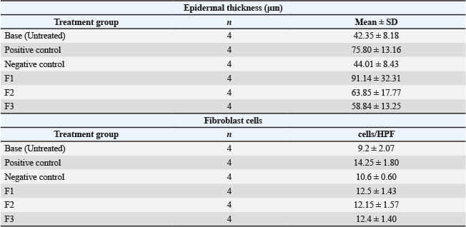
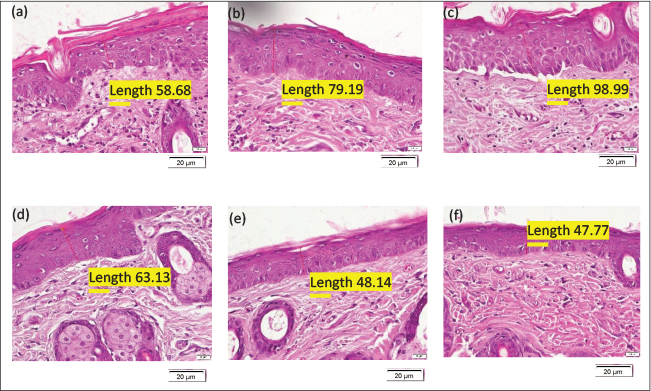
In vivo mouse skin tissue histology and SPSS dataThe effects of the collagen lotion formulations on skin regeneration were evaluated through histological examination of epidermal thickness and fibroblast cell counts after 14 days of treatment (Fig. 4 and Table 7 ). Histological measurements were obtained from five randomly selected microscopic fields for each animal, and the mean value for each mouse (n=4 per group) was used for statistical analysis. The F1 formulation produced the greatest mean epidermal thickness (91.14 ± 32.31 μm), followed by the positive control (75.80 ± 13.16 μm), F2 (63.85 ± 17.77 μm), F3 (58.84 ± 13.25 μm), the negative control (44.01 ± 8.43 μm), and the untreated base group (42.35 ± 8.18 μm) (Table 7). Representative histological sections (Fig. 4) showed a visibly thicker epidermal layer in the F1-treated group compared with the untreated and negative control groups. Statistical analysis demonstrated significant differences among treatment groups (p < 0.05). Post hoc analysis indicated that the epidermal thickness of the F1 group was significantly greater than that of the untreated and negative control groups, whereas no significant difference was observed between F1 and the positive control. Fibroblast cell counts also differed significantly among treatment groups (p < 0.05). The positive control exhibited the highest fibroblast count (14.25 ± 1.80 cells), followed by F1 (12.50 ± 1.43 cells), F3 (12.40 ± 1.40 cells), F2 (12.15 ± 1.57 cells), the negative control (10.60 ± 0.60 cells), and the untreated base group (9.20 ± 2.07 cells). The collagen lotion formulations (F1–F3) resulted in significantly higher fibroblast counts than the untreated group, while the fibroblast count in F1 was not significantly different from that of the positive control.
Fig. 3. Tropical lotion formulated with sea cucumber collagen (S. herrmanni). Table 6. Organoleptic properties and stability evaluation of lotion formulations.
Table 7. Histological analysis of epidermal thickness.
DiscussionMarine collagen, derived from marine organisms such as fish, jellyfish, starfish, and sea cucumbers, has been extensively developed as a natural biomaterial for cosmetic and dermatological applications. In this study, S. herrmanni was selected because its body wall is rich in collagen and contains other bioactive compounds, such as glycosaminoglycans, flavonoids, and hyaluronic acid, which have the potential to support skin regeneration and exhibit therapeutic properties, including anti-inflammatory, antioxidant, and antimicrobial activities (Hossain et al., 2022; Aulia et al., 2024). The collagen yield obtained was 1.26%, which is comparatively lower than that reported in several previous studies, such as Saallah et al. (2021), who reported a yield of 5.15% for Holothuria scabra. This discrepancy is likely influenced by the species, raw material conditions, and extraction methods employed. In this study, extraction was conducted using acetic acid without enzymatic assistance; thus, the solubility of collagen with strong crosslinking may have been more limited. Furthermore, deproteinization using NaOH for an extended duration may have affected the amount of collagen. Marine collagen is widely recognized for its superior bioactive properties, such as high biocompatibility and biodegradability, making it safe for use in cosmetics and dermatological applications. In addition, marine collagen possesses a high water retention capacity, which is closely related to the presence of hydrophilic groups, such as hydroxyl and amide groups, which can bind water molecules through hydrogen bonding and play a crucial role in maintaining skin moisture and enhancing hydration function in topical preparations. Collagen quality was also reflected in the water content test results, with sea cucumber collagen exhibiting a lower water content (3.1%) than commercial collagen (4.6%), and both values remained below the maximum threshold set by the SNI (SPI-kan/02/29/1987). Low water content indicates better stability and potentially a longer shelf life for collagen. The initial characterization of collagen was performed using qualitative tests and physicochemical parameters to facilitate the identification of the extracted collagen. The Hopkins–Cole test yielded negative results for both sea cucumber and commercial collagen, as evidenced by the absence of a purple ring, indicating no detectable tryptophan residues. This observation aligns with the general characteristics of collagen, which possesses very low or undetectable tryptophan content (Hosokawa et al., 2023). In addition, the ninhydrin test yielded positive results for both samples, as indicated by a color change. Sea cucumber collagen exhibited a deep purple color, indicating the presence of free amino acids, whereas commercial collagen resulted in a pale yellow color, suggesting the presence of specific amino acids such as proline, hydroxyproline, and glycine. The variation in color intensity is likely attributable to differences in the amino acid compositions of each collagen sample. The amino acid content is crucial for maintaining the stability of the triple helix structure of collagen, as reflected in the FTIR spectra (Senadheera et al., 2020). FTIR analysis of S. herrmanni and commercial collagen revealed the presence of collagen-specific amide bands, namely amide A, B, I, II, and III, indicating the successful extraction and preservation of the primary collagen structure. The amide A band at 3547–3500 cm⁻1 is associated with N-H stretching vibrations, whereas the OH stretching band at 3401–3407 cm⁻1 indicates hydrogen bonds contributing to the hydrophilic properties of collagen. The amide B band at 2923–2927 cm⁻1 indicates an asymmetric CH₂ stretching vibration, whereas the amide I band at 1651 cm⁻1 serves as the primary marker of the secondary structure of collagen through C=O stretching vibration. The amide II and III bands in the range of 1480–1491 cm⁻1 and 1242–1244 cm⁻1 are related to CN stretching and NH bending vibrations, reflecting the stability of the collagen helical structure (Sadat and Joye, 2020). Although there were slight shifts in the absorption band positions between sea cucumber and commercial collagen, the amide I band remained at 1651 cm⁻1, and the amide III band remained within a similar range, indicating that the secondary and helical structures of collagen were relatively well preserved in both samples.
Fig. 4. Histological appearance of mouse skin epidermis after topical treatment: (a) positive control; (b) negative control; (c) base (untreated); (d) collagen lotion formulation 1; (e) collagen lotion formulation 2; and (f) collagen lotion formulation 3. According to the physicochemical characterization results, the collagen extracted from sea cucumbers demonstrated favorable properties and was subsequently incorporated into a lotion formulation. This formulation utilizes a combination of auxiliary ingredients to achieve a stable and easily applicable topical preparation. Cetyl alcohol served as an emollient and viscosity enhancer, lanolin constituted the oil phase, and stearic acid and triethanolamine functioned as the emulsification system. Glycerol was used as a humectant, methylparaben as a preservative, and distilled water as the aqueous phase. Collagen from S. herrmanni was incorporated as a biofunctionally active ingredient with potential benefits for skin hydration and tissue regeneration. Marine collagen is acknowledged for its potential applications in cosmeceuticals, particularly in maintaining skin moisture and elasticity (Chen et al., 2022). The evaluation results indicated that all formulations satisfied the quality parameters for lotions, including organoleptic properties, homogeneity, pH, spreadability, adhesiveness, and washability. The organoleptic evaluation conducted by the panelists yielded scores ranging from 7 to 9, signifying a high level of acceptance. The lotion exhibited a cream color, non-pungent marine odor, and semi-solid texture that facilitated easy application. The homogeneity test confirmed the absence of clumps or phase separation, indicating a uniform collagen distribution within the lotion base. The pH of the lotion was between 7.19 and 7.93, which is slightly higher than the normal skin pH range of 4.5–6.5 (Proksch, 2018). This condition is likely influenced by the inclusion of basic triethanolamine (Gao et al., 2025). Although no signs of irritation were observed in the test animals, long-term safety evaluation in humans remains necessary. The spreadability parameters ranged from 5 to 7 cm, indicating suitable viscosity for topical application and effective distribution of the active ingredients. Adhesiveness testing revealed contact times of 7–10 s for the sea cucumber collagen lotion and 12 s for the commercial lotion, supporting the potential absorption of active ingredients through the skin. In the washability test, the sea cucumber collagen lotion required 14–20 ml of water, whereas the commercial lotion required 17 ml to completely remove the preparation. These results indicate variations in the retention of the preparation on the skin surface (Lubis et al., 2023). Overall, the test results demonstrated that the sea cucumber collagen lotion formulation possessed good physical stability and had potential for use as a marine collagen-based topical preparation. All sea cucumber collagen lotion formulations met the quality parameters and exhibited good physical stability. This study utilized three collagen concentrations: 1 g (F1), 2 g (F2), and 3 g (F3), to evaluate the effect of concentration on the formulation characteristics and biological response. The results indicated that higher collagen concentrations did not produce a linear increase in the biological response, as F1 actually showed the highest epidermal thickness compared to F2 and F3 formulations. These findings suggest that the effectiveness of a topical preparation is influenced not only by the collagen content but also by formulation stability, homogeneity, spreadability, and active ingredient distribution across the skin surface. The F1 formulation was selected as the most optimal because it displayed the most balanced characteristics, including a pH of 7.19, spreadability of 5.9 cm, adhesiveness of 10 s, washability of 15 ml, and stability, homogeneity, and non-irritating nature. These characteristics support application comfort and sustained contact between the preparation and skin surface. Biological evaluation was performed through in vivo testing on Mus musculus for 14 days. Histological analysis revealed significant variations in epidermal thickness and fibroblast counts among the treatment groups. The F1 formulation exhibited the most pronounced response, as evidenced by increased epidermal thickness, suggesting enhancement of the epidermal structure following collagen lotion application. This increase is attributed to collagen's capacity to support skin hydration and improve the microenvironment of the tissue. Collagen is recognized for its biocompatibility and role in facilitating cell adhesion, proliferation, and differentiation during tissue regeneration (Mbese et al., 2021). Although the F1 formulation demonstrated the highest epidermal thickness, its fibroblast count was lower than that of the positive control, indicating that it did not exceed the positive control in stimulating dermal fibroblast activity. This discrepancy suggests that epidermal thickness and fibroblast counts are likely governed by distinct biological mechanisms. The increase in epidermal thickness in F1 is believed to be associated with collagen's ability to enhance skin hydration, improve barrier function, and support epidermal cell proliferation. Conversely, fibroblast counts more accurately reflect extracellular matrix remodeling activity and collagen synthesis in the dermal layer (Zou et al., 2022; Barzkar et al., 2023). Thus, formulation F1 tended to elicit a more pronounced response in enhancing the epidermal layer than in stimulating the dermal fibroblasts. Nonetheless, the fibroblast count in F1 remained higher than that in the negative control and untreated groups, indicating a regenerative response in the dermis. Furthermore, the positive control likely contained other active compounds that were more effective in activating fibroblast proliferation than the sea cucumber collagen formulation used in this study (Lesmana et al., 2023). Although marine collagen holds promise as a topical biomaterial because of its biocompatibility and bioactivity, safety aspects and potential allergenicity must be considered. Marine collagen and several excipients, such as lanolin and preservatives, may induce hypersensitivity reactions in sensitive individuals (Holmes et al., 2017; Geahchan et al., 2022). In addition, this study has limitations, including the relatively small number of experimental animals and limited biological parameters and is further constrained by the absence of long-term stability testing (shelf-life stability) and evaluation of microbial contamination of the samples. Consequently, the storage stability and microbiological safety of the formulation during use and long-term storage cannot be confirmed. Further studies are warranted to evaluate the safety and efficacy of this formulation. ConclusionThe collagen extract from the sea cucumber yielded a concentration of 1.26%. Preliminary characterization revealed a negative result in the Hopkins–Cole test, indicating the absence of tryptophan residues, while the ninhydrin test confirmed the presence of free amino acids. The extract exhibited a pH of 7.11 and water content of 3.1%, and FTIR analysis revealed characteristic amide bands of collagen, suggesting that the extracted collagen possesses physicochemical properties consistent with those of collagen. Collagen was successfully incorporated into a lotion, which exhibited organoleptic properties such as cream color, marine odor, and semi-solid texture. The lotion demonstrated satisfactory homogeneity, pH, spreadability, adhesiveness, and washability, in accordance with the quality standards for lotion formulations. In vivo testing revealed significant differences in epidermal thickness among the groups, with formulation F1 exhibiting the highest value, whereas the fibroblast count in F1 and F2 did not significantly differ from that of the positive control. This indicates the preliminary potential of sea cucumber collagen lotion to promote skin regenerative responses. These findings underscore the potential of S. herrmanni collagen as a natural topical active ingredient, although the current research is limited to initial physicochemical evaluation and histological parameters in a mouse model. Consequently, further studies are warranted to conduct a more comprehensive characterization of collagen, clinically validate its efficacy and safety, assess long-term formulation stability, and optimize the development of pharmaceutical and cosmeceutical products based on marine collagen. AcknowledgmentsThe authors would like to express their sincere gratitude to the Laboratory of the Faculty of Fisheries and Marine Sciences, Diponegoro University, and the Integrated Laboratory of Diponegoro University for providing research facilities and technical support. The authors also acknowledge the Biology Laboratory of Universitas Negeri Semarang for facilitating the in vivo experiments. Conflict of interestThe authors declare that they have no conflict of interest. FundingThis research was funded by the Faculty of Fisheries and Marine Sciences, Diponegoro University, through the Faculty Research Grant Program (Non-APBN funding source) for the Fiscal Year 2026, under contract number 28/UN7.F10/PP/II/2026. Authors' contributionsConceptualization: R.P. and W.A.S.; Methodology: S., R.P., and W.A.S.; Investigation: B.A.W.; Data Curation: S., B.A.W., J.G.L.G., and H.I.; Formal Analysis: R.P., W.A.S., and S.; Writing – Original Draft Preparation: B.A.W. and J.G.L.G.; Writing – Review & Editing: R.P., W.A.S., and S. All authors contributed to the discussion of the research ideas and concepts and approved the final version of the manuscript. Data availabilityAll data supporting the findings of this study are available within the manuscript. ReferencesAfifah, A., Suparno, O., Haditjaroko, L., Tarman, K., Setiyono, A. and Nugraha, A.W. 2023. Isolation and characterization of collagen from salmon (Salmo salar) skin using acid method. Squalen Bull. Mar. Fisheries Postharvest Biotechnol. 18, 139–147; doi:10.15578/squalen.756 Aguirre-Cruz, G., León-López, A., Cruz-Gómez, V., Jiménez-Alvarado, R. and Aguirre-Álvarez, G. 2020. Collagen hydrolysates for skin protection: oral administration and topical formulation. Antioxidants 9, 181; doi:10.3390/antiox9020181 Anitha, M.G., Pooja, H., Divya, K. and Abhiram, P. 2023. A survey study on the adverse effects of synthetic cosmetics. Int. J. Community Med. Public Health, 10, 4147–4152; doi: 10.18203/2394-6040.ijcmph20233442 Aulia, A.A., Astuti, A.R., Wulandari, A.A., Nugroho, G.D. and Setyawan, A.D. 2024. Chemical profiles and therapeutic potentials of golden sea cucumber (Stichopus hermanii). Cell. Biol. Dev. 8, 58–67; doi:10.13057/cellbioldev/v080202 Barzkar, N., Sukhikh, S., Babich, O., Venmathi Maran, B.A. and Tamadoni Jahromi, S. 2023. Marine collagen: purification, properties and application. Front. Mar. Sci. 10, 1245077; doi:10.3389/fmars.2023.1245077 Chen, H.J., Lee, P.Y., Chen, C.Y., Huang, S.L., Huang, B.W., Dai, F.J., Chau, C.F., Chen, C.S. and Lin, Y.S. 2022. Moisture retention of glycerin solutions with various concentrations: a comparative study. Sci. Rep. 12, 10232; doi:10.1038/s41598-022-13452-2 Coates, J. 2000. Interpretation of infrared spectra, a practical approach. In Encyclopedia of Analytical Chemistry. Ed., Meyers, R.A. pp: 22–31; doi: 10.1097/00010694-197107000-00005 Dvoretsky, A.G. and Dvoretsky, V.G. 2021. Cucumaria in Russian waters of the Barents Sea: biological aspects and aquaculture potential. Front. Mar. Sci. 8, 613453; doi:10.3389/fmars.2021.613453 FAO. 2024. Global Fisheries and Aquaculture Production Reaches a New Record High. Gaikwad, S. and Kim, M.J. 2024. Fish by-product collagen extraction using different methods and their application. Mar. Drugs 22, 60; doi:10.3390/md22020060 Gao, M., Luo, Z., Zhang, M., Liu, X., Liu, L., Ye, J., Zhang, X., Yang, Y., Ge, M., Yang, H., Rong, S. and Wang, Z. 2025. Effect of triethanolamine on the properties and hydration of fly ash-based geopolymer foam. Waste. Manage. 198, 106–116; doi:10.1016/j.wasman.2025.02.045 Geahchan, S., Baharlouei, P. and Rahman, A. 2022. Marine collagen: a promising biomaterial for wound healing, skin anti-aging, and bone regeneration. Mar. Drugs 20, 61; doi:10.3390/md20010061 Holmes, R., Kirk, S., Tronci, G., Yang, X. and Wood, D. 2017. Influence of telopeptides on the structural and physical properties of polymeric and monomeric acid-soluble type I collagen. Mater. Sci. Eng. 77, 823–827; doi:10.1016/j.msec.2017.03.267 Hosokawa, S., Morinishi, T., Ohara, K., Yamaguchi, K., Tada, S. and Tokuhara, Y. 2023. A spectrophotometric method for the determination of tryptophan following oxidation by the addition of sodium hypochlorite pentahydrate. PLoS One 18(1), e0279547; doi: 10.1371/journal.pone.0279547 Hossain, A., Dave, D. and Shahidi, F. 2022. Antioxidant potential of sea cucumbers and their beneficial effects on human health. Mar. Drugs 20, 521; doi:10.3390/md20080521 Indriastuti, M., Wahlanto, P. and Utami, D.S. 2023. Formulation and evaluation of Moringa leaves extract (Moringa Oleifera L.) lotion with variation concentration of triethanolamin. Ad-Dawaa. J. Pharm. 1(1), 18–28; doi:10.52221/dwj.v1i1.222 Jadach, B., Mielcarek, Z. and Osmałek, T. 2024. Use of collagen in cosmetic products. Curr. Issues. Mol. Biol. 46, 2043–2070; doi:10.3390/cimb46030132 Ji, Y., Yang, X., Ji, Z., Zhu, L., Ma, N., and Chen, D. 2020. DFT-calculated IR spectrum amide I, II, and III band contributions of N-methylacetamide fine components. ACS Omega 5(15), 8572–8578; doi: 10.1021/acsomega.9b04421 Khan, A., Ullah, S., Ramzan, F., Rehman, A.U., Rehman, S., Kholik, K., Sukri, A., Munawaroh, M. and Sucipto, T.H. 2025. Combined effects of Aloe vera leaf extract and vitamin E on wound healing in Oryctolagus cuniculus (Rabbits). Open Vet. J. 15, 700; doi:10.5455/OVJ.2025.v15.i2.18 Klein, M., Tacker, M. and Apprich, S. 2025. Product waste resulting from insufficient emptiability of cosmetic packaging and its economic and environmental implications. Sustainability 17, 1056; doi:10.3390/su17031056 Kong, J. and Yu, S. 2007. Fourier transform infrared spectroscopic analysis of protein secondary structures. Acta Biochimica et Biophysica Sinica. 39(8), 549–559; doi: 10.1111/j.1745-7270.2007.00320.x Lesmana, M.A., Amri, I.A., Cahyanissa, A.R., Geoputri, K.M., Anisa, A.K. and Hardian, A.B. 2023. Efficacy of oregano extract ointment on fibroblast cells and epidermis in albino rats with excisional wound model. J. Medik. Veterinar. 6(2), 262–270; doi:10.20473/jmv.vol6.iss2.2023.262-270 Lubis, M.S., Rani, Z., Wahyuni, W. and Arlian, R.Y. 2023. Test of sunscreen activity of pineapple weevil ethanol extract (ananas comosus (L.) merr.) in gel and lotion preparations. AMCA. J. Sci. Technol. 3(1), 7–12; doi:10.51773/ajst.v3i1.196 Mbese, Z., Alven, S. and Aderibigbe, B. A. 2021. Collagen-based nanofibers for skin regeneration and wound dressing applications. Polymers 13(24), 4368; doi: doi: 10.3390/polym13244368 Montemayor, B.P., Price, B.B. and van Egmond, R.A. 2013. Accounting for intended use application in characterizing the contributions of cyclopentasiloxane (D5) to aquatic loadings following personal care product use: Antiperspirants, skin care products and hair care products. Chemosphere 93(5), 735–740; doi: 10.1016/j.chemosphere.2012.10.043 Nirmaan, A.M.C., Rohitha Prasantha, B.D. and Peiris, B.L. 2020. Comparison of microwave drying and oven-drying techniques for moisture determination of three paddy (Oryza sativa L.) varieties. Chem. Technol. Agriculture 7, 1–7; doi:10.1186/s40538-019-0164-1 Prajaputra, V., Isnaini, N., Maryam, S., Ernawati, E., Deliana, F., Haridhi, H.A., Fadli, N., Karina, S., Agustina, S., Nurfadillah, N., Arisa, I.I., Desiyana, L.S. and Bakri, T.K. 2024. Exploring marine collagen: sustainable sourcing, extraction methods, and cosmetic applications. S. Afr. J. Chem. Eng. 47, 197–211; doi:10.1016/j.sajce.2023.11.006 Pramesti, R., Setyati, W.A., Soenardjo, N. and Ariyanto, D. 2025. Antioxidant, antibacterial, and antifungal characteristics on crude collagen extracts of sea cucumber (Stichopus hermanii). Int. J. Design. Nature. Ecodynamics. 20, 523–528; doi:10.18280/ijdne.200307 Proksch, E. 2018. PH in nature, humans and skin. J. Dermatology 45, 1044–1052; doi:10.1111/1346-8138.14489 Rajagukguk, Y.L., Pringgenies, D., Setyati, W.A. and Istiadi, H. 2025. Moisturizing efficacy of Stichopus hermanii-derived collagen in Balb/c mice (Mus musculus). J. Fish. Environ. 49(3), 159–176. Available via https://li01.tci-thaijo.org/index.php/JFE/article/view/265182 Rinwa, P., Eriksson, M., Cotgreave, I. and Bäckberg, M. 2024. 3R-Refinement principles: elevating rodent well-being and research quality. Lab. Anim. Res. 40(11), 11; doi:10.1186/s42826-024-00198-3 Saallah, S., Roslan, J., Julius, F.S., Saallah, S., Mohamad Razali, U.H., Pindi, W., Sulaiman, M.R., Pa’Ee, K.F. and Mustapa Kamal, S.M. 2021. Comparative study of the yield and physicochemical properties of collagen from sea cucumber (Holothuria scabra), obtained through dialysis and the ultrafiltration membrane. Molecules 26, 2564; doi:10.3390/molecules26092564 Sadat, A. and Joye, I.J. 2020. Peak fitting applied to fourier transform infrared and raman spectroscopic analysis of proteins. Appl. Sci. 10, 5918; doi:10.3390/app10175918 Senadheera, T.R.L., Dave, D. and Shahidi, F. 2020. Sea cucumber derived type I collagen: a comprehensive review. Mar. Drugs 18, 471; doi:10.3390/md18090471 Zhang, J., Sun, Y., Zhao, Y., Wei, B., Xu, C., He, L., Oliveira, C.L.P. and Wang, H. 2017. Centrifugation-induced fibrous orientation in fish-sourced collagen matrices. Soft Matter 13, 9220–9228; doi:10.1039/C7SM01871A Zou, Q., Zhang, M., Yuan, R., Wang, Y., Gong, Z., Shi, R., Li, Y., Fei, K., Luo, C., Xiong, Y., Zheng, T., Zhu, L., Tang, G., Li, M., Li, X. and Jiang, Y. 2022. Small extracellular vesicles derived from dermal fibroblasts promote fibroblast activity and skin development through carrying miR-218 and ITGBL1. J. Nanobiotechnol. 20, 296; doi:10.1186/s12951-022-01499-2 | ||



| How to Cite this Article |
| Pubmed Style Pramesti R, Setyati WA, Subagiyo S, Wibowo BA, Gaol JGL, Istiadi H. In vitro and in vivo evaluation of a topical marine collagen lotion derived from sea cucumber (Stichopus herrmanii). Open Vet. J.. 2026; 16(8): 5230-5241. doi:10.5455/OVJ.2026.v16.i8.18 Web Style Pramesti R, Setyati WA, Subagiyo S, Wibowo BA, Gaol JGL, Istiadi H. In vitro and in vivo evaluation of a topical marine collagen lotion derived from sea cucumber (Stichopus herrmanii). https://www.openveterinaryjournal.com/?mno=314864 [Access: August 08, 2026]. doi:10.5455/OVJ.2026.v16.i8.18 AMA (American Medical Association) Style Pramesti R, Setyati WA, Subagiyo S, Wibowo BA, Gaol JGL, Istiadi H. In vitro and in vivo evaluation of a topical marine collagen lotion derived from sea cucumber (Stichopus herrmanii). Open Vet. J.. 2026; 16(8): 5230-5241. doi:10.5455/OVJ.2026.v16.i8.18 Vancouver/ICMJE Style Pramesti R, Setyati WA, Subagiyo S, Wibowo BA, Gaol JGL, Istiadi H. In vitro and in vivo evaluation of a topical marine collagen lotion derived from sea cucumber (Stichopus herrmanii). Open Vet. J.. (2026), [cited August 08, 2026]; 16(8): 5230-5241. doi:10.5455/OVJ.2026.v16.i8.18 Harvard Style Pramesti, R., Setyati, . W. A., Subagiyo, . S., Wibowo, . B. A., Gaol, . J. G. L. & Istiadi, . H. (2026) In vitro and in vivo evaluation of a topical marine collagen lotion derived from sea cucumber (Stichopus herrmanii). Open Vet. J., 16 (8), 5230-5241. doi:10.5455/OVJ.2026.v16.i8.18 Turabian Style Pramesti, Rini, Wilis Ari Setyati, Subagiyo Subagiyo, Belinda Aureliawati Wibowo, Josua Gabriel Lumban Gaol, and Hermawan Istiadi. 2026. In vitro and in vivo evaluation of a topical marine collagen lotion derived from sea cucumber (Stichopus herrmanii). Open Veterinary Journal, 16 (8), 5230-5241. doi:10.5455/OVJ.2026.v16.i8.18 Chicago Style Pramesti, Rini, Wilis Ari Setyati, Subagiyo Subagiyo, Belinda Aureliawati Wibowo, Josua Gabriel Lumban Gaol, and Hermawan Istiadi. "In vitro and in vivo evaluation of a topical marine collagen lotion derived from sea cucumber (Stichopus herrmanii)." Open Veterinary Journal 16 (2026), 5230-5241. doi:10.5455/OVJ.2026.v16.i8.18 MLA (The Modern Language Association) Style Pramesti, Rini, Wilis Ari Setyati, Subagiyo Subagiyo, Belinda Aureliawati Wibowo, Josua Gabriel Lumban Gaol, and Hermawan Istiadi. "In vitro and in vivo evaluation of a topical marine collagen lotion derived from sea cucumber (Stichopus herrmanii)." Open Veterinary Journal 16.8 (2026), 5230-5241. Print. doi:10.5455/OVJ.2026.v16.i8.18 APA (American Psychological Association) Style Pramesti, R., Setyati, . W. A., Subagiyo, . S., Wibowo, . B. A., Gaol, . J. G. L. & Istiadi, . H. (2026) In vitro and in vivo evaluation of a topical marine collagen lotion derived from sea cucumber (Stichopus herrmanii). Open Veterinary Journal, 16 (8), 5230-5241. doi:10.5455/OVJ.2026.v16.i8.18 |


