





| Short Communication | ||
Open Vet. J.. 2026; 16(8): 5486-5491 !!
Open Veterinary Journal, (2026), Vol. 16(8): 5486–5491 Short Communication An accelerated transition to the late inflammatory phase in ovariohysterectomy wounds in stray cats by high-power class IV laser therapyPhinidda Cha-umphol1*, Nutthawadee Aeimsaard1, Atchara Dokkhaoram1, Bussayamas Pakasamith1, Wilin Khonchaiyaphom1, Chanisara Khongphibarn1 and Chapkit Charnsamorn21Faculty of Veterinary Medicine, Mahanakorn University of Technology, Nongchok, Thailand 2Faculty of Science, Mahanakorn University of Technology, Nongchok, Thailand *Corresponding Author: Phinidda Cha-umphol. Faculty of Veterinary Medicine, Mahanakorn University of Technology, Nongchok, Thailand. Email: phinidda [at] mut.ac.th Submitted: 17/03/2026 Revised: 11/06/2026 Accepted: 22/06/2026 Published: 08/08/2026 © 2025 Open Veterinary Journal
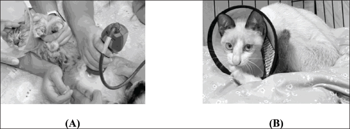
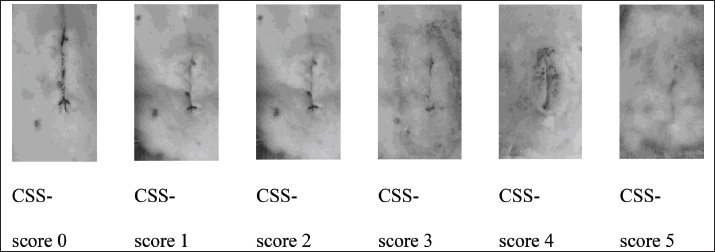
AbstractBackground: Stray cat population control programs commonly rely on ovariohysterectomy (OVH); however, prolonged postoperative confinement is often impractical under field conditions. Therefore, rapid healing is essential to shorten hospitalization periods, allowing for an earlier reintroduction of stray cats. High-power class IV laser therapy has been increasingly used to enhance wound healing, but its effect on the progression of inflammatory phases in feline surgical wounds remains insufficiently characterized, and thus, this pilot study was conducted. Aim: This pilot study aimed to evaluate whether high-power class IV laser therapy could shorten the time required to reach the late inflammatory phase in OVH wounds in female stray cats, assessed using a Clinical Scar Scale (CSS) and the wound size measurements. Methods: Thirteen clinically healthy female stray cats undergoing OVH were included and allocated to a control group (n=4) and a laser-treated group (n=9). Laser therapy was applied at 3, 24, 33, and 48 hours postoperatively. Wound healing progression was assessed using standardized photographic evaluation every 3 hours for 85 hours based on a CSS, along with wound length measurements at 7 and 14 days postoperatively. Cytological examination of wound exudate was performed during the early postoperative period to confirm the presence. Results: The laser-treated group demonstrated greater wound contraction compared to the control group at 7 days (38.24% vs. 17.24%) and 14 days (39.58% vs. 20.31%). Macroscopic observation indicated earlier progression to the late inflammatory phase in the laser-treated group (approximately 35 hours) compared to the control group (approximately 50 hours). Statistical analysis confirmed a significantly shorter time to reach CSS score 3 in the laser-treated group (53.36 ± 26.79 hours) than in the control group (67.05 ± 28.43 hours; p=0.039). Cytological findings revealed neutrophil predominance during the early inflammatory phase. Conclusion: The class IV laser therapy may accelerate wound healing by shortening the inflammatory phase and promoting earlier transition to subsequent healing stages. This may allow a clinically significant reduction of postoperative confinement time for field-based sterilization programs, improving both animal welfare and operational efficiency. Further controlled studies are recommended. Keywords: Class IV laser, Clinical Scar Scale, Late inflammation, Photo-biomodulation, Surgical wound healing. IntroductionStray cats represent an important animal welfare and population management challenge in many countries, including Thailand. Surgical sterilization, particularly ovariohysterectomy (OVH), is widely used to control stray cat populations (Joyce and Yates, 2011). However, free-roaming or community-fed cats cannot usually be confined for extended postoperative care; achieving rapid wound recovery is essential prior to their release. Wound healing is a complex biological process consisting of inflammatory, proliferative, and remodelling phases (Lopez, 2017). Among these stages, the inflammatory phase plays a key role in determining the overall healing timeline. In practical clinical settings, postoperative wound progression is often monitored using simple indicators, such as redness, swelling, and wound site temperature. Laser therapy has increasingly been used in veterinary medicine to reduce inflammation and stimulate tissue repair (Wardlaw et al., 2019; Fesseha , 2020). High-power class IV laser therapy has shown potential benefits in promoting postoperative healing (Prouza et al., 2013). However, limited information is available regarding its effect on the timing of inflammatory phase progression in feline surgical wounds. Therefore, this pilot study aimed to evaluate whether high-power class IV laser therapy promoted a faster transition to the late inflammatory phase in OVH wounds in female stray cats, assessed by using the Clinical Scar Scale (CSS) and the wound size measurements. Materials and MethodsThis prospective pilot study was conducted as part of a community stray cat population control program in a suburban area near Bangkok, Thailand. Thirteen clinically healthy female stray cats, 4–5 months old, undergoing OVH by the same veterinary surgeon, were enrolled and allocated to a control group (n=4) and a laser treatment group (n=9). The unequal group sizes resulted from logistical constraints and the limited number of stray cats captured during the community sterilization campaign. To achieve a uniform biostimulator effect across all subjects, only cats with normal body condition and non-heavily pigmented skin around the incision were included. These criteria were established to reduce variations in photon absorption and to facilitate clear, longitudinal visual evaluation of the wound. All cats underwent OVH via a ventral midline incision of approximately 2 cms under aseptic surgical conditions (Coe et al., 2006). The post-operative incisions in all cat subjects were maintained at a length not exceeding 3 cms and were treated using the Mphi Orange model, which is a class IV Multiwave Locked System (MLS; 808 and 905 nm) laser. This device was classified as a class IV laser (maximum output power 75 W, 1.2 Watts average). This machine offers preset protocols (equine, canine, feline; anti-inflammation, and analgesic), manual mode (energy level, pulse rate, and treatment time), and point-by-point mode. We use a preset protocol with feline, post-surgery mode. This setting gives pulse output power of 4 J/cm2. By dividing each incision into three segments of 1 cm each, each segment received a targeted irradiation for a duration of 25 seconds with an energy density set at 4/25=0.16 W/cm2. Cats in the treatment group received laser therapy at 3, 24, 33, and 48 hours postoperatively (Fig. 1). The CSS score (0–5) was assessed according to the protocols described by Wardlaw et al. (2019) and Prouza et al. (2013). A score of 0 indicated a fresh surgical incision, while a score of 1 represented a fresh incision without haemorrhage. A score of two was assigned when mild scabbing, swelling, or bruising was present. A score of three indicated visible healing with ongoing skin remodelling and resolving inflammation. A score of four represented advanced healing with a visible scar remaining. A score of five indicated a completely healed surgical incision. Postoperative wound healing was documented using standardized photographs taken every 3 hours during the first 85 hours after surgery. This observation interval (every 3 hours) and monitoring duration (85 hours) were determined based on preliminary pilot observations in two cats. That data indicate that the wound progression reached a plateau beyond the 85-hour mark. At each time point, clinical signs associated with the inflammatory phase of wound healing, including redness, swelling, and wound site temperature, were recorded (Lopez, 2017). Over 250 photographs were obtained by the same operator using a standardized photographic protocol with consistent positioning and lighting conditions to minimize human error. Wound appearance was blindly evaluated using a Clinical Scar Scale (0–5) based on photographic images assessed independently in three separate sessions by five veterinary nursing students and two veterinarians following previously described (Wardlaw et al., 2019).
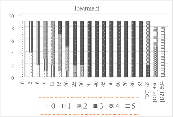
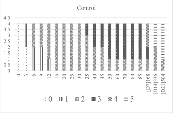
Fig. 1. Application of class IV laser therapy and postoperative care of stray cats. (A) Laser treatment at the surgical site. (B) Routine wound care is performed twice daily (08:00 and 18:00), with additional photographs taken every 3 hours without disturbing the wound. Wound length was measured immediately after surgery, 7 and 14 days postoperatively using a vernier calliper. To confirm the presence of inflammatory exudate during the early postoperative period, direct impression smears were collected between 6 and 24 hours postoperatively, stained, and examined microscopically for inflammatory cells by existence of neutrophils. Independent sample t-tests were used to compare wound length and the time required to reach specific Clinical Scar Scale scores 3 (late inflammatory stage) between groups, with statistical significance set at p < 0.05. All procedures were approved by the Animal Care and Use Committee of Mahanakorn University of Technology (ACUC-MUT-2025). Ethical approvalAll procedures were approved by the Animal Care and Use Committee of Mahanakorn University of Technology (ACUC-MUT-2025). Results and DiscussionDuring the early postoperative period, wound contraction in the laser-treated group progressed more rapidly than in the control group. Within the first 7 days after surgery, wound length reduction reached 38.24% in the laser-treated group, whereas the control group showed a reduction of 17.24%, as illustrated in Figure 2. By 14 days postoperatively, the treatment group demonstrated a total wound reduction of 39.58%, while the control group showed a reduction of 20.31%. These findings suggest that laser therapy may enhance tissue repair and accelerate wound contraction during the early phase of healing. Previous studies (Wardlaw et al., 2019; Fesseha, 2020) have reported that photo-biomodulation can stimulate mitochondrial activity, increase ATP production, and promote fibroblast proliferation, which contribute to improved collagen synthesis and tissue repair. Observation of postoperative inflammatory signs also revealed differences between the two groups. In both groups, the haemostasis phase occurred during the first 0–3 hours after surgery. Based on macroscopic observation, the early inflammatory phase was detected between approximately 3–30 hours in the laser-treated group and 3–35 hours in the control group. The transition to the late inflammatory phase appeared earlier in the laser-treated group at approximately 35 hours postoperatively (Fig. 3), whereas in the control group it occurred at around 50 hours, as illustrated in Figure 4. To further confirm these observations, wound photographs obtained every 3 hours were evaluated using the Clinical Scar Scale (CSS; Fig. 5). Statistical analysis demonstrated that the mean time required for wounds to reach CSS score 3, representing the late inflammatory stage characterized by a clinically stable wound with no further tissue deterioration, reduced exudate, and absence of granulation tissue formation, was 53.36 ± 26.79 hours in the laser-treated group and 67.05 ± 28.43 hours in the control group. Independent t-test analysis revealed a significant difference between groups. (t=−2.093, p=0.039). These results support the visual observation that laser therapy may promote earlier resolution of inflammation and facilitate faster progression toward the proliferative stage of wound healing. Direct impression smear examination of wound exudate during the early postoperative period revealed the presence of red blood cells and inflammatory cells, particularly neutrophils. Neutrophils are the predominant leukocytes during the early inflammatory phase and play an essential role in phagocytosis, debris removal, and microbial defence in surgical wounds (Lopez, 2017). The gradual reduction of red blood cells and inflammatory exudate observed after approximately 20 hours postoperatively is consistent with the normal physiological progression of wound healing following OVH procedures (Kiani et al., 2014).
Fig. 2. Wound contraction percentage during postoperative days 0, 7, and 14.
Fig. 3. Distribution of Clinical Scar Scale (CSS) scores over time in the laser-treated group. (Data represent serial wound images (approximately 20 images per case) obtained at 3-hour intervals from 9 cats in the treatment group, categorized according to Clinical Scar Scale scores).
Fig. 4. Distribution of Clinical Scar Scale (CSS) scores over time in the control group. (Data represent serial wound images (approximately 20 images per case) obtained at 3-hour intervals from 4 cats in the control group, categorized according to Clinical Scar Scale scores). The accelerated wound healing observed in the laser-treated group may be explained by the biological mechanisms of photo-biomodulation. Laser therapy has been reported to improve cellular metabolism, enhance ATP production, and stimulate fibroblast proliferation, thereby promoting collagen synthesis and tissue regeneration (Guo and Dipietro, 2010; Fesseha, 2020; Sukeri et al., 2025). In addition, laser irradiation may improve local microcirculation and modulate inflammatory responses, contributing to earlier resolution of inflammation and faster transition to the proliferative phase of wound healing (Prouza et al., 2013; Fesseha, 2020). Similar improvements in surgical wound healing following laser therapy have been reported in veterinary patients, supporting the potential benefits of laser-assisted postoperative management (Wardlaw et al., 2019).
Fig. 5. Representative images of postoperative wounds corresponding to Clinical Scar Scale (CSS) scores 0, 1, 2, 3, 4, and 5. Although this pilot study was conducted under field conditions as part of a community stray cat sterilization program, this study reveals the preliminary evidence that class IV laser therapy may enhance postoperative wound healing in cats undergoing OVH. However, as this was a pilot study, the relatively small sample size and unequal group distribution should be considered when interpreting the results. Increasing the sample size would provide the statistical power necessary to characterize the influence of specific covariates, such as age or breed, on healing rates. ConclusionHigh-power class IV laser therapy appears to promote a more efficient progression of wound healing following OVH in cats, particularly by shortening the time required to reach the late inflammatory stage. This earlier transition reflects improved resolution of inflammation and a faster shift toward subsequent healing phases, consistent with the biological effects of photo-biomodulation. Importantly, this may represent a potentially clinically meaningful reduction in postoperative confinement time in field-based sterilization programs, helping to minimize stress in stray cats and enable earlier return to their original environment. Such benefits may improve both animal welfare and the overall efficiency of population control efforts. Further controlled studies are warranted to confirm these findings and support broader clinical application. AcknowledgmentsThe authors would like to thank the Small Animal Hospital, Mahanakorn University of Technology, for providing the laser equipment. The authors also sincerely thank Dr. Natnaree Kaewsiri for her assistance in the Clinical Scar Scale (CSS) evaluation, together with the research team. FundingThis study was supported by Mahanakorn University of Technology. Authors’ contributionsPhinidda Cha-umphol conceptualized and designed the study, performed data collection, and drafted the manuscript. Nutthawadee Aeimsaard, Atchara Dokkhaoram, Bussayamas Pakasamith, Wilin Khonchaiyaphom, and Chanisara Khongphibarn performed data collection and wound evaluation. Chapkit Chansamorn supervised the study and critically revised the manuscript. All authors read and approved the final manuscript. Conflict of interestThe authors declare no conflict of interest. Data availabilityThe datasets generated and/or analysed during this study are not publicly available but are available from the corresponding author upon reasonable request. ReferencesCoe, R.J., Grint, N.J., Tivers, M.S., Hotston Moore, A. and Holt, P.E. 2006. Comparison of flank and midline approaches to the ovariohysterectomy of cats. Vet. Record. 159, 309–313. Fesseha. and H. 2020. Laser therapy and its potential application in veterinary practice: a review. J. Light Laser. Curr. Trends 3, 1–9. Guo, S. and Dipietro, L.A. 2010. Factors affecting wound healing. J. Dental Res. 89, 219–229. Joyce, A. and Yates, D. 2011. Help stop teenage pregnancy! Early-age neutering in cats. J. Feline. Med. Surg. 13, 3–10. Kiani, F.A., Kachiwal, A.B., Shah, M.G., Khan, M.S., Lochi, G.M., Manan, A., Ul Haq, I. and Khan, F.M. 2014. Histological characterization of wound healing of flank versus midline ovariohysterectomy in different age groups of cats. J. Clin. Pathol. Forensic Med. 5, 6–16. Lopez, D.J. 2017. Physiology of wound healing and clinical considerations.In Laser therapy in veterinary medicine: photobiomodulation. Godbold. and Riegel John Wiley & Sons, Inc., pp: 37–51. Prouza, O., Jenicek, J. and Procházka, M. 2013. Class 4 non-invasive laser therapy in clinical rehabilitation. J. Light. Laser. Curr. Trends. 20, 113–119. Sukeri, S.F., Azlina, A. and Zainal, S.A. 2025. Photobiomodulation therapy for wound healing: a narrative review. IMJM 24(1), 31–39; doi:10.31436/imjm.v24i01.2545 Wardlaw, J.L., Gazzola, K.M., Wagoner, A., Brinkman, E., Burt, J., Butler, R., Gunter, J.M. and Senter, L.H. 2019. Laser therapy for incision healing in 9 dogs. Front. Vet. Sci. 5, 349. | ||

| How to Cite this Article |
| Pubmed Style Cha-umphol P, Aeimsaard N, Dokkhaoram A, Pakasamith B, Khonchaiyaphom W, Khongphibarn C, Charnsamorn C. An accelerated transition to the late inflammatory phase in ovariohysterectomy wounds in stray cats by high-power class IV laser therapy. Open Vet. J.. 2026; 16(8): 5486-5491. doi:10.5455/OVJ.2026.v16.i8.40 Web Style Cha-umphol P, Aeimsaard N, Dokkhaoram A, Pakasamith B, Khonchaiyaphom W, Khongphibarn C, Charnsamorn C. An accelerated transition to the late inflammatory phase in ovariohysterectomy wounds in stray cats by high-power class IV laser therapy. https://www.openveterinaryjournal.com/?mno=314256 [Access: August 08, 2026]. doi:10.5455/OVJ.2026.v16.i8.40 AMA (American Medical Association) Style Cha-umphol P, Aeimsaard N, Dokkhaoram A, Pakasamith B, Khonchaiyaphom W, Khongphibarn C, Charnsamorn C. An accelerated transition to the late inflammatory phase in ovariohysterectomy wounds in stray cats by high-power class IV laser therapy. Open Vet. J.. 2026; 16(8): 5486-5491. doi:10.5455/OVJ.2026.v16.i8.40 Vancouver/ICMJE Style Cha-umphol P, Aeimsaard N, Dokkhaoram A, Pakasamith B, Khonchaiyaphom W, Khongphibarn C, Charnsamorn C. An accelerated transition to the late inflammatory phase in ovariohysterectomy wounds in stray cats by high-power class IV laser therapy. Open Vet. J.. (2026), [cited August 08, 2026]; 16(8): 5486-5491. doi:10.5455/OVJ.2026.v16.i8.40 Harvard Style Cha-umphol, P., Aeimsaard, . N., Dokkhaoram, . A., Pakasamith, . B., Khonchaiyaphom, . W., Khongphibarn, . C. & Charnsamorn, . C. (2026) An accelerated transition to the late inflammatory phase in ovariohysterectomy wounds in stray cats by high-power class IV laser therapy. Open Vet. J., 16 (8), 5486-5491. doi:10.5455/OVJ.2026.v16.i8.40 Turabian Style Cha-umphol, Phinidda, Nutthawadee Aeimsaard, Atchara Dokkhaoram, Bussayamas Pakasamith, Wilin Khonchaiyaphom, Chanisara Khongphibarn, and Chapkit Charnsamorn. 2026. An accelerated transition to the late inflammatory phase in ovariohysterectomy wounds in stray cats by high-power class IV laser therapy. Open Veterinary Journal, 16 (8), 5486-5491. doi:10.5455/OVJ.2026.v16.i8.40 Chicago Style Cha-umphol, Phinidda, Nutthawadee Aeimsaard, Atchara Dokkhaoram, Bussayamas Pakasamith, Wilin Khonchaiyaphom, Chanisara Khongphibarn, and Chapkit Charnsamorn. "An accelerated transition to the late inflammatory phase in ovariohysterectomy wounds in stray cats by high-power class IV laser therapy." Open Veterinary Journal 16 (2026), 5486-5491. doi:10.5455/OVJ.2026.v16.i8.40 MLA (The Modern Language Association) Style Cha-umphol, Phinidda, Nutthawadee Aeimsaard, Atchara Dokkhaoram, Bussayamas Pakasamith, Wilin Khonchaiyaphom, Chanisara Khongphibarn, and Chapkit Charnsamorn. "An accelerated transition to the late inflammatory phase in ovariohysterectomy wounds in stray cats by high-power class IV laser therapy." Open Veterinary Journal 16.8 (2026), 5486-5491. Print. doi:10.5455/OVJ.2026.v16.i8.40 APA (American Psychological Association) Style Cha-umphol, P., Aeimsaard, . N., Dokkhaoram, . A., Pakasamith, . B., Khonchaiyaphom, . W., Khongphibarn, . C. & Charnsamorn, . C. (2026) An accelerated transition to the late inflammatory phase in ovariohysterectomy wounds in stray cats by high-power class IV laser therapy. Open Veterinary Journal, 16 (8), 5486-5491. doi:10.5455/OVJ.2026.v16.i8.40 |


