





| Case Report | ||
Open Vet. J.. 2026; 16(8): 5525-5530 !
Open Veterinary Journal, (2026), Vol. 16(8): 5525–5530 Case Report Laryngeal paralysis associated with esophageal foreign body in a dogLuca Ciammaichella1 Jessica Campanerut1 Stefania Golinelli1 Veronica Cola1 Stefano Zanardi1 Armando Foglia1* Marco Pietra1 Luciano Pisoni1 Sara Del Magno11Department of Veterinary Medical Sciences, Alma Mater Studiorum University of Bologna, Via Tolara di Sopra, Ozzano Dell’Emilia (Bologna), Italy *Corresponding Author: Armando Foglia, Department of Veterinary Medical Sciences, Alma Mater Studiorum University of Bologna, Via Tolara di Sopra, Ozzano Dell’Emilia (Bologna), Italy. Email: armando.foglia2 [at] unibo.it Submitted: 16/03/2026 Revised: 22/06/2026 Accepted: 07/07/2026 Published: 08/08/2026 © 2025 Open Veterinary Journal
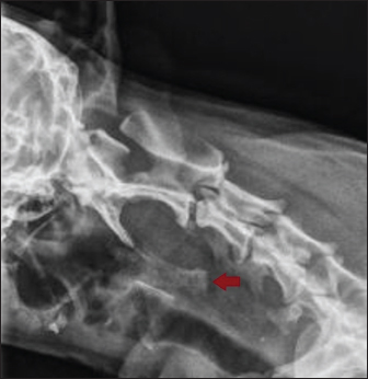
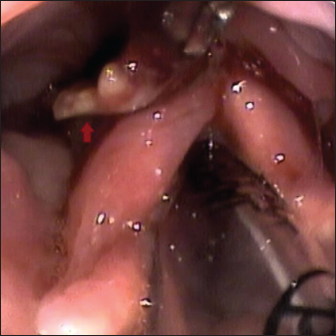
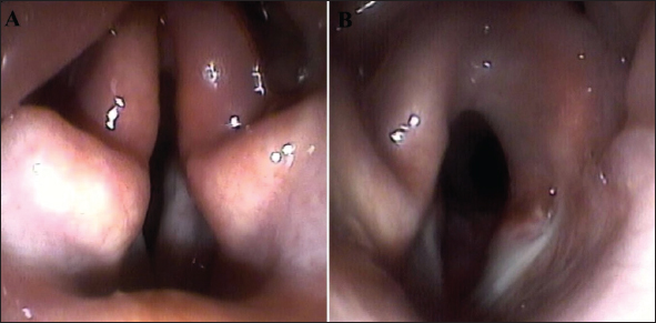
AbstractBackground: Laryngeal paralysis is a major cause of dyspnea in dogs and can be related to several conditions, including trauma; however, it has never been described as a consequence of an esophageal foreign body. Case description: A 14-year-old spayed female mixed-breed dog was presented for acute dyspnea and a bony foreign body in the proximal esophageal sphincter detected on cervical radiographic survey. After foreign body retrieval, the clinical signs persisted, and bilateral laryngeal paralysis was observed endoscopically. A left arytenoid lateralization was performed. Clinical signs promptly resolved without complications, and the dog remained asymptomatic for 18 months postoperatively. Conclusion: Although other etiologies were considered in this dog, inflammation and damage caused by the esophageal foreign body to the laryngeal structures and innervation were considered the most probable cause of the laryngeal paralysis described in this case. Esophageal foreign bodies should therefore be considered a possible contributing cause of laryngeal paralysis in dogs. Keywords: Dog, Foreign body, Laryngeal paralysis IntroductionLaryngeal paralysis is a major cause of upper airway dyspnea in dogs and is often associated with damage to the laryngeal innervation, ranging from the nucleus ambiguous to the vagus and recurrent laryngeal nerve, ultimately failing abduction of the cricoarytenoid dorsalis muscle (Millard and Tobias, 2009; Monnet, 2023). This condition can be congenital or acquired and may be classified as idiopathic or secondary to several diseases, such as polyneuropathy of various etiologies, lead or organophosphate toxicity, retropharyngeal infection, rabies, polyradiculoneuritis, systemic lupus erythematosus, bronchogenic carcinoma, brainstem lesions, and laryngeal myopathy (Salisbury et al., 1990; Burbidge, 1995; Millard and Tobias, 2009; Kvitko-White et al., 2012; Monnet, 2023). The most common acquired form is typical of old, often large-breed dogs, and it seems to be related to a generalized peripheral polyneuropathy, known as geriatric onset laryngeal paralysis polyneuropathy (GOLPP) (Burbidge, 1995; Monnet, 2023). Clinical signs usually occur in bilateral forms in dogs and may vary depending on disease severity. Diagnosis is often based on history and clinical signs and is confirmed by laryngoscopy, while treatment commonly involves arytenoid cartilage lateralization (MacPhail and Monnet, 2001; Monnet, 2023). The long-term outcome for dogs treated surgically is generally good, but it depends on the underlying cause. Aspiration pneumonia is a common and sometimes life-threatening complication that may develop in the perioperative period or long term after surgery (MacPhail and Monnet, 2001; Wilson and Monnet, 2016; Monnet, 2023). Although traumatic events and penetrating injuries have been reported as causes of laryngeal paralysis, to the author’s knowledge, it has never been reported in association with an esophageal foreign body (Camargo et al., 2022; Picavet et al., 2022; Monnet, 2023; Allen et al., 2025). This case report aims to describe the presentation, the diagnosis, the therapeutic approach, and the prognosis of a dog with laryngeal paralysis induced by an esophageal foreign body. Case DetailsA 14-year-old spayed female mixed breed dog, weighing 3.7 kg, was presented to the emergency unit for progressive dyspnea, associated with impaired food prehension and dysphagia, which had developed 7 days earlier. The dog had previously been evaluated at another clinic with suspicion of tracheitis, and nebulization therapy together with force syringe feeding had been prescribed. The dog subsequently went missing for a few days and was later showing severe dyspnea and an inability to take food. Remote medical history was unremarkable, and no dysphonia, exercise intolerance, or hindlimb weakness were reported. Upon clinical examination, mixed inspiratory and expiratory dyspnea was evident, accompanied by stridor and congested mucous membrane; the remainder of the physical examination was within normal limits, and no neurological deficits (e.g., cranial nerves, gait, postural reaction, or spinal reflex abnormalities) were identified. Thoracic radiographs showed neither pulmonary disease nor other abnormalities, such as a mediastinal mass; however, a 2.5-cm-long calcific foreign body was observed in the proximal cervical region, dorsal and caudal to the larynx, presumably located in the proximal esophagus, immediately distal to the upper esophageal sphincter (Figure 1). Blood work was consistent with non-specific systemic inflammation, including neutrophilic leukocytosis and increased C-reactive protein; thyroid profile (total T4, TSH) was within normal limits. The dog was subsequently placed under general anesthesia, intubated, and an endoscopic examination of the esophagus was performed (Pentax EG-2970K). Abundant grass material was observed in the pharynx, straddling the upper esophageal sphincter and extending into the esophageal lumen (Figure 2). After removal of the grass material, a non-obstructive Y-shaped foreign body was identified a few centimeters distal to the sphincter, lodged in the cervical esophagus. It was later presumed to be a chicken sternal keel. Endoscopic removal was attempted but unsuccessfully; therefore, under endoscopic guidance, Carmalt forceps were introduced, allowing partial fragmentation of the foreign body and facilitating its removal. After gradually lightening the anesthetic plane and administering a bolus of doxapram (Respipram®; Uvefarma S.r.l., Italy) at 1.5 mg/kg IV, the patient was awakened and extubated; however, mixed dyspnea and stridor persisted. The dog therefore underwent laryngoscopy, which revealed bilateral laryngeal paralysis with paradoxical movement of the arytenoid cartilages, along with severe mucosal edema and hyperemia (Figure 3-A). Attempts were made to gradually awaken the patient to reassess clinical status and laryngeal motion; however, the dog consistently developed dyspnea whenever awakening was attempted. After discussing with the owner, the dog was placed under general anesthesia, and left arytenoid lateralization by cricoarytenoid suture was performed. Briefly, with the dog in right lateral recumbency, a 2-cm-long skin incision was made ventral to the left jugular vein. During dissection, a subcutaneous hematoma and severe inflammation of the surrounding tissue were observed. After retracting the left thyroid cartilage wing laterally, the cricothyroid junction was separated, and the cricoarytenoid dorsalis muscle was transected, exposing the cricoarytenoid articulation. A 3-0 USP nonabsorbable monofilament suture was placed between the cricoid cartilage and the muscular process of the arytenoid cartilage. Adequate laryngeal opening was verified before tightening the knot by temporarily extubating the patient for laryngoscopic inspection (Figure 3-B). The knot was then secured, and the surgical wound was closed routinely. The dog recovered from anesthesia uneventfully, and no respiratory symptoms were observed after recovery. The patient was discharged after a 24-hour observation period, with instructions for routine wound care. Postoperative clinical rechecks revealed mild wound inflammation, which rapidly resolved with topical honey ointment (Vetramil®; Bfactory Health Products B.V., Rhenen, The Netherlands). No other complication or recurrence of respiratory signs was observed. At 18 months from surgery, the dog remained bright, alert, and asymptomatic, with no evidence of respiratory dysfunction.
Fig 1. Lateral cervical radiographic view of the bony foreign body dorsal to the larynx (red arrow).
Fig 2. Endoscopic view of the larynx with the foreign body (red arrow) before retrieval.
Fig 3. Endoscopic view of the larynx before (A) and after (B) the cricoarytenoid lateralization. DiscussionThis is the first report describing the clinical presentation of a dog with acute laryngeal paralysis, probably caused by a concurrent esophageal foreign body. The main literature databases (e.g., Google Scholar®, Pubmed®) were searched without date range limitations and until 2026, using specific keywords (e.g., “dog” or “canine” or “small animal” and “oesophageal” or “pharyngeal” or “ingestion” and “foreign body” and “laryngeal paralysis”), to identify previous reports of this condition: to the authors’ knowledge, foreign body ingestion has never been reported as a direct cause of laryngeal paralysis in dogs (Camargo et al., 2022; Picavet et al., 2022; Monnet, 2023; Allen et al., 2025). Laryngeal paralysis results from a lack of contraction of the cricoarytenoid dorsalis muscle, leading to impaired abduction of the arytenoid cartilage and subsequent airway obstruction, resulting from damage to the muscle or to its innervation (Monnet, 2023). In small animals, laryngeal paralysis may be congenital (e.g., inherited degeneration) or acquired, and male dogs are reported to be more predisposed to the disease (Millard and Tobias, 2009; Monnet, 2023). Several contributing causes have been described, including chronic endocrine, infectious, or immune-mediated polyneuropathy, trauma, lead or organophosphate toxicity, retropharyngeal infection, rabies, polyradiculoneuritis, systemic lupus erythematosus, bronchogenic carcinoma, brainstem lesions, and laryngeal myopathy. When no cause is identified, the condition may be defined as part of generalized idiopathic polyneuropathy; this is actually the most common form, also known as GOLPP, and it is typical of old large-breed dogs (Salisbury et al., 1990; Burbidge, 1995; Kvitko-White et al., 2012; Monnet, 2023). Among these causes, surgical or traumatic injuries may induce laryngeal paralysis. The recurrent laryngeal nerves run along the trachea in the cervical region, and traumatic injuries at this level may potentially result in nerve damage and subsequent laryngeal paralysis of varying severity (Millard and Tobias, 2009; Camargo et al., 2022; Picavet et al., 2022; Monnet, 2023). In addition to penetrating wounds, both internal and external compression (e.g., endotracheal tube cuff, cervical or mediastinal masses) may impair the microvasculature of the nerve and ultimately cause nerve injury, from neuropraxia, characterized by transient functional loss, to axonotmesis and neurotmesis, which result in severe and potentially irreversible neurologic impairment (Burnett and Zager, 2004; Kikura et al., 2007). Esophageal dysfunctions are reported in dogs with laryngeal paralysis, due to the proximity of the respective innervations and thus trauma to the esophagus and cervical region might cause, in reverse, damage to larynx function (Stanley et al., 2010). The wide hematoma found intraoperatively at the muscle level demonstrates the severity of the trauma caused by the foreign body: in fact, the nerve could have been both directly compressed by the foreign body through the intervening tissues and indirectly involved in the subsequent inflammation and edema. While both hypotheses are possible, direct trauma to the nerve is less probable due to the presence of surrounding tissues that would have protected it; therefore, indirect trauma caused by inflammation and edema is a more realistic hypothesis. Finally, local acute inflammation and edema directly caused by the foreign body in the laryngeal region might have also enhanced the cartilage muscle damage, thus promoting laryngeal paralysis (Millard and Tobias, 2009; Monnet, 2023). Except for the age, which made GOLPP a reasonable differential diagnosis, the signalment (i.e., small size), history, presentation, and clinical findings supported the exclusion of chronic contributing causes, particularly in the absence of chronic symptoms (dysphonia, exercise intolerance, or hindlimb weakness), previous respiratory distress, or hypothyroidism (Millard and Tobias, 2009; Monnet, 2023). In addition, no abnormalities were detected on physical or neurological examination, such as muscle atrophy or deficits in cranial nerves, proprioception, or spinal reflexes, which might have suggested an underlying polyneuropathy and GOLPP. However, no further diagnostic tests (i.e., electromyography) were performed to investigate generalized polyneuropathy or to confirm laryngeal paralysis (Millard and Tobias, 2009). Indeed, the acute presentation in the emergency and visualization of the foreign body guide the clinicians toward emergency management, avoiding time-consuming procedures. Nevertheless, a definitive causal relationship between the esophageal foreign body and the subsequent laryngeal paralysis cannot be established with certainty. Although less likely, since neither risk factors nor signs of idiopathic laryngeal paralysis were evident in the history or at presentation, the patient may have had a pre-existing asymptomatic form of laryngeal paralysis, and the foreign body injury might have simply triggered the worsening of the disease, unmasking the acute clinical signs. As mentioned before, not all potential causes or triggering factors for laryngeal paralysis were completely excluded, both because they were considered less probable and because of the need to spare time in the emergency treatment. Furthermore, laryngeal edema might sometimes clinically mimic laryngeal paralysis and may also impair endoscopic evaluation by limiting arytenoid movement (Monnet, 2023). In addition, the mechanical obstruction caused by the presence of the foreign body within the pharyngeal region, or its indirect compression of the laryngeal airway, might alone have caused the acute dyspnea. However, the persistence of respiratory signs after foreign body removal, the findings on endoscopic evaluation rather than direct visualization, and the presence of paradoxical arytenoid movement supported the diagnosis of laryngeal paralysis rather than simple mucosal edema. Finally, the complete resolution of clinical signs following surgical treatment, even in the long term, suggests that trauma associated with the foreign body was likely the primary predisposing cause of the laryngeal paralysis observed in this case. Surgical treatment was deemed necessary because of the severity and persistence of dyspnea. Unilateral arytenoid cartilage lateralization was performed using a cricoarytenoid suture, which represents the standard surgical technique, as described by White (White, 1989). However, given the acute presentation and the presence of laryngeal edema, less invasive options could have been considered, such as the placement of a laryngeal stent. This approach might have avoided emergency surgery while providing temporary airway stabilization and allowing the laryngeal tissues to recover as edema subsided (Théron and Lahuerta-Smith, 2022). Nevertheless, it is impossible to determine whether temporary stenting would have resulted in the resolution of the laryngeal paralysis, and the presence of a stent might have exacerbated mucosal edema. Another possible option would have been temporary tracheostomy, allowing time for inflammation to decrease and for reassessment of the patient. However, this procedure is associated with a relatively high complication rate, even when intensive management is provided (Nicholson and Baines, 2012). Complications following surgical treatment of laryngeal paralysis range from wound seroma formation (10%) to aspiration pneumonia (8–21%) and recurrence of clinical signs (approximately 33%), with a higher incidence reported in small-breed dogs (MacPhail and Monnet, 2001; Snelling and Edward, 2003; Hammel et al., 2006; Wilson and Monnet, 2016). Despite the risks, both quality of life and overall prognosis are generally good, particularly in the absence of progressive neurological decline in polyneuropathic patients (MacPhail and Monnet, 2001; Hammel et al., 2006; Monnet, 2023). The present case further supports the effectiveness of surgical treatment in trauma-induced laryngeal paralysis, in which an underlying chronic disease is typically absent, and the prognosis may therefore be excellent. As previously mentioned, although alternative causes appeared unlikely in this case, not all differential diagnoses were certainly excluded, and specific tests (e.g., electromyography/electroneurography, magnetic resonance imaging) were not performed because of the emergency setting. Moreover, laryngeal paralysis was diagnosed following general anesthesia for foreign body removal, which might theoretically have impaired arytenoid movement, even though the plane of anesthesia was considered fairly light and doxapram was used to increase arytenoid excursion (Tobias et al., 2004). Attempts were made to gradually awaken the patient before proceeding with surgery; however, no improvement in respiratory distress or arytenoid movement was observed. Nevertheless, given the potential for overdiagnosis of laryngeal paralysis due to laryngeal edema and inflammation, a more conservative approach might alternatively have been considered. Such an approach could have involved medical management of upper airway obstruction (i.e., orotracheal intubation and mechanical ventilation) and close monitoring for clinical improvement, reserving surgery for persistent long-term symptoms. Although theoretically feasible, this strategy might have exposed the patient to life-threatening complications associated with untreated upper airway obstruction and prolonged intensive care, with expensive treatments. Finally, temporary stabilization techniques such as laryngeal stenting or temporary tracheostomy might have allowed clinicians additional time to determine whether laryngeal inflammation and edema were exacerbating a previously subclinical condition or whether true paralysis was the primary cause of respiratory distress. ConclusionThis report describes a case of laryngeal paralysis probably induced by an esophageal foreign body, likely through direct tissue damage and laryngeal inflammation. Retrieval of the foreign body alone was not sufficient to resolve the condition, and surgical arytenoid lateralization was required and proved effective in completely relieving respiratory distress, including in the long term. Foreign body ingestion should therefore be considered as a potential differential cause of laryngeal paralysis. Conflict of interestThe authors declare there is no conflict of interest. FundingThis research received no specific grant. Authors’ contributionsLuca Ciammaichella: case management, manuscript – original draft, manuscript – review and editing. Jessica Campanerut: case management, manuscript – original draft, manuscript – review and editing. Stefania Golinelli: case management, manuscript review, and editing. Veronica Cola: case management, manuscript review, and editing. Stefano Zanardi: case management, manuscript review, and editing. Armando Foglia: case management, manuscript review, and editing. Marco Pietra: case management, manuscript review, and editing. Luciano Pisoni: case management, manuscript review, and editing. Sara Del Magno: case management, supervision, manuscript review, and editing. Data availabilityAll data supporting the findings of this study are available within the manuscript. Ethical statementThe study did not fall within the application areas of Italian Legislative Decree 26/2014, which governs the protection of animals used for scientific or educational purposes; therefore, ethical approval was waived for this report. The dog was treated according to the current standards. The owner signed a written informed consent. ReferencesAllen, A., Rozanski, E., Destefano, I., Kent, M. and Kudej, R. 2025. Bilateral laryngeal paralysis following single‐staged, bilateral total ear canal ablation and lateral bulla osteotomy in a cocker spaniel. Vet. Rec. Case Rep. 13(1), e1095. Burbidge, H.M. 1995. A review of laryngeal paralysis in dogs. Br. Vet. J. 151, 71–82. Burnett, M.G. and Zager, E.L. 2004. Pathophysiology of peripheral nerve injury: a brief review. Neurosurg. Focus 16(5), 1–7. Camargo, J.F., Teixeira, L.G., Trindade-Gerardi, A.B., Dos Santos, B.S., Da Rosa, M.P., Paim, M.G. and Contesini, E.A. 2022. Laryngeal paralysis following endotracheal intubation in a dog: a case report. Top. Companion Anim. Med. 48, 100635. Hammel, S.P., Hottinger, H.A. and Novo, R.E. 2006. Postoperative results of unilateral arytenoid lateralization for treatment of idiopathic laryngeal paralysis in dogs: 39 cases (1996-2002). J. Am. Vet. Med. Assoc. 228, 1215–1220. Kikura, M., Suzuki, K., Itagaki, T., Takada, T. and Sato, S. 2007. Age and comorbidity as risk factors for vocal cord paralysis associated with tracheal intubation. Br. J. Anaesth. 98(4), 524–530. Kvitko-White, H., Balog, K., Scott-Moncrieff, J.C., Johnson, A. and Lantz, G.C. 2012. Acquired bilateral laryngeal paralysis associated with systemic lupus erythematosus in a dog. J. Am. Anim. Hosp. Assoc. 48(1), 60–65. MacPhail, C.M. and Monnet, E. 2001. Outcome of and postoperative complications in dogs undergoing surgical treatment of laryngeal paralysis: 140 cases (1985–1998). J. Am. Vet. Med. Assoc. 218(12), 1949–1956. Millard, R.P. and Tobias, K.M. 2009. Laryngeal paralysis in dogs. Compend. Contin. Educ. Vet. 31(5), 212–219. Monnet, E. 2023. Laryngeal paralysis.In Small Animal Soft Tissue Surgery. Monnet. and E Hoboken, NJ: John Wiley and Sons, Inc, pp: 459–72. Nicholson, I. and Baines, S. 2012. Complications associated with temporary tracheostomy tubes in 42 dogs (1998 to 2007). J. Small. Anim. Pract. 53(2), 108–114. Picavet, P., Hamon, M., Etienne, A.L., Guieu, L.V., Claeys, S., Billen, F. and Noël, S. 2022. Laryngeal paralysis secondary to cervical bite injuries in five dogs. New. Zeal. Vet. J. 70(2), 109–118. Salisbury, S.K., Forbes, S. and Blevins, W.E. 1990. Peritracheal abscess associated with tracheal collapse and bilateral laryngeal paralysis in a dog. J. Am. Vet. Med. Assoc. 196, 1273–1275. Snelling, S.R. and Edward, G.A. 2003. A retrospective study of unilateral arytenoid lateralization in the treatment of laryngeal paralysis in 100 dogs (1992-2000). Aust. Vet. J. 81, 464. Stanley, B.J., Hauptman, J.G., Fritz, M.C., Rosenstein, D.S. and Kinns, J. 2010. Esophageal dysfunction in dogs with idiopathic laryngeal paralysis: a controlled cohort study. Vet. Surg. 39(2), 139–149. Théron, M.L. and Lahuerta-Smith, T. 2022. Laryngeal silicone stent as a treatment option for laryngeal paralysis in dogs: a preliminary study of 6 cases. J. Vet. Sci. 23(4), 58. Tobias, K.M., Jackson, A.M. and Harvey, R.C. 2004. Effects of doxapram hydrochloride on laryngeal function in healthy dogs and dogs with laryngeal paralysis. Vet. Surg. 33(1), 77–81. White, R.A.S. 1989. Arytenoid lateralization: an assessment of technique, complications and long-term results in 62 dogs with laryngeal paralysis. Vet. Surg. 18, 72. Wilson, D. and Monnet, E. 2016. Risk factors for the development of aspiration pneumonia after unilateral arytenoid lateralization in dogs with laryngeal paralysis: 232 cases (1987–2012). J. Am. Vet. Med. Assoc. 248, 188–194. | ||
| How to Cite this Article |
| Pubmed Style Ciammaichella L, Campanerut J, Golinelli S, Cola V, Zanardi S, Foglia A, Pietra M, Pisoni L, Magno SD. Laryngeal paralysis associated with esophageal foreign body in a dog. Open Vet. J.. 2026; 16(8): 5525-5530. doi:10.5455/OVJ.2026.v16.i8.45 Web Style Ciammaichella L, Campanerut J, Golinelli S, Cola V, Zanardi S, Foglia A, Pietra M, Pisoni L, Magno SD. Laryngeal paralysis associated with esophageal foreign body in a dog. https://www.openveterinaryjournal.com/?mno=314121 [Access: August 08, 2026]. doi:10.5455/OVJ.2026.v16.i8.45 AMA (American Medical Association) Style Ciammaichella L, Campanerut J, Golinelli S, Cola V, Zanardi S, Foglia A, Pietra M, Pisoni L, Magno SD. Laryngeal paralysis associated with esophageal foreign body in a dog. Open Vet. J.. 2026; 16(8): 5525-5530. doi:10.5455/OVJ.2026.v16.i8.45 Vancouver/ICMJE Style Ciammaichella L, Campanerut J, Golinelli S, Cola V, Zanardi S, Foglia A, Pietra M, Pisoni L, Magno SD. Laryngeal paralysis associated with esophageal foreign body in a dog. Open Vet. J.. (2026), [cited August 08, 2026]; 16(8): 5525-5530. doi:10.5455/OVJ.2026.v16.i8.45 Harvard Style Ciammaichella, L., Campanerut, . J., Golinelli, . S., Cola, . V., Zanardi, . S., Foglia, . A., Pietra, . M., Pisoni, . L. & Magno, . S. D. (2026) Laryngeal paralysis associated with esophageal foreign body in a dog. Open Vet. J., 16 (8), 5525-5530. doi:10.5455/OVJ.2026.v16.i8.45 Turabian Style Ciammaichella, Luca, Jessica Campanerut, Stefania Golinelli, Veronica Cola, Stefano Zanardi, Armando Foglia, Marco Pietra, Luciano Pisoni, and Sara Del Magno. 2026. Laryngeal paralysis associated with esophageal foreign body in a dog. Open Veterinary Journal, 16 (8), 5525-5530. doi:10.5455/OVJ.2026.v16.i8.45 Chicago Style Ciammaichella, Luca, Jessica Campanerut, Stefania Golinelli, Veronica Cola, Stefano Zanardi, Armando Foglia, Marco Pietra, Luciano Pisoni, and Sara Del Magno. "Laryngeal paralysis associated with esophageal foreign body in a dog." Open Veterinary Journal 16 (2026), 5525-5530. doi:10.5455/OVJ.2026.v16.i8.45 MLA (The Modern Language Association) Style Ciammaichella, Luca, Jessica Campanerut, Stefania Golinelli, Veronica Cola, Stefano Zanardi, Armando Foglia, Marco Pietra, Luciano Pisoni, and Sara Del Magno. "Laryngeal paralysis associated with esophageal foreign body in a dog." Open Veterinary Journal 16.8 (2026), 5525-5530. Print. doi:10.5455/OVJ.2026.v16.i8.45 APA (American Psychological Association) Style Ciammaichella, L., Campanerut, . J., Golinelli, . S., Cola, . V., Zanardi, . S., Foglia, . A., Pietra, . M., Pisoni, . L. & Magno, . S. D. (2026) Laryngeal paralysis associated with esophageal foreign body in a dog. Open Veterinary Journal, 16 (8), 5525-5530. doi:10.5455/OVJ.2026.v16.i8.45 |


