





| Research Article | ||
Open Vet. J.. 2026; 16(8): 5256-5265
Open Veterinary Journal, (2026), Vol. 16(8): 5256–5265 Research Article Comparative evaluation of flank laparotomy approaches in sheep and goatsWafi Adel Al-Antaif1, Mohamed Karam Zabady1, Zakriya Ali Al Mohamad1, Ahmed M. A. Meligy1, Sayed Fathi El-Hawari1*, Sohaila F. El-Hawary2, Nabil Misk3 and Amal Galal Abouelella41Department of Clinical Sciences, College of Veterinary Medicine, King Faisal University, Al Ahsa, Saudi Arabia 2Department of Biology, College of Science, Jazan University, Jazan, Kingdom of Saudi Arabia 3Department of Surgery, Anesthesiology, and Radiology, Faculty of Veterinary Medicine, Assiut University, Asyut, Egypt 4Department of Surgery, Faculty of Veterinary Medicine, Sadat City University, Menofia, Egypt *Corresponding Author: Sayed Fathi El-Hawari. Department of Clinical Sciences, College of Veterinary Medicine, King Faisal University, Al Ahsa, Saudi Arabia. Email: selhawari [at] kfu.edu.sa Submitted: 15/03/2026 Revised: 15/06/2026 Accepted: 28/06/2026 Published: 08/08/2026 © 2025 Open Veterinary Journal
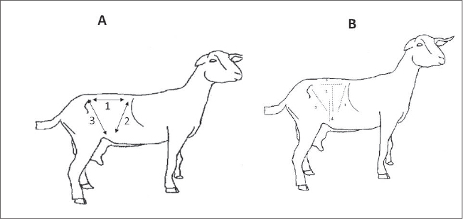
AbstractBackground: Surgical approaches in small ruminants are commonly performed in daily clinical practice. During laparotomy, the gold standard for each surgeon is the proper approach for each abdominal organ through an accurate site of incision with minimal traumatic injuries. Aim: To describe the morphometric character of the flank region and to investigate different sites of incision at both right and left flank regions, with detection of abdominal organ accessibility from each incision site. Methods: We selected 15 sheep and 15 goats for the investigations. Five sheep and five goats were used to determine the flank region dimensions. Five incisions were performed at the right and left flank regions: cranial, middle, caudal, low, and transverse paravertebral flank incisions. At each laparotomy incision, organs directly observed at the site of incision, exteriorized and manipulated from the incision site, and organs palpated from the incision were recorded. Differences between sheep and goats were also recorded. Results: Morphometric measurements of the flank regions were higher in sheep than in goats. The spleen may be approached through a left cranial flank incision, the rumen may be accessed through a left middle flank incision. Low right and left flank incisions provided visualization of the gravid uterus. Left and right transverse paravertebral flank incisions may provide a better approach for the left and right kidneys, respectively. Free access to the duodenum may be achieved from a right cranial flank incision, whereas the caecum, jejunum, and colon may be directly seen following right middle flank laparotomy. Conclusion: Choosing an accurate site for incision in the current study may provide higher visualization with less tissue manipulation. Sheep differ from goats in the size of the abdominal cavity and in access to abdominal organs from right and left flank laparotomy. Keywords: Flank region, Goats, Laparotomy, Sheep, Surgical incision. IntroductionSheep and goats are significant ruminants in temperate and tropical agriculture. They provide fibers, meat, hides, and milk. Sheep and goats are better adapted to arid tropics than cattle, probably because of their superior water and nitrogen economy (Nair et al., 2021; Mahgoub et al., 2025). Numerous digestive complications, such as traumatic reticulitis, ruminal impaction, omasal impaction, ruminal distension, bezoar formation, abomasal displacement, gastrointestinal displacement correction, vagal indigestion, and abomasal ulcers, can be resolved via laparotomy (Navarre et al., 2012; Fubini and Ducharme, 2016; Bayne and Edmondson, 2021; Martin et al., 2021). Moreover, laparotomy could be used to treat some intestinal conditions (Fubini and Ducharme, 2016). In addition, laparotomy can resolve obstetrical problems in ruminants, such as dystocia, pregnancy toxemia, uncorrectable uterine torsion, and uterine tear (Kumar et al., 2013; Fubini and Ducharme, 2016; Hasan et al., 2017; Cowley et al., 2023). Several factors, such as species, facilities for restraint, available anesthesia, type of surgery, location of the affected organ within the abdomen, and surgeon preference, govern the selection of a particular site in the flank regions for laparotomy incisions. Flank laparotomy is generally performed on standing animals under the effect of local analgesia and sedatives, but it can be performed in small ruminants with lateral recumbency (Abubakar et al., 2014; Kabkia et al., 2021; Elane et al., 2023). During laparotomy and laparoscopy, the gold standard for each surgeon is a proper approach for each abdominal organ through an accurate site of incision and minimal traumatic injuries. This study aimed to comprehensively evaluate different right and left flank laparotomy incision sites in sheep and goats, with particular emphasis on organ accessibility, surgical field visualization, and the degree of tissue manipulation required. The clinical relevance of this work lies in providing evidence-based guidance for selecting the most appropriate flank incision in small ruminants to facilitate abdominal surgical procedures, improve operative access, and minimize unnecessary tissue trauma. Materials and MethodsAnimalsThirty crossbreed sheep (n=15) and crossbreed goats (n=15) were selected for the investigations. Animals were randomized using the RAND function in Microsoft Excel. The ages of the animals ranged between 2 and 4 years, and their average body weights were 41.9 ± 4.2 kg (sheep) and 26.3 ± 2.9 kg (goats). Before the experimental surgical incisions were made, 10 animals (five sheep and five goats) were used to determine the dimensions of the flank region (Fig. 1) by measuring three lines (cm) with a measuring tape. The first line was horizontal, extending from the head of the last rib to the tuber coxae point. The second line was oblique, extending downward and backward from 1st lumbar transverse process to the stifle joint level ventrally. The third line was oblique, extending downward and forward from the tuber coxae to the stifle joint level. All thirty animals underwent two operations in a crossover design with a 1-month interval. At the first time, they were subjected to different forms of left flank laparotomies, while at the second occasion, they were subjected to different forms of right flank laparotomies.
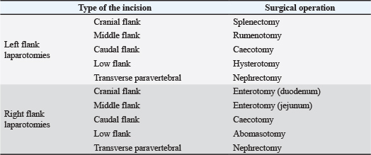
Fig. 1. Anatomic description of the flank region (A). Whereas 1: Line extending from the last rib to the tuber coxae, 2: Oblique line extending from the 1st lumbar transverse process to the level of the stifle joint, and 3: Oblique line extending from the tuber coxae and directed cranioventrally to the level of the stifle joint. (B) Displaying the sites of incisions in the flank region. 1: Cranial flank incision; 2: Middle flank incision; 3: Caudal flank incision; 4: Low-flank incision; 5: Transverse paravertebral flank incision. Flank laparotomiesFive incisions were made at the right and left flank regions, whereas each incision was conducted on six females (three sheep and three goats) as follows (Fig. 1) (Table 1). Cranial flank incisionThe site of the incision was 2 cm ventral to the lumbar transverse process and 2–3 cm caudal to the last rib and directed downward with a slightly caudal direction. The incision length was 15 cm. Middle flank incisionThe incision was made at equidistance between the tuber coxae and the last rib, 3 cm ventral to the transverse processes of the lumbar vertebrae, and directed ventrally. The length of the skin incision was 15 cm. Caudal flank incisionThe incision site started 2 cm ventral to the tuber coxae and extended cranioventrally with a length of 15 cm. Low flank incisionThe skin incision was started 6 cm ventral to the lumbar transverse processes and 9 cm caudal to the last rib. The incision was directed downward with a length of 15–20 cm. Table 1. Various surgical operations performed at the left and right flank regions.
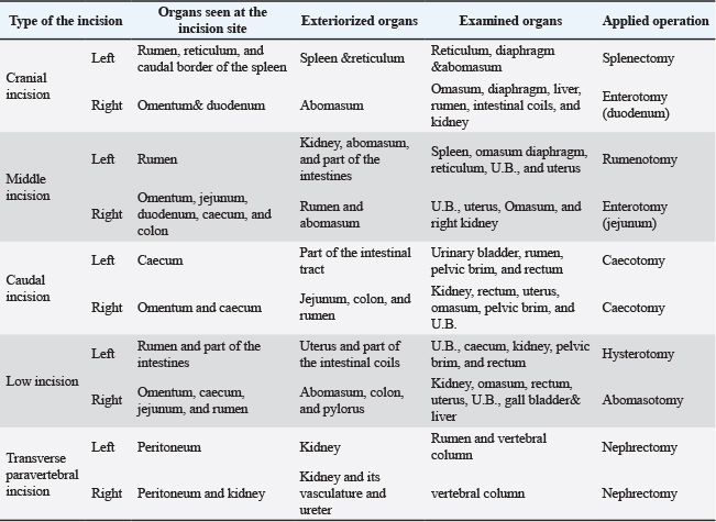
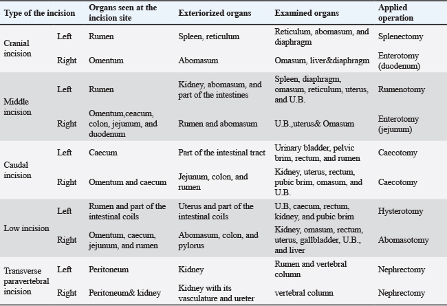
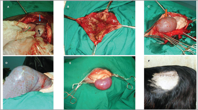
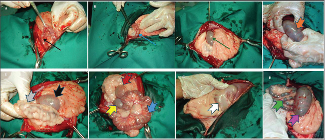
Transverse paravertebral flank incisionThis incision started 3 cm ventral to the transverse process of the 3rd lumbar vertebra and was directed caudally in a horizontal manner with a length of 10 cm. Surgical proceduresAll animals were fasted for 24 hours before surgery. They were sedated using 2% xylazine HCL (Adwia company- Xylaject) at a dose of 0.2 mg/kg in sheep (Mekonnen et al., 2023) and 0.02 mg/kg in goats (Biobaku et al., 2016). The right and left flank regions were blocked using 1% lidocaine HCL (30 ml) in an inverted L manner. The right and left flank regions were prepared aseptically, and the animals were kept in lateral recumbency during the operation. The skin and abdominal muscles were incised routinely, and the peritoneum was punctured using a scalpel. The incision was widened upward and downward using scissors. At each laparotomy incision, organs could be directly observed at the incision site, surgically exteriorized and easily manipulated from the incision site, and organs palpated from the incision site without exteriorization were recorded. One operation was conducted through each laparotomy incision (Table 1). The current study was qualitative, and all the observations were reported in a descriptive manner. Abdominal organs and abdominal muscles were sutured using Catgut No. 0 and 1, respectively, with a rounded atraumatic needle. The skin was closed using silk No. 1 with a cutting needle and horizontal mattress suture pattern. Each animal was given specific postoperative care (Hexasol LA, 300 mg oxytetracycline/ml + 20 mg flunixine/ml, single IM injection 1 ml/10 kg B.Wt, Norbrook, UK), and the follow-up period was extended up to 6 weeks. Skin stitches were removed 10 days after surgery. Ethical approvalThe institutional ethical committee previously approved all procedures in the current study (approval no. KFU - ETHICS3657). ResultsMorphometric measurements for the flank regions revealed that line one was 22.4 ± 3.6 and 20.4 ± 1.6 cm in sheep and goats, respectively; the second oblique line was 29.6 ± 3.6 and 26.9 ± 2.0 cm in sheep and goats, respectively; and the third oblique line was 32.3 ± 3.3 and 29.6 ± 2.8 cm in sheep and goats, respectively. No significant differences were observed in the morphometric measurements of the flank region between sheep and goats. Organs seen at the site of incision, exteriorized, and examined at each incision site, in addition to the applied operation from both left and right flank laparotomies, were tabulated in goats (Table 2) and sheep (Table 3). Cranial flank incisionAt left laparotomy, the organs seen at the site of incision in goats were the parietal surface of the dorsal sac of the rumen, the parietal surface of the reticulum, and the caudal border of the spleen (about 3 cm from its caudal border) (Fig. 2). However, gentle pressure on the rumen toward the abdominal cavity provided good spleen visualization. In sheep, only the parietal surface of the dorsal sac of the rumen was seen. The organs that can be exteriorized in both small ruminants were the spleen from the cranial part of the incision and the reticulum from the cranioventral part of the incision. The organs that can be examined in both animals were the reticulum cranioventrally, the diaphragm cranially, and the abomasum ventrally. Splenectomy was performed without any postoperative complications. At right laparotomy, the organs seen at the site of incision in goats were the omentum, duodenum dorsally, and part of the abomasum ventrally (Fig. 3). In sheep, the omentum was the only structure seen at this site of incision. In both animals, the only organ that could be exteriorized was the abomasum from the ventral aspect of the incision site. The organs which can be examined were the omasum (hard handball structure), liver cranially, and diaphragm at the most cranial part of the incision in both animals. However, in goats, the surgeon’s hand can be extended caudoventrally to examine the rumen, intestinal coils caudally, and the kidney dorsally. In sheep, the organs cannot be recognized well due to the fatty omentum. Enterotomy was conducted on the duodenum, and no postoperative complications were recorded. Table 2. Organs seen, exteriorized, examined, and applied operation at different incisions of left and right flank laparotomies in goats.
Middle flank incisionIn both sheep and goats, the organ visualized at left laparotomy was the parietal surface of the rumen only. The exteriorized organs in both animals were the left kidney from the caudo-dorsal part of the incision, part of the intestine from the caudal part of the incision, and the abomasum from the ventral part of the incision. Organs that could be examined in both animals were the spleen cranio-dorsally, the diaphragm cranially, the omasum cranially and somewhat to the midline, the reticulum cranioventrally, and the urinary bladder and uterus caudo-dorsally. Rumenotomy was easily performed in both animals. At right laparotomy, the organs noticed at the site of incision were the omentum, duodenum, and caecum dorsally, and the jejunum and colon ventrally in goats. The presence of a highly fatty omentum at the incision site in sheep caused some type of hindrance, but the previously described organs in goats can also be located after retraction of the thick omentum in sheep. Organs that could be exteriorized in both animals were the abomasum from the cranio-ventral aspect of the incision and the rumen ventrally. Organs palpated in goats were the urinary bladder and uterus caudo-dorsally, omasum cranially, and right kidney cranio-dorsally, while in sheep, the same organs could be palpated except the right kidney. Enterotomy was performed with no obvious complications. Caudal flank incisionAt left laparotomy, only the caecum was viewed in both sheep and goats. The exteriorized organs were intestinal coils and the caecum from the caudo-ventral part of the incision. The organs investigated in both animals were rumen cranially, urinary bladder and pelvic brim caudally, and rectum caudo-dorsally. Typhlotomy was easily performed after proper exteriorization of the caecum. The only recorded complication was the presence of skin stretching at the dorsal part of the incision site after the removal of skin stitches. Table 3. Organs seen, exteriorized, examined, and applied operation at different incisions of left and right flank laparotomies in sheep.
At right laparotomy, the omentum and caecum were the organs spotted at the site of incision in both animals. The organs that could be projected to sit outside the incision were the rumen from the cranio-ventral aspect and the jejunum and colon from the ventral aspect of the incision site. Organs palpated were the kidney cranio-dorsally, urinary bladder, uterus, rectum, and pelvic brim caudally, and omasum cranially and toward the midway. Caecotomy was performed as in the left caudal flank laparotomy. Low flank incisionAt left laparotomy, the parietal surface of the ventral sac of the rumen and part of the intestinal coils (jejunum) were observed in both sheep and goats. Outward organs included the uterus and parts of the intestinal coils from the caudal aspect of the incision. The organs checked out were the caecum dorsally, urinary bladder caudally, pelvic brim and rectum caudo-dorsally, and kidney cranio-dorsally. Hysterotomy was easily performed through this incision. At right laparotomy, organs marked through this approach in both animals were omentum, caecum cranially, jejunum in the middle, and rumen ventrally. The organs that can be exteriorized were the abomasum and pylorus from the cranial aspect of the incision and the colon from the caudal aspect of the incision site. The right kidney was examined cranio-dorsally, the omasum cranially and to the midline, the liver and gall bladder cranially and adhering to the costal arch, and the rectum, uterus, and urinary bladder caudo-dorsally. Abomasotomy was performed without any postoperative complications. Transverse paravertebral flank incisionAt left laparotomy, the peritoneum was distinguished. The left kidney from the medial site of incision, with its vasculature and left ureter, was exteriorized. The dorsal sac of the rumen was checked ventro-medially and the vertebral column dorsally. Nephrectomy was completed through this incision without any postoperative complications. At right laparotomy, the peritoneum and the right kidney were observed. The organs exteriorized through incision were the right kidney with its vasculature and the right ureter. The ventral aspect of the vertebral column was the organ surveyed. Right nephrectomy was maneuvered more easily than left nephrectomy. DiscussionLaparotomy is indicated for resolving several digestive, obstetrical, and urinary dilemmas. In the current study, the dimensions of the flank regions revealed that sheep have larger dimensions than goats. In addition, previous comparative morphometric and osteometric studies have demonstrated that sheep (Ovis aries) generally exhibit larger metric dimensions of various body regions and skeletal elements than goats (Capra hircus), although they do not specifically measure flank width; they documented measurable differences in body size structure between sheep and goats (Lloveras et al., 2022; Jeanjean et al., 2023). In the current study, all laparotomies were performed in lateral recumbency under the effect of local analgesia and a tranquilizer (Abdullah et al., 2025).
Fig. 2. Left flank laparotomy approaches in sheep and goats and surgical operation from each incision site. A: The dorsal sac of the rumen (black arrow), the reticulum (green arrow), and the caudal border of the spleen (blue arrow) were displayed at the site of left cranial flank incision in goats. B: Rumenotomy from the left middle flank incision. C: Left caudal flank incision with caecum at the site of incision (orange arrow). D: Left low flank incision from which the gravid uterus was exteriorized for cesarean section. E: Left paravertebral transverse flank incision and exteriorized left kidney. F: Sutured left paravertebral transverse flank incision in a goat. Five laparotomy incisions were made at the left and right flank regions. Four of them were described collectively in ruminants by Singh et al. (2019) and the 5th transverse paravertebral incision was described previously by El-Hawari et al. (2010) to visualize the kidney. The left cranial flank incision was performed parallel and caudal to the last rib, 2 cm apart, and ventral to the lumbar transverse processes by 2 cm and was convenient for splenectomy. Das et al. (2023) previously mentioned a similar surgical approach. Moreover, the performance of the incision 2 cm posterior to the last rib retains enough muscles and skin for proper incision closure and provides good visualization for spleen handling with less trauma. The caudal border of the spleen can be noticed in goats through the left cranial flank incision, but it is difficult to observe in sheep. This may be because goats generally have a narrower abdominal cavity and a slightly different rumen contour, which can result in a less cranially emphasized splenic position compared with sheep. Organs exteriorized or examined at this site of incision in sheep and goats followed the same anatomical considerations mentioned by Dyce et al. (2017). A right cranial flank incision was made to access the liver, gall bladder, hepatic ducts, and portal vein (Singh et al., 2019). In sheep, a high fatty omentum hindered the surgeon from localizing the underlying organs, but the omentum and duodenum were easily seen in goats (König and Liebich, 2014; Dyce et al., 2017). In addition, the descending duodenum was horizontally observed through the incision site. Belge et al. (2002) previously performed experimental duodenal cannulation in sheep from a right cranial flank incision. The liver could be palpated and examined in goats but could not be exteriorized due to its strong ligaments and diaphragmatic attachment (Dyce et al., 2017). The rumen and intestinal coils were also recognized easily in goats, but it was difficult in sheep due to the fatty omentum. Abomasum is located in the abdominal floor (Smith and Sherman, 2022). Omasum examined and felt a hard, handball-sized structure between ribs VII and X through the incision’s ventral half.
Fig. 3. Right flank laparotomy in small ruminants. A: Right cranial flank incision showing the duodenum dorsally (black arrow). B: Right middle flank incision with the omentum (under the surgeon’s left hand), duodenum (blue arrow), and caecum (pointed by the tissue forceps) appeared dorsally. C: Right caudal flank incision and organs checked were omentum and caecum (green arrow). D: Visualization of the caecum (orange arrow) from the right middle flank incision after fatty omentum retraction in sheep. E: Right caudal flank incision with caecum (black arrow) and colon (gray arrow) exteriorized from the incision site. F: Right caudal flank incision with caecum (yellow arrow), rumen (red arrow), and jejunum (blue arrow) exteriorized from the site of incision. G and H: Right low-flank incision in which the caecum is seen directly at the site of incision (rose arrow) while the abomasum and pylorus (white arrow) are exteriorized from the cranial aspect of the incision, and the colon from the caudal aspect of the incision (green arrow). In small ruminants, rumenotomy was performed by left middle flank incision, and exploratory laparotomy could be accomplished. However, many authors have done the incision cranial to the midway point to facilitate easy access to the reticulum in large ruminants (Hendrickson and Baird, 2013; Weaver et al., 2018). While in the present study in small ruminants, the reticulum may be examined through a middle left flank approach due to the smaller size of the animal’s body cavity than in large ruminants. Foreign body reticulitis is less common in small ruminants due to their selective feeding (Zakir and Abdo, 2022). Only the rumen was seen at the middle left flank incision in sheep and goats (Hendrickson and Baird, 2013; Dyce et al., 2017). In general, the middle left flank approach provides sufficient visualization and a good approach to the rumen (Singh et al., 2019). The right middle flank incision was performed for enterotomy as described previously by Sylvain and Fecteau (2018) and Lozier and Niehaus (2025). Also, Green and Husband (1996) used a 30-cm laparotomy incision in the mid-right flank of a dairy cow to access the caecum. The current study in small ruminants revealed the presence of omentum, caecum dorsally, and jejunum and colon at the site of incision. These findings were consistent with those previously mentioned by Hendrickson and Baird (2013). Exteriorization of the omasum was performed as it is located cranioventrally to the incision site, limited by the costal arch cranially and the ventral wall of the abdominal cavity ventrally. However, omasal surgical exteriorization was previously described by Abdullah et al. (2025) through right pre-umbilical paramedian incision in goats. In the present study, the omasum, liver, right kidney, urinary bladder, uterus, and rumen were felt and examined for any abnormalities through this incision. In sheep, the presence of a large amount of peri-renal fat may interfere with its palpation and examination. The left caudal flank incision was previously described by Singh et al. (2019) for cystotomy operation in male large ruminants. However, in the current study, we could not exteriorize the empty urinary bladder to the incision site (Dyce et al., 2017). In addition, Fubini and Ducharme (2016) stated that ruminal gas distension may hinder bladder visualization in lateral recumbency. The intestinal coils were easily exteriorized through this site of incision, as described previously by Weaver et al. (2018). The observed stretching of the dorsal skin region at this site of incision after healing may be due to the presence of the tuber coxae, which causes skin distraction in this area. The caudal right flank incision was performed as a second trial to reach the urinary bladder. However, due to the same previously mentioned reasons in the caudal left flank incision, the trials from the right caudal flank incision also failed. On the contrary, Singh et al. (2019) stated that the urinary bladder may be reached through the caudal flank incision. Organs seen at the site of incision were the omentum and the caecum, through which a caecotomy was performed (Steiner, 2004). The recognition of the omasum was marked by its hard consistency and considerable size. Despite the description of this site of incision by many authors (Misk et al., 2006; Hendrickson and Baird, 2013; Singh et al., 2019) for the conduction of cesarean section, they preferred the left side over the right side to avoid evisceration of the intestinal coils. However, this type of approach was performed on non-pregnant animals. Low right and left flank incisions were previously described in ruminants for cesarean section (Misk et al., 2006; Hendrickson and Baird, 2013; Fubini and Ducharme, 2016; Singh et al., 2019). However, the current study revealed that the left low flank incision was preferred over the right low flank incision to avoid intestinal coil evisceration at the right flank side. The organs seen, exteriorized, and examined in the current study at these two incisions follow the arrangement of organs in the abdominal cavity of small ruminants described previously by Dyce et al. (2017). Moreover, cesarean delivery in small ruminants is better done in lateral recumbency than in standing position, as ewe and doe will not stand as quietly as most cows (Matthews, 2002). The left transverse paravertebral flank incision was previously described by Mulon and Desorchers (2005)for nephrectomy using a retroperitoneal technique. Peritoneum wasn’t incised by this technique, so the incidence of peritonitis was not expected by this approach. The left kidney (floating kidney) was exteriorized as described by Dyce et al. (2017). However, it required a lot of manipulation to be grasped (Mulon and Desorchers, 2005). The right transverse paravertebral flank incision was performed as in the left side approach, but access to the right kidney was found easier than the left kidney through an incision starting 3 cm ventral to the 2nd lumbar vertebra. This may be due to the normal anatomical location of the right kidney in the liver impression between the last rib and 2nd lumbar vertebra. In addition, the right kidney was superficially located compared to the left one, which was found more medially. This result is parallel to that mentioned by Mulon and Desrochers (2005). ConclusionThe current descriptive study in small ruminants revealed that the spleen can be approached from a left cranial flank incision with a subsequent splenectomy operation, while the rumen and rumenotomy may be conducted from a left middle flank incision. Low right and left flank incisions appeared to provide better visualization of the gravid uterus for cesarean delivery. The left and right transverse paravertebral flank incisions appeared to provide a better approach for the left and right kidney, respectively. An easy access to the duodenum may be achieved from a right cranial flank incision, while the caecum, jejunum, and colon may be directly seen after a right middle flank laparotomy with exteriorization of the omasum from such incision. Moreover, a caecotomy operation may be performed easily from the right caudal flank incision. As the current work is a preliminary study, further investigations for access to different abdominal viscera via laparoscopy from the same aforementioned sites of incisions are required in future studies. AcknowledgmentThis work was supported by the Deanship of Scientific Research, Vice Presidency for Graduate Studies and Scientific Research, King Faisal University, Saudi Arabia [Grant No. KFU262949]. Conflict of interestThe authors have no conflicts of interest to declare regarding the publication of this article. FundingThis work was supported by the Deanship of Scientific Research, Vice Presidency for Graduate Studies and Scientific Research, King Faisal University, Saudi Arabia [Grant No. KFU262949]. Authors’ contributionsAll authors equally contributed to this study. Data availabilityAll data are valid upon reasonable request from the corresponding author. ReferencesAbdullah, Z., El-Hawari, S.F., Almubarak, A.I., Abdel- Raheem, S.M., Marzok, M. and Abouelella, A.G. 2025. Investigation of different pre-umbilical surgical incisions for ventral abdominal celiotomy in sheep and goats. World. Vet. J. 15(4), 989–996; doi:10.54203/scil.2025.wvj100 Abubakar, A.A., Andeshi, R.A., Yakubu, A.S., Lawal, F.M. and Adamu, U. 2014. Comparative evaluation of midventral and flank laparotomy approaches in goats. J. Vet. Med. 920191; doi:10.1155/2014/920191 Bayne, J.E. and Edmondson, M.A. 2021. Diseases of the gastrointestinal system. In Sheep, goat, and cervid medicine. Elsevier, pp: 63–96; doi:10.1016/B978-0-323-62463-3.00014-1 Belge, A., Bakir, B. and Özcelik, A. 2002. A technique of duodenal cannulation in sheep. Small Ruminant Res. 44, 167–171; doi:10.1016/S0921-4488(02)00052-4 Biobaku, K.T., Agaie, B.M., Onifade, K.I., Odetokun, I.A., Okediran, B.S., Ameen, S.A., Ismaila, M.S., Adam, M. and Raji, L.O. 2016. Effects of xylazine on physiological and biochemical parameters of Sahel bucks exposed to twenty-eight hours road transportation. Sokoto J. Vet. Sci. 14(2), 43–52; doi:10.4314/sokjvs.v14i2.6 Cowley, J., Stockler, J. and Maxwell, H. 2023. A review of small ruminant cesarean section: case selection, surgical techniques, care of the neonates, and postoperative care of the dam. Clin. Theriogenol. 15, 9620; doi:10.58292/CT.v15.9620 Das, B.C., Das, A., Paul, T., Sarker, D., Sutradhar, B.C. and Siddiki, A.M. 2023. Anaesthetics and surgical evaluation of splenectomy in calves. Bangladesh. J. Vet. Anim. Sci. 11(2), 18–23; doi:10.60015/bjvas.v11i2.217 Dyce, K.M., Sack, W.O. and Wensing, C.J.G. 2017. Textbook of veterinary anatomy, Elane, G.L., Portela, D.A., Hobbs, K.J., Bauck, A.G. and Biedrzycki, A.H. 2023. Sedated cesarean sections are associated with increased kid survival compared with general anesthesia in goats: a retrospective cohort of 45 cases (2011–2021). J. Am. Vet. Med. Assoc. 261(3), 391–396; doi:10.2460/javma.22.10.0466 El-Hawari, S.F., Misk, N.A. and Abou-El-Ella, A.G. 2010. Surgical exposure of some abdominal organs through left flank laparotomy in small ruminants. In Proceedings of the 14th Scientific Congress, Faculty of Veterinary Medicine, Assiut University, Assiut, Egypt. Fubini, S.L. and Ducharme, N.G. 2016. Farm animal surgery, Green, M.J. and Husband, J.A. 1996. Sigmoid caecal volvulus in a dairy cow treated by total typhlectomy. Vet. Rec. 139(7), 233–235; doi:10.1136/vr.139.10.233 Hasan, T., Parvez, A., Hossain, M.A. and Barman, T.R. 2017. Left oblique laparotomy for caesarean section in a cow due to dystocia. Asian. J. Med. Biol. Res. 3(2), 282–289; doi:10.3329/ajmbr.v3i2.33581 Hendrickson, D.A. and Baird, A.N. 2013. Surgical procedures of the gastrointestinal system. In Turner and McIlwraith’s techniques in large animal surgery, Jeanjean, M., Mcgrath, K., Valenzuela-Lamas, S., Nieto-Espinet, A., Schafberg, R., Parés-Casanova, P.M., Jiménez-Manchón, S., Guintard, C., Tekkouk, F., Ridouh, R., Mureau, C. and Evin, A. 2023. ZooMS confirms geometric morphometrics species identification of ancient sheep and goat. Roy. Soc. Open Sci. 10, 230672; doi:10.1098/rsos.230672 Kabkia, D., Sahidi, A., Bilkiss, A., Kadja, M. and Kondi, A. 2021. Surgical anatomy and approach to the abdominal wall of domestic animals. Asian. J. Med. Health. 19, 11–21; doi:10.9734/ajmah/2021/v19i530325 König. and Liebich. 2014. Veterinary anatomy of domestic animals, Kumar, V., Talekar, H., Ahmad, R., Mathew, D. and Zama, M.M.S. 2013. Delayed cases of dystocia in small ruminants: Etiology and surgical management. Indian J. Vet. Sci. 1, 47–54. Available via https://www.researchgate.net/profile/Vineet-Kumar/publication/292565213_Delayed_cases_of_dystocia_in_small_ruminants_Etiology_and_Surgical_management/links/56b07bca08ae9f0ff7b47085/Delayed-cases-of-dystocia-in-small-ruminants-Etiology-and-Surgical-management.pdf Lloveras, L., Rissech, C., Davis, S. and Parés-Casanova, P.M. 2022. Morphological differences between sheep and goat calcanea using two-dimensional geometric morphometrics. Animals 12(21), 2945; doi: 10.3390/ani12212945 Lozier, J.W. and Niehaus, A.J. 2025. Surgical diseases of the gastrointestinal tract. In: Noordsy’s food animal surgery, 6th ed. Wiley-Blackwell; doi:10.1002/9781119930372.ch18 Mahgoub, O., Osman, N.E.H.I.E. and Lu, C.D. 2025. Nutrient utilization and requirements in sheep and goats raised under different systems and fed low-nutrient novel feeds for meat production. Animals 15, 2658; doi:10.3390/ani15182658 Martin, S., López, A.M., Morales, M., Morales, I., Tejedor-Junco, M.T. and Corbera, J.A. 2021. Rumenotomy in small ruminants: a review. J. Appl. Anim. Res. 49(1), 104–108; doi:10.1080/09712119.2021.1894156 Matthews, J. 2002. Diseases of the goat, 2nd ed. Chichester, UK: Blackwell Science. Mekonnen, A., Abderehman, K. and Seyaka, F. 2023. Review on commonly used veterinary anesthetic drugs in small ruminants. Vet. Sci. Res. Rev. 9(2), 103–113; doi: 10.17582/journal.vsrr/2023/9.2.103.113 Misk, N.A., Semieka, M.A., Ahmed, A.F. and Misk, T.N. 2006. Atlas of veterinary surgery: urogenital system. Assiut, Egypt: Assiut University Press. Mulon, P.Y. and Desrochers, A. 2005. Surgical abdomen of the calf. Vet. Clin. North Am. Food Anim. Pract. 21, 101–132. Available via https://www.vetfood.theclinics.com/article/S0749-0720(04)00089-1/abstract Nair, M.R.R., Sejian, V., Silpa, M.V., Fonsêca, V.F.C., de Melo Costa, C.C., Devaraj, C., Krishnan, G., Bagath, M., Nameer, P.O. and Bhatta, R. 2021. Goat as the ideal climate-resilient animal model in tropical environment: revisiting advantages over other livestock species. Int. J. Biometeorol, 65(12), 2229–2240; doi: 10.1007/s00484-021-02179-w Navarre, C.B., Baird, A.N. and Pugh, D.G. 2012. Diseases of the gastrointestinal system. In Sheep and goat medicine. Elsevier, pp: 71–105; doi:10.1016/B978-1-4377-2353-3.10005-8 Singh, J., Singh, S. and Tyagi, R.P.S. 2019. Ruminant surgery: a textbook of the surgical diseases of cattle, buffaloes, camels, sheep and goats, 2nd ed. CBS Publishers & Distributors. Available via https://www.scribd.com/document/724851684/Ruminant-Surgery-by-R-P-S-Tyagi-and-Jit-Singh Smith, M.C. and Sherman, D.M. 2022. Goat medicine, 3rd ed. Wiley-Blackwell. Available via https://www.wiley.com/en-us/shop/general-introductory-veterinary-medicine/goat-medicine-3rd-edition-p-9781119382744 Steiner, A. 2004. Caecal dilatation/dislocation: medical and surgical approach. In Proceedings of the 23rd World Buiatrics Congress, Quebec, Canada. Available via https://www.ivis.org/library/wab/wbc-congress-canada-2004/cecal-dilatation/dislocation-medical-and-surgical-approaches Sylvain, N. and Fecteau, G. 2018. Surgical management of abomasal and small intestinal disease. Vet. Clin. North Am. Food Anim. Pract. 34(1), 55–81; doi:10.1016/j.cvfa.2017.10.007 Weaver, A.D., St Jean, G. and Steiner, A. 2018. Bovine surgery and lameness, 3rd ed. Wiley-Blackwell. Available via https://rexresearch1.com/CattleLibrary/BovineSurgeryLameness.pdf Zakir, S. and Abdo, S. 2022. Review of gastrointestinal foreign bodies in bovine cattle. Healthcare Rev. 2(1), 12–18. Available via https://www.researchgate.net/publication/358387685_Review_on_Gastrointestinal_Foreign_Bodies_in_Bovine_Cattle | ||
| How to Cite this Article |
| Pubmed Style Al-antaif WA, Zabady MK, Mohamad ZAA, Meligy AMA, El-hawari SF, El-hawary SF, Misk N, Abouelella AG. Comparative evaluation of flank laparotomy approaches in sheep and goats. Open Vet. J.. 2026; 16(8): 5256-5265. doi:10.5455/OVJ.2026.v16.i8.20 Web Style Al-antaif WA, Zabady MK, Mohamad ZAA, Meligy AMA, El-hawari SF, El-hawary SF, Misk N, Abouelella AG. Comparative evaluation of flank laparotomy approaches in sheep and goats. https://www.openveterinaryjournal.com/?mno=313963 [Access: August 08, 2026]. doi:10.5455/OVJ.2026.v16.i8.20 AMA (American Medical Association) Style Al-antaif WA, Zabady MK, Mohamad ZAA, Meligy AMA, El-hawari SF, El-hawary SF, Misk N, Abouelella AG. Comparative evaluation of flank laparotomy approaches in sheep and goats. Open Vet. J.. 2026; 16(8): 5256-5265. doi:10.5455/OVJ.2026.v16.i8.20 Vancouver/ICMJE Style Al-antaif WA, Zabady MK, Mohamad ZAA, Meligy AMA, El-hawari SF, El-hawary SF, Misk N, Abouelella AG. Comparative evaluation of flank laparotomy approaches in sheep and goats. Open Vet. J.. (2026), [cited August 08, 2026]; 16(8): 5256-5265. doi:10.5455/OVJ.2026.v16.i8.20 Harvard Style Al-antaif, W. A., Zabady, . M. K., Mohamad, . Z. A. A., Meligy, . A. M. A., El-hawari, . S. F., El-hawary, . S. F., Misk, . N. & Abouelella, . A. G. (2026) Comparative evaluation of flank laparotomy approaches in sheep and goats. Open Vet. J., 16 (8), 5256-5265. doi:10.5455/OVJ.2026.v16.i8.20 Turabian Style Al-antaif, Wafi Adel, Mohamed Karam Zabady, Zakriya Ali Al Mohamad, Ahmed M. A. Meligy, Sayed Fathi El-hawari, Sohaila F. El-hawary, Nabil Misk, and Amal Galal Abouelella. 2026. Comparative evaluation of flank laparotomy approaches in sheep and goats. Open Veterinary Journal, 16 (8), 5256-5265. doi:10.5455/OVJ.2026.v16.i8.20 Chicago Style Al-antaif, Wafi Adel, Mohamed Karam Zabady, Zakriya Ali Al Mohamad, Ahmed M. A. Meligy, Sayed Fathi El-hawari, Sohaila F. El-hawary, Nabil Misk, and Amal Galal Abouelella. "Comparative evaluation of flank laparotomy approaches in sheep and goats." Open Veterinary Journal 16 (2026), 5256-5265. doi:10.5455/OVJ.2026.v16.i8.20 MLA (The Modern Language Association) Style Al-antaif, Wafi Adel, Mohamed Karam Zabady, Zakriya Ali Al Mohamad, Ahmed M. A. Meligy, Sayed Fathi El-hawari, Sohaila F. El-hawary, Nabil Misk, and Amal Galal Abouelella. "Comparative evaluation of flank laparotomy approaches in sheep and goats." Open Veterinary Journal 16.8 (2026), 5256-5265. Print. doi:10.5455/OVJ.2026.v16.i8.20 APA (American Psychological Association) Style Al-antaif, W. A., Zabady, . M. K., Mohamad, . Z. A. A., Meligy, . A. M. A., El-hawari, . S. F., El-hawary, . S. F., Misk, . N. & Abouelella, . A. G. (2026) Comparative evaluation of flank laparotomy approaches in sheep and goats. Open Veterinary Journal, 16 (8), 5256-5265. doi:10.5455/OVJ.2026.v16.i8.20 |


