





| Research Article | ||
Open Vet. J.. 2026; 16(8): 5294-5308
Open Veterinary Journal, (2026), Vol. 16(8): 5294–5308 Research Article Topical Peperomia pellucida ethanolic extract ointment on wound healing in diabetic rats: Biochemical and histopathological evaluationTri Wijayanto1,2*, Ida Nurwati1,3, Soetrisno Soetrisno1,4, Tatar Sumandjar1,5, Ratih Puspita Febrinasari1,6, Vitri Widyaningsih1,7 and Paramasari Dirgahayu1,81Doctoral Program of Medical Sciences, Faculty of Medicine, Universitas Sebelas Maret, Surakarta, Indonesia 2Nursing Program, Faculty of Health Science, Universitas Muhammadiyah Pringsewu, Lampung, Indonesia 3Department of Biochemistry, Faculty of Medicine, Universitas Sebelas Maret, Surakarta, Indonesia 4Department of Obstetrics and Gynecology, Faculty of Medicine, Hospital Universitas Sebelas Maret, Surakarta, Indonesia 5Department of Internal Medicine, Faculty of Medicine, Universitas Sebelas Maret, Surakarta, Indonesia 6Department of Pharmacology, Faculty of Medicine, Universitas Sebelas Maret, Surakarta, Indonesia 7Department of Public Health, Faculty of Medicine, Universitas Sebelas Maret Surakarta, Indonesia 8Department of Parasitology and Mycology, Faculty of Medicine, Universitas Sebelas Maret, Surakarta, Indonesia *Corresponding Author: Tri Wijayanto. Doctoral Program of Medical Sciences, Faculty of Medicine, Universitas Sebelas Maret, Surakarta, Indonesia. Email: triwijayanto781 [at] gmail.com Submitted: 27/02/2026 Revised: 01/06/2026 Accepted: 17/06/2026 Published: 08/08/2026 © 2025 Open Veterinary Journal
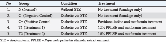
AbstractBackground: Delayed wound healing is a significant complication of diabetes mellitus (DM). In Indonesia, Peperomia pellucida leaves exhibit anti-inflammatory activity due to its phenolic and flavonoid compounds. Aim: This study aimed to evaluate the efficacy of P. pellucida leaf ethanol extract (PPLEE) Ointment for healing acute wounds through modulation of angiogenesis, neovascularization, and epithelialization in an acute streptozotocin–nicotinamide (STZ–NA)-induced diabetic rat. Methods: A total of 30 rats were randomized into five groups: normal (N); negative control (C?); positive control (C+); treatment 1, DM rats with 12% ointment (T1); and treatment 2, DM rats with 16% ointment (T2). Groups C?, C+, T1, and T2 received STZ–NA injections for 5 days. Five groups underwent excision wounds on the backs of rats. All groups received wound care according to the procedure for 15 days. Rats were sacrificed, and tissue was collected for ELISA (TNF-α, IL-6, TGF-β, MDA, and VEGF), while neovascularization and epithelialization were assessed histopathologically. Results: The study clearly showed that topical application of PPLEE at a 16% dose can reduce inflammation, as indicated by markers TNF-α, IL-6, TGF-β, and MDA (p-value < 0.001). The angiogenesis process was better characterized by VEGF (p-value < 0.001), by mature neovascularization tissue (p-value < 0.001), and by epithelialization tissue in the intervention group, which showed almost complete epithelialization (p-value < 0.001). The wound closure fraction showed that the T2 group showed nearly the same wound closure as the N group (p value < 0.001), while the T1 group showed no difference with C+. Conclusion: Topical PPLEE may reduce inflammation, oxidative stress, and support vascular and epithelial regeneration in diabetic rats. Keywords: Angiogenesi, Epithelialization, Neovascularization, Peperomia pellucidaleafextract ointment, Diabetic wound. IntroductionWound healing involves a complex series of events that restore the integrity of injured skin, beginning with inflammation, followed by proliferation (including angiogenesis and epithelialization), and tissue remodelling (Dwivedi et al., 2024). Wounds in diabetes mellitus (DM) heal more slowly than under normal conditions due to specific changes in inflammatory cytokines (Mukai et al., 2025). The pathophysiology of DM wound conditions involves prolonged inflammation that increases oxidative stress and inhibits angiogenesis, resulting in poor epithelial regeneration (Chan, 2020; Ahmad et al., 2025; Elajaili et al., 2025). Understanding this mechanism is very important for developing targeted therapies to improve wound-healing outcomes in diabetic wounds (Elajaili et al., 2025). The accumulation of advanced glycation end products in DM disrupts fibroblast function, reducing their ability to produce and regulate collagen. Glycation dysregulates inflammatory phases, such as tumor necrosis factor-α (TNF-α) and Interleukin-6 (IL-6), which are essential for initiating defense and recruiting wound-healing cells (Guillon et al., 2021; Solfaine et al., 2024). During angiogenesis, vascular endothelial growth factor (VEGF) acts as a specific mitogen for endothelial cells, promoting the formation of new blood vessels. VEGF works together with other growth factors such as fibroblast growth factor and transforming growth factor-β (TGF-β), to regulate angiogenesis and wound healing. Excessive TGF-β activity can lead to fibrosis, characterized by excessive deposition of extracellular matrix, resulting in scarring and tissue thickening (Chandra et al., 2025). In hyperglycemic conditions, DM wounds trigger systemic and tissue oxidative stress, characterized by increased malondialdehyde (MDA), which causes cell damage and lipid peroxidation (Mukai et al., 2025). Dysregulation of TNF-α, IL-6, and MDA prolongs the inflammatory phase, inhibiting the transition to the proliferative phase in DM wound healing. High inflammation is associated with abnormal VEGF overexpression and ineffective angiogenesis. In addition, the expression of VEGF and TGF-β is impaired, thereby worsening neovascularization and tissue regeneration, leading to chronic diabetic ulcers (den Dekker and Gallagher, 2020). This understanding is essential for the simultaneous modulation of inflammation, oxidative stress, angiogenesis, and epithelial regeneration, which are molecularly interconnected and may accelerate, and thus improve, the healing process (Rangwala et al., 2023). Although various synthetic topical agents such as silver sulfadiazine, povidone-iodine, and topical insulin, have been used to treat DM wounds, their effectiveness in chronic hyperglycemic conditions remains limited (Rangwala et al., 2023; Hu et al., 2025). Furthermore, long-term use of synthetic compounds is often associated with skin irritation, microbial resistance, high costs, and limited access in developing countries. The need to develop accessible, affordable, nature-based treatments remains especially urgent in resource-limited settings. Peperomia pellucida has traditionally been used to treat wounds, ulcers, and skin infections in various cultures, including in Southeast Asia and South America, due to its ease of cultivation. Phytochemically, this plant contains flavonoids, tannins, and phenolic compounds, all of which have been scientifically proven to have anti-inflammatory, antioxidant, and angiogenic activities. These three key pillars are impaired in DM wounds (Ahmad et al., 2023). Recent studies have demonstrated the efficacy of P. pellucida in wound healing. For example, a study using a P. pellucida nanoemulsion showed significant improvement in burn wound healing in rats, with increased wound closure and reduced inflammation (Prananda et al., 2024). Another study found that the ethanol extract of P. pellucida leaves supported burn wound healing in rats, with a 50% concentration being the most effective (Putri and Puspitasari, 2022). While P. pellucida shows promise for wound healing, further research is needed to fully understand its mechanisms and potential side effects. There has been no in vivo study that comprehensively evaluates the topical effects of PPLEE ointment in DM wound healing with a multi-target pathway-based biomolecular approach in acute wound rat models with DM induction, such as (STZ–NA). Previous P. pellucida studies have tended to be limited to general wound models, thus not sufficiently addressing the potential of this plant in chronic metabolic wound conditions (Putri and Puspitasari, 2022). Peperomia pellucida is used in traditional medicine to treat inflammation, rheumatic pain, gout, skin diseases, acne, and abscesses. Previous studies have demonstrated its antioxidant, antibacterial, anti-inflammatory, and anti-diabetic properties. However, despite these pharmacological potentials, few studies have comprehensively evaluated its effects on diabetic wound healing, particularly using an integrated approach that includes biochemical markers and histopathological assessment. Therefore, it is essential to further investigate this plant as a potential candidate for scientifically competitive herbal-based wound therapy, supported by evidence from biomarker analysis, including TNF-α, IL-6, MDA, VEGF, and TGF-β, as well as histological parameters. This study aimed to evaluate the topical effects of PPLEE ointment on wound healing in STZ–NA-induced rats by assessing inflammation (TNF-α, IL-6), proliferation (TGF-β), angiogenesis (VEGF), and oxidative stress (MDA). Materials and MethodsMaterials preparation of ointmentPeperomia pellucida leaves were collected from yards and around oil palm trees during the rainy season in Pringsewu Regency, Lampung, Indonesia. Sampling was carried out in the morning, from 6:00 to 9:00 AM. After the P. pellucida leaves were collected, they were selected/sorted, washed, dried, packaged, and stored. The determination test for Chinese betel leaf was carried out at the Functional Service Unit of Tawangmangu Traditional Health Services, Dr. Sarjito General Hospital, Central Java, followed by drying and processing into 50-mesh simplicia with result number: 912/UN1/LPPT/TR/2025. An ethanol extract of Chinese betel leaves was prepared in the Biology Unit of the Pharmaceutical Laboratory at Gadjah Mada University, Yogyakarta, through maceration (Priamsari et al., 2024). A total of 1,000 g of dried P. pellucida leaves were macerated with 70% ethanol solvent. The ratio of powdered simplicia to solvent was 1:10. It was left in a tightly closed container at room temperature for 5 days to prevent solvent evaporation. The process was continued by evaporating the filtrate until the extract was constant. Phytochemical examination of PPLEE was carried out at the Integrated Testing Research Laboratory of Gadjah Mada University. The compounds examined in PPLEE included total alkaloids, phenols, tannins, saponins, and flavonoids, which were determined using a spectrophotometer, with certificate number 911/UN1/LPPT/TR/2025 (Ibe-Diala and Igwe, 2022). The PPLEE, as an active ingredient of the ointment, was added to vaseline album, liquid paraffin, a preservative, and sterile distilled water (Hasan et al., 2024). The ointment was prepared according to a procedure to obtain concentrations of 12% and 16%, as reported in previous research (Sari, 2023). The concentrations of 12% and 16% were selected based on preliminary studies, which indicated optimal physical stability and potential wound-healing activity. The concentrations of 12% and 16% were selected based on preliminary studies that indicated optimal physical stability and potential wound-healing activity. Ointment concentration 16%: 16 grams of extract in 100 grams of total ointment with a base of 76.36 g vaseline album, and 7.64 ml liquid paraffin. The packaging and storage of PPLEE ointment were performed in a clean, sterile pot at the specified concentrations (Garay et al., 2025). Researchers conducted organoleptic, pH, and spreadability tests over 30 days, with measurements taken every 10 days. MethodsResearch with a quantitative design using a posttest-only control group design approach to evaluate the effect of PPLEE on the acute wound rat model of DM. Preparation of experimental animals and treatmentThe sample size was determined using the resource equation method for one-way ANOVA experimental designs. An acceptable error degree of freedom (E) of 10–20 was used to ensure adequate statistical validity. Based on five experimental groups and an anticipated dropout adjustment of 20%, six rats were included in each group (total n=30) (Zhang and Hartmann, 2023). Researchers used 30 healthy male Rattus norvegicus rats, divided into five groups, weighing 200–220 g and aged 2–3 months, raised in the Experimental Animal Laboratory of the Center for Food and Nutrition Studies at Universitas Gadjah Mada, Yogyakarta. All experimental animals were selected within a body weight range of 200–220 g to ensure homogeneity at baseline. The rats were acclimatized for 7 days in a 40 ? 30 ? 24 cm cage at 24°C–25°C, 45%–50% humidity, and a 12:12-hour light-dark cycle. Researchers randomized them into five groups. In this study, after the rats had adapted to their cages for 7 days, they were induced with STZ 45 mg/kg body weight/day and NA 110 mg/kg body weight/day intraperitoneally for 5 days for DM induction (Oktavia et al., 2025; Razoki et al., 2025), except for the rats in the normal group (N). Before excision, the researchers anesthetized the back with an IM injection of ketamine 60 mg/kgBW. Excisional wounds were created in two locations on the backs of the rats, measuring 1.5 cm in length and 1.5 cm in width, with a depth that reached the hypodermis/subcutaneous layer (Celepli, 2022). In the normal control group (N) and negative control (C?), after excision, the excision wound was covered with a foam bandage without any topical treatment. In the positive control group (C+), after excision, the excision wound was smeared with povidone-iodine ointment. In Treatment group 1 (T1) after excision, the excision wound was smeared with 12% PPLEE ointment. In Treatment group 2 (T2) after excision, the excision wound was smeared with 16% PPLEE. The entire excision wound was covered with a foam bandage. Wound care was performed 4 times over 15 days; the wound was cleaned with saline and treated according to the assigned group. In addition, groups C+, T1, and T2 received oral metformin treatment at a dose of 45 mg/kg body weight per day (Table 1). Sample collection and marker measurementsSampling was conducted using blood and wound tissue samples. Blood sampling was carried out three times during the research period: pre-induction, on day 5 of induction, and on day 20. Researchers fasted for 12 hours overnight. Blood glucose was measured from the orbital vein according to standard procedures, and fasting blood glucose levels ≥ 250 mg/dl were considered hyperglycemia (Chanu et al., 2023). On day 20, the collected samples were then injected with ketamine and xylazine to be sacrificed according to ethical research procedures. The samples were then prepared in a 10% formalin buffer solution for enzyme-linked immunosorbent assay (ELISA), tissue, and histopathology examination (Fine Test, Wuhan, China). Tissue ELISA examination was used to measure TNF-α, IL-6, TGF-β, VEGF, and MDA markers, and Histopathological examination of wound tissue was conducted by the Anatomical Pathology Laboratory of Gadjah Mada University, Yogyakarta. Histopathological examination with hematoxylin and eosin (H&E) staining for the microtome, embedding center, and tissue processor, using the Sakura brand (Accu-Cut SRM) from Japan. Reading of H&E-stained preparations using the Olympus CX33 digital camera microscope from Japan. Table 1. Experimental groups and treatment protocol in the excision wound model.
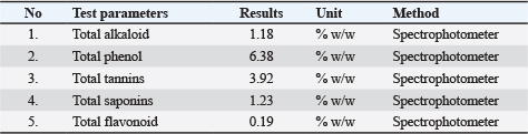
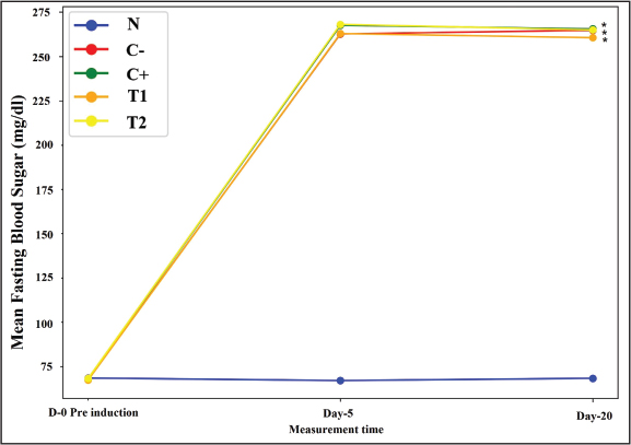
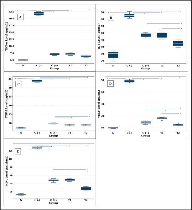
Statistical data analysisData analysis was performed using the Statistical Package for the Social Sciences (SPSS) version 26. Numerical data were presented in the form of averages and standard deviations, while categorical data were presented in the form of frequency distributions. Before statistical analysis, normality of the data was assessed using the Shapiro–Wilk test, and homogeneity of variance was evaluated using Levene’s test. Data that were normally distributed were analyzed using one-way ANOVA and repeated measures ANOVA, followed by the Tukey HSD post hoc test. Data that did not meet these assumptions were analyzed using an appropriate non-parametric statistical tests. Categorical data were analyzed using the chi-square test. Ethical approvalThe authors adhered to the ethical principles of research on experimental animals throughout the study, in accordance with the guidelines of the Ethics Commission of the Faculty of Medicine, Public Health, and Nursing, Universitas Gadjah Mada, Yogyakarta. This study received approval with registration number: KE/FK/0651/EC/2025. ResultsContent of PPLEE ointmentThese results were evaluated from the phytochemical content of PPLEE ointment, anti-inflammatory effects through TNF-α, IL-6, wound proliferation process evaluation of TGF-β values, angiogenesis process seen from VEGF, and DM severity through MDA oxidative stress, neovascularization, and epithelialization processes are essential to see complete wound healing, which is characterized by macroscopic wound-healing reports. Table 2 shows the phytochemical results of PPLEE containing alkaloids, phenols, tannins, saponins, and flavonoids. Among these compounds, phenols showed the highest, followed by tannins and saponins in moderate amounts, while flavonoids and alkaloids were present in lower concentrations. The results of making ointments in two formulations with 12% and 16% content. Based on preliminary studies, the ointment preparation exhibited no changes in organoleptic properties, including a brownish-green color, a distinctive herbal odor, and a homogeneous semi-solid consistency. The pH values of both ointment formulas ranged from 5.2 to 6.5; the spreadability showed fluctuations in the range (5–7 cm2) over a period of 30 days in room temperature (25?C ± 3?C) under ambient light conditions, without exposure to direct sunlight. Identification of DM models in ratsFasting blood glucose levels are presented in Fig. 1, demonstrating sustained hyperglycemia in diabetic groups throughout the experimental period. A DM rat model was created by administering STZ and NA intraperitoneally for 5 days. Fig. 1 illustrates that, before model creation, all rats had normal blood sugar levels. On the fifth day until the study was completed, the rats’ blood sugar levels were examined in the C?, C+, T1, and T2 groups, showing an increase in blood sugar levels > 250 mg/dl, where there was a significant increase in blood sugar (p-value < 0.001), except in the N group where blood sugar levels remained within the normal range (p-value > 0.05). The effect of topical PPLEE ointment on inflammation, proliferation, angiogenesis, and oxidative stressTable 3 shows that the protein levels of TNF-α, IL-6, TGF-β, VEGF, and MDA were highest in the C? group and lowest in the N group. The C+ and T1 groups showed almost identical values, whereas the T2 group demonstrated lower levels, although not fully restored to normal. The statistics were significantly different (p-value < 0.001), with very large effect sizes (η2=0.953–0.999). Post hoc analysis in Fig. 2 shows significant differences (p < 0.05–p < 0.001) between groups in all analyzed biomolecular parameters. In rats with DM experiencing acute wounds, group C? showed elevated inflammatory markers, with cytokines still elevated on day 15 (Fig. 2A and B). This is in line with the still high levels of TGF-β, indicating that the proliferation process is ongoing (Fig. 2C). VEGF levels, as a marker of angiogenesis, increased sharply in the C? group (p < 0.001) (Fig. 2D). Administration of PPLEE ointment significantly reduced VEGF levels in the T2 group compared to T1 and C+ (p < 0.05–0.001), indicating a regulatory effect on abnormal angiogenesis in DM wounds. This is because DM conditions are associated with high oxidative stress at the protein level in wound tissue (Fig. 2E). The PPLEE ointment significantly reduced MDA levels in both the T1 and T2 groups, with the greatest decrease in T2, which was statistically significant compared to C−, C+, and T1 (p < 0.001). Table 2. Content of active substances in P. pellucida extract.
Fig. 1. Graph of fasting blood sugar levels during study in diabetic rats. Table 3 Differences in TNF-α, IL-6, TGF-β, VEGF, and MDA given P. pellucida extract on tissue.
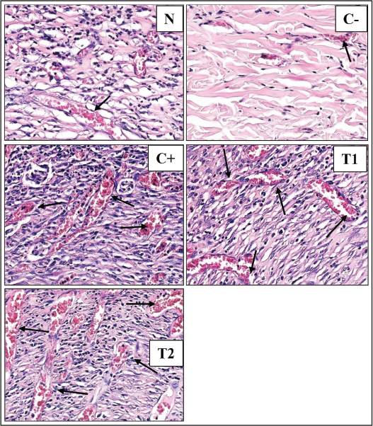
The effect of the topical PPLEE ointment on neovascularization and epithelialization of wound tissueThe neovascularization in group C? was entirely highly immature, whereas in the normal group, the histopathological picture was more mature, indicating that all were mature. In the C+ group intervention, the results were similar to those of T1. In contrast, group T2 consisted of half mature subjects and the other half highly mature subjects. These results indicate that administering ointment containing PPLEE significantly increases neovascularization in acute wound tissue in DM conditions (p < 0.05). These findings indicate that PPLEE encourages angiogenesis and stabilizes neovascular structures during the proliferative phase of DM wound healing (Table 4).
Fig. 2. Effect of administering PPLEE ointment on improving inflammation (TNF-α, IL-6, TGF-β), angiogenesis (VEGF), and oxidative stress (MDA) in a rat model of diabetes mellitus. N; normal, C?; negative control, C+; positive control, T1; treatment 1 and T2; treatment 2. In Figure 3, histopathology of the tissue reveals that in group N, dermal neovascularization consists of a few new blood vessels with relatively wide lumens. In the C? group, neovascularization in the dermal layer appears characterized by a low number of new blood vessels with narrow lumens. In the C+ in the dermis layer, the number of new blood vessels is quite large, and the lumen is quite large. In group T1, in the dermis layer, new blood vessels appear more numerous, and the lumen is quite large. In group T2 of the dermis, multiple new blood vessels appear, and the lumen is larger. Table 4. Neovascularization and epithelialization in wound tissue in each group.
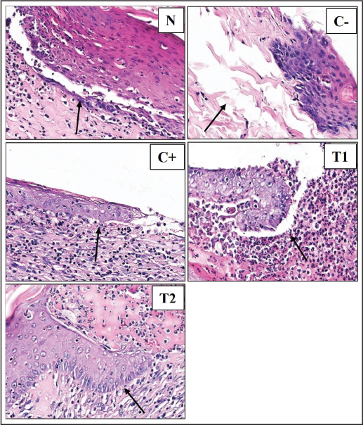
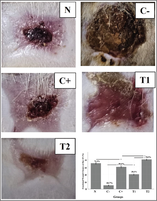
Histopathological features of epithelialization showed that group C showed no epithelial closure (“no epithelialization”), whereas groups N and C+ showed a majority with near-complete epithelialization. The T2 group showed greater epithelial regeneration, with 50% of samples exhibiting complete epithelialization (p-value < 0.001). These results indicate that PPLEE ointment significantly enhances neovascular maturation and epithelial regeneration in DM wounds (Table 3). Figure 4 shows that in group (N) epithelialization, the stratum basale appeared thick and regular with a clear dermo-epidermal junction. However, the healing process was incomplete. In the C? group, the epithelial layer was completely absent, and the wound surface had not closed. In group (C+), epithelialization, the stratum basale was clearly visible and dense, but there were still gaps in the wound, and the wound surface had not closed. In group (T1), epithelialization, the stratum basale was clearly visible, but not dense; there were still gaps in the wound. In group (T2) epithelialization, the stratum basale was dense and thick; there were no gaps in the wound, there were signs of keratinocyte maturation (the stratum basale and spinosum were visible), and the wound surface was well closed. Figure 5 shows representative wounds from each group. On Day 0, all groups exhibited full-thickness excision wounds of similar size and depth. On Day 15, the wound area in the T2 group showed substantial closure with minimal crust formation, whereas T1 and C+ showed partial healing and residual crust. The C? group retained extensive necrotic tissue and minimal contraction, indicating delayed wound healing. Quantitative analysis of Fractional Wound Closure (FWC) revealed significantly higher closure in T2 across almost all groups (p < 0.001). DiscussionThe results of the quantitative phytochemical test of the PPLEE showed that the highest compound content was total alkaloids, followed by total phenols, tannins, saponins, and flavonoids. The ointment was made according to the standards for topical treatment. These results indicate that hyperglycemic conditions in STZ–NA-induced rat models constitute a successful diabetes model. The decrease in TNF-α, IL-6, and MDA levels may be due to PPLEE ointment modulating inflammation during the DM wound-healing process. However, administration of PPLEE ointment with a concentration of 16% (T2) was more effective in reducing TNF-α, IL-6, and MDA levels compared to the group given povidone-iodine ointment (C+) and PPLEE ointment with a concentration of 12% (T2) in diabetic rat models. Histopathology of the wound tissue with H&E staining showed that in the T2 Group, neovascularization was very abundant in the dermis, with larger lumens. While the epithelialization of the basal stratum was dense and thick, there was no wound gap; there were signs of keratinocyte maturation (the basal and spinous strata were visible), and the wound surface closure was good. The above results could be due to the phytochemicals in PPLEE, which contain phenols, tannins, saponins, alkaloids, and flavonoids, and may play a critical role in repairing DM tissue wounds. Among these compounds, phenols showed the highest followed by tannins and saponins in moderate amounts, while flavonoids and alkaloids were present in lower concentrations. Several factors, such as polarity, extraction duration, and possible degradation during drying, can explain the low flavonoid content. However, we followed extraction standards. Therefore, plant geography and harvesting methods may influence these results. These findings are consistent with a previous study reporting that alkaloids and saponins are among the predominant constituents of PPLEE. In contrast, flavonoids were present in lower levels but remained pharmacologically relevant (Alfi, 2024).
Fig. 3. Histopathological examination (H&E) to assess neovascularization in Rattus norvegicus rat excision wound tissue on day 15 after different topical interventions. HE in photo 5 has a field of view at 400? magnification. N; normal, C?; negative control, C+; positive control, T1; treatment 1 and T2; treatment 2. Phenols and tannins each possess antioxidant and anti-inflammatory properties, contributing to the plant’s therapeutic potential (Oktavia et al., 2025). The presence of saponins in PPLEE contributes to its potential use in treating inflammatory conditions and supporting overall health (Razoki et al., 2025). Although flavonoids are present in relatively low amounts, they can reduce the expression of pro-inflammatory cytokines such as IL-6 and TNF-α and increase the expression of anti-inflammatory cytokines such as IL-10, thereby modulating the inflammatory response (Song et al., 2022; Alfi, 2024). Flavonoids scavenge reactive oxygen species (ROS) and increase endogenous antioxidants to reduce oxidative stress in diabetic wounds (Yang et al., 2024). This indicates that the preparation has good stability in storage during the study at room temperature (25?C ± 3?C) under ambient light conditions, without exposure to direct sunlight. The pH range is still within the normal skin pH range (4.5–6.5). The spreadability showed mild fluctuations, remaining within the optimal range (5–7 cm2), indicating no significant changes in viscosity. The stability of the ointment formulations over 30 days indicates they are suitable for storage without significant changes in their physical properties.
Fig. 4. Histopathological examination (H&E) to assess epithelialization in the excision wound tissue of Rattus norvegicus rats on day 15 under the intervention of each group. HE in photo 5 has a field of view at 400? magnification. N; normal, C?; negative control, C+; positive control, T1; treatment 1 and T2; treatment 2. The DM rat model was developed to mimic human conditions, characterized by indicators of hyperglycemia. Although this induction still cannot accurately describe the DM metabolic syndrome as it occurs in humans, this study can provide insight into the healing process in the DM model (Wahyuningsih et al., 2024). DM wounds often exhibit elevated levels of inflammatory cytokines, such as TGF-β, which prolong the inflammatory phase and delay healing. This study showed that cytokines remained elevated in DM rats even on day 15, indicating a prolonged inflammatory response (Mukai et al., 2025). Intraperitoneal induction of DM in a rat model resulted in a hyperglycemic state that persisted for 20 days (Lolok et al., 2021). There was no significant difference in glucose levels between the treatment groups, indicating that the wound healing in groups T1 and T2 was not due to reduced blood glucose levels, but rather to the local effects of the PPLEE ointment. Metformin therapy as a glucose-lowering agent cannot be excluded and may have contributed to the observed wound-healing effect (Fig. 1). PPLEE ointment may support wound healing in diabetic conditions through local effects, modulating inflammation, oxidative stress, and angiogenesis. This finding aligns with research indicating that certain plant extracts, such as Cymbopogon proximus, exhibit wound-healing benefits through immunomodulatory properties and local effects. Metformin administration controls blood sugar in rats, although it does not consistently significantly alter systemic glucose levels (Balykova et al., 2023). Metformin administration is a widely used antidiabetic drug known for its glucose-lowering effects. However, its impact on wound healing is less clear. Metformin, a widely used antidiabetic medication, is recognized for its ability to lower glucose levels and reduce inflammation. The use of metformin has also been shown to support the wound-healing process, including reducing inflammation and proliferation in DM (Kominato et al., 2022).
Fig. 5. Macroscopic appearance of wound closure in DM rats treated with topical ointment Peperomia pellucida ethanol extract compared with the control group on day 15. The results showed that the wound closure fraction in the T2 group was significantly better (p-value < 0.001). N=normal, C− (control ?), C+ (control +), T1 (treatment 1) and T2 (treatment 2). DM wounds are characterized by chronic inflammation, with elevated levels of pro-inflammatory cytokines such as TNF-α and IL-6, which inhibit healing (Strang et al., 2020; Mukai et al., 2025). PPLEE ointment appears to reduce these cytokines, as evidenced by lower levels in the T2 group compared to the C? group, suggesting its potential anti-inflammatory effects (Oktavia et al., 2025). TGF-β is essential for cell proliferation and tissue regeneration, while VEGF is essential for angiogenesis, both of which are impaired in diabetic wounds (Strang et al., 2020). In the C? group VEGF overexpression in diabetic wounds reflects of dysfunctional angiogenesis rather than effective vascular regeneration (den Dekker and Gallagher, 2020). The C group showed high MDA. Oxidative stress is a significant barrier to effective wound healing in DM. Excessive ROS levels cause oxidative stress that impairs the healing process, leading to cell damage, prolonged inflammation, and inhibition of angiogenesis, and disrupting vascular endothelial function, neovascularization, and epithelialization in diabetic lesions (Yan et al., 2025). The decrease in MDA levels in the T2 group suggests that PPLEE ointment can reduce oxidative stress, contributing to a more favorable healing environment (Strang et al., 2020; Oktavia et al., 2025). PPLEE content modulates this response by reducing cytokine levels, thereby aiding the transition from inflammation to proliferation. Saponins promote cell proliferation and angiogenesis by activating the VEGF pathway (Lei et al., 2022; Yang et al., 2025). PPLEE contains significant levels of antioxidants such as flavonoids, phenols, and saponins, which are known to combat oxidative stress by neutralizing free radicals (Asiwe et al., 2024). Alkaloids also increase the activity of antioxidant enzymes, reduce oxidative markers, and help mitigate oxidative stress that impairs wound healing. Alkaloids increase levels of growth factors, such as TGF-β and VEGF, for tissue regeneration and wound healing (Alamre et al., 2024; Xiao et al., 2024). In a previous study involving rats, the plant extract not only reduced MDA levels but also overall antioxidant status, further supporting its efficacy in reducing oxidative damage (Okoro et al., 2025). Similar studies have shown that natural antioxidant astaxanthin can promote wound healing in DM rat models with oral wounds (Andriani et al., 2025). The results in Fig. 2 showed that there was no difference between the administration of povidone-iodine ointment (C+) and the administration of PPLEE ointment with a concentration of 12% (T1) on the levels of TNF-α, IL-6, and MDA in healing excision wounds in DM rats. The results closest to normal were observed with the 16% PPLEE ointment (T2), where TNF-α, IL-6, TGF-β, VEGF, and MDA levels were nearly back to normal. Traditionally used for its antiseptic properties, povidone-iodine is effective in reducing microbial load in wounds, which is very important for preventing infection and improving healing (Gryson et al., 2025; Harun et al., 2025). The decrease in MDA levels in the 16% PPLEE ointment group indicates an anti-inflammatory effect, which is beneficial in healing DM wounds where oxidative stress is a significant barrier. The greater effect in the T2 group with 16% PPLEE ointment suggests that higher concentrations of bioactive compounds contribute to the biological effects. This finding is consistent with previous studies reporting the potential role of PPLEE in wound healing (Sari, 2023). DM wounds are known to be challenging to heal due to impaired inflammatory responses and increased oxidative stress. The findings suggest that PPLEE ointment, particularly at higher concentrations, may be a promising natural alternative to traditional treatments, such as povidone-iodine, offering anti-inflammatory and antioxidant benefits. Peperomia pellucida normalizes inflammatory and oxidative stress markers in DM wound healing, demonstrating its potential as a phytotherapeutic agent. These results demonstrate its potential as a phytotherapeutic agent for mitigating inflammation, supporting proliferation, angiogenesis and reducing oxidative stress in acute DM wound tissue. The use of plant compounds, such as those found in P. pellucida, can enhance the wound-healing process by promoting angiogenesis, reducing inflammation, and increasing collagen synthesis. Previous studies have shown that P. pellucida is efficacious in improving burn wound healing in animal models. Its PPLEE ointment formulation significantly accelerated wound closure, increased re-epithelialization, and increased collagen deposition, indicating increased angiogenesis in the healing process. The ability of plants to reduce inflammatory cytokine levels and increase antioxidant enzyme activity suggests that plants can modulate the inflammatory environment, which is crucial for angiogenesis during wound healing (Prananda et al., 2024). This compound exhibits anti-inflammatory, antioxidant, and antimicrobial properties, which are crucial in reducing inflammation and oxidative stress in wounds, thereby creating a conducive environment for neovascularization (Prananda et al., 2024). Previous research has shown that P. pellucida, particularly in the form of an ethanol extract, significantly accelerates wound closure and enhances granulation tissue formation, indicating effective neovascularization (Putri and Puspitasari, 2022). Epithelialization is a critical phase in wound healing, involving the migration and proliferation of keratinocytes to restore the epidermis (Yan et al., 2025). The presence of neuropathy and poor blood circulation further complicates the healing process, making DM wounds particularly challenging to treat. Standard treatments often fail to address these underlying issues, leading to prolonged healing times and an increased risk of complications (Jesús et al., 2022). PPLEE contains bioactive compounds, including flavonoids, alkaloids, saponins, tannins, steroids, and triterpenoids, which are known to contribute to its wound-healing properties. These compounds help reduce inflammation, promote cell proliferation, and enhance collagen synthesis, all of which are essential for effective re-epithelialization (Gulo and Maulana, 2024). Compounds in natural extracts, such as flavonoids, stimulate the proliferation of fibroblasts and keratinocytes, aiding the remodeling phase of wound healing by increasing tissue strength and elasticity (Nandhini et al., 2024). In line with other studies, this study’s promising results suggest that natural extracts may serve as a cost-effective and accessible alternative to conventional wound-healing treatments, especially in low-resource settings (Nandhini et al., 2024; Garay et al., 2025). Alkaloid compounds, tannins, saponins, phenols, and flavonoids in P. pellucida have the potential to provide therapeutic effects, including anti-inflammatory and antioxidant properties, increased angiogenesis, increased blood supply to the wound area, increased collagen synthesis, and enhanced epithelialization in diabetic wound healing. The PPLEE ointment significantly affects wound healing. This study shows that the administration of povidone-iodine ointment, PPLEE ointment with a concentration of 12%, and 16%, can reduce the levels of TNF-α, IL-6, TGF-β, VEGF, and MDA in wound tissue on day 15, as well as increase neovascularization and epithelialization in excisional wound healing in diabetic rats. However, the administration of PPLEE ointment with a concentration of 16% has a more effective impact on wound healing in diabetic rats compared to the administration of povidone-iodine ointment and the administration of PPLEE ointment with a concentration of 12%. The PPLEE ointment at 16% concentration can be an alternative therapy to accelerate the healing of diabetic excision wounds. PPLEE ointment shows promise in modulating key pathways involved in DM wound healing; it is essential to consider the broader context of wound-healing research. The complexity of cytokine interactions and the need for proper regulation of the inflammatory response are crucial for effective healing (Mahmoud et al., 2024). Furthermore, while natural treatments offer potential benefits, their integration into clinical practice requires further validation and a better understanding of their mechanisms of action compared to standard therapies. Although the present study demonstrated the potential of the PPLEE ointment in supporting wound healing, several limitations should be considered. Phytochemical analysis was limited to total spectrophotometric counting methods. Detailed characterization of antioxidant content and specific quantification of antioxidants, such as HPLC, were not performed and should be considered in future studies. Metformin, which was administered to maintain glycemic control in diabetic wounds, may have modulated inflammation and oxidative stress, factors that could not be controlled in this study. In addition, this research did not test the ointment’s toxicity, long-term safety, blinding, or stability at different temperatures, underscoring the challenges associated with its application. Furthermore, exploratory studies are necessary to enhance the stability and efficacy of bioactive compounds in natural extracts. ConclusionThis study demonstrates that PPLEE ointment can support wound healing as an alternative treatment for excision wounds in DM rat models induced by STZ and NA. The potential of PPLEE ointment for wound healing is characterized by reduced levels of inflammatory markers, oxidative stress, and angiogenesis, as well as improved histopathological parameters. PPLEE ointment at a dose of 16% (T2) showed a greater effect than C+ and T1. However, this finding should be considered, as metformin administration in the C+, T1, and T2 groups may have contributed to the observed effects. Furthermore, toxicity, pharmacokinetics, and long-term effects have not been evaluated in this study. Future research is needed to unravel the mechanism of action of the DM healing system relative to other pathways and to evaluate the clinical efficacy of PPLEE ointment until this intervention model is recognized as a standard therapy for acute wound healing in DM. AcknowledgmentsNone. FundingThe author would like to thank the Indonesian Education Scholarship, Center for Higher Education Funding and Assessment, and Indonesian Endowment Fund for Education, with number: 02337/J5.2.3./BPI.06/9/2022 in the Doctoral Program of Medical Sciences, Faculty of Medicine, Universitas Sebelas Maret, Indonesia Authors’ contributionsTri Wijayanto: Conceptualization, Methodology, Writing, original draft preparation. Ida Nurwati: Supervision, Writing, Data curation and validation. Soetrisno: Supervision, Methodology, Data curation and Validation. Tatar Sumandjar: Supervision, Investigation and Visualization. Ratih Puspita Febrinasari: Formal Analysis, Investigation and Validation. Vitri Widyaningsih: Writing, Methodology, Formal analysis and Investigation. Paramasari Dirgahayu: Writing, Validation, Reviewing and Editing Conflict of interestThe Authors declare that there is no conflict of interest. Data availabilityAll data supporting the findings of this study are available within the manuscript ReferencesAhmad, I., Hikmawan, B.D., Mun’Im, A. and Sulistiarini, R. 2023. Peperomia pellucida (L.) Kunth herbs: a comprehensive review on phytochemical, pharmacological, extraction engineering development, and economic promising perspectives. J. Appl. Pharm. Sci. 13(1), 1–9; doi:10.7324/JAPS.2023.130201 Ahmad, U., Hanaffi, W.N.W., Islam, A., Salman, A., Khan, M.M., Shakeel, F., Cai, Q., Cai, X. and Shubhra, Q.T.H. 2025. Cutting edge strategies for diabetic wound care: nanotechnology, bioengineering, and beyond. BMEMat 4(1), 2751–7438; doi: 10.1002/bmm2.70033 Alamre, J.B., Alkreathy, H., Ali, A.S., Alfadil, A. and Alghamdi, S.S. 2024. Effect of 3-hydrazinylquinoxaline-2-thiol hydrogel on skin wound healing process in diabetic rats. Scientific Rep. 14(1), 1–14; doi: 10.1038/s41598-024-70849-x Alfi, T.F. 2024. Determination of total alkaloid content of Chinese betel (Peperomia pellucida) based on differences in ethanol concentration using UV-VIS spectrophotometry method. J. Pharm. Care Sci. 5(1), 158–167; doi:10.33859/jpcs.v5i1.673 Andriani, D., Roestamadji, R.I., Rahayu, R.P., Luthfi, M., Narmada, I.B., Notobroto, H.B., Siswandono, S. and Rohmaniar, P.D. 2025. Hyaluronic acid-astaxanthin gel : anti-inflammatory effects in oral ulcer diabetes mellitus rats. Trends Sci. 22(7), 1–10. Asiwe, J.N., Yovwin, G.D., Igben, V.O., Abe, B., Akoko, S., Oritsemuelebi, B., Chimezie, J., Ojieh, A.E. and Dagogo, G. 2024. Chemical characterization, in vivo anti-oxidant, anti-inflammatory and anti-apoptotic properties of Peperomia pellucida in modulating vascular tone in Swiss mice exposed to aluminium chloride. Pharmacol. Res. 2, 1–14; doi:10.1016/j. prenap.2024.100034 Balykova, L.A., Inchina, V.I., Tarasova, T.V., Khaydar, D.A., Mosina, L.M., Saushev, I.V., Kuchuk, A.V., Mutvakel, A., Begoulov, I.V., Tarasov, R.S., Ageev, P.V. and Ovchenkov, D.S. 2023. Regeneration of an infected skin wound under conditions of experimental steroid hyperglycemia. Res. Practical Med. J. 10(1), 90–99. Celepli, S. 2022. The effects of Capparis ovata seed oil on the healing of traumatic skin wounds. Türk. J. Trauma Emerg Surg. 28(9), 1205–1213; doi:10.14744/tjtes.2021.31526 Chan, Y.C.C. 2020. Chapter 6—Angiogenic response in wound healing. In Roy Tissue Repair, and Regeneration in Diabetes. Eds., D. Bagchi, A. Das and S.B.T.W.H. Academic Press, pp: 97–120; doi: 10.1016/B978-0-12-816413-6.00006-X Chandra, P., Faizan, M., Porwal, M., Sharma, H. and Sachan, N. 2025. An overview and review of growth factors in wound healing: emerging trends and innovations. Curr. Diabetes Rev. 22(1), 1–27; doi: 10.2174/0115733998332692241202072249 Chanu, N.R., Gogoi, P., Barbhuiya, P.A., Dutta, P.P., Pathak, M.P. and Sen, S. 2023. Natural flavonoids as potential therapeutics in the management of diabetic wound: a review. Curr. Topics MedChem. 23(8), 690–710; doi:10.2174/1568026623666230419102140 denDekker, A.D. and Gallagher, K.A. 2020. Chapter 5—Dysregulated inflammation in diabetic wounds. In Roy Tissue Repair, and Regeneration in Diabetes. Eds., D. Bagchi, A. Das and S.B.T.W. H. Academic Press, pp: 81–95; doi: 10.1016/B978-0-12-816413-6.00005-8 Dwivedi, J., Sachan, P., Wal, P., Wal, A. and Rai, A.K. 2024. Current state and future perspective of diabetic wound healing treatment: present evidence from clinical trials. Curr. Diabetes. Rev. 20(5), 45–65; doi:10.2174/1573399820666230828091708 Elajaili, H., Lyttle, B.D., Lewis, C.V., Bardill, J.R., Dee, N., Seal, S., Nozik, E.S., Liechty, K.W. and Zgheib, C. 2025. Increased ROS and persistent pro-inflammatory responses in a diabetic wound healing model (db/db): implications for delayed wound healing. Int. J. Mol. Sci. 26(10), 26; doi:10.3390/ijms26104884 Garay, J.G.L., Manga, J.S.C.E., Mapantas, N.A., Mindalano, J.I., Montuya, J.P. and Remulta, R.L. 2025. Wound healing potential of citrus maxima (Rutaceae) methanolic leaf extract cream and ointment formulation on excised cutaneous wounds in mice. Int. J. Innov. Sci. Res. Technol. 10(7), 1754–1760; doi:10.38124/ijisrt/25jul950 Gryson, L., Gryson, T. and Gryson, T. 2025. The role of antiseptics in wound healing, wound disinfection, and biofilm management with a focus on povidone iodine (PVP-I) BT—principles of nursing infection prevention control: enhancing preparedness: implementation across diverse fields. In Principles of Nursing Infection Prevention Control. Eds., Oomen. and Gastaldi. Springer Nature Switzerland, 2, pp: 47–63; doi: 10.1007/978-3-032 01446-7_4 Guillon, C., Ferraro, S., Clément, S., Bouschbacher, M., Sigaudo-Roussel, D. and Bonod, C. 2021. Glycation by glyoxal leads to profound changes in the behavior of dermal fibroblasts. BMJ. Open Diabetes Res. Care 9(1), 1–11; doi:10.1136/ BMJDRC-2020-002091 Gulo, T.E.J. and Maulana. 2024. Formulation and evaluation of burns gel preparations from Herba Suruhan (Peperomia pellucida) leaf extract in Bogor City. Jurnal. Farmasi. Kryonaut. 3(1), 94–100; doi:10.59969/jfk.v3i1.87 Harun, H., Haroen, H., Mirwanti, R., Apriani, N., Akuoko, C.P., Harun, H., Haroen, H. and Mirwanti, R. 2025. Uncovering the benefits of povidone iodine compared to other therapeutic agents in wound infection prevention and healing outcomes. J. Multidiscip. Healthc. 25(17), 3605–3616; doi:10.2147/JMDH.S469037 Hasan, D., Faradika Abdullah, A.Y., Agustina, S. and Idrus, I. 2024. A cream formulation herba ethanol extract Peperomia pellucida and antibacterial activity of Propionibacterium acnes. Jurnal Kesehatan 16(2), 133–140; doi:10.24252/kesehatan.v16i2.33315 Hu, S., Gong, Y., Zhang, H., Guo, X. and Li, P. 2025. The effect of topical insulin therapy on diabetic foot ulcers: a systematic review and meta-analysis. J. Tissue. Viability. 34(3), 100932; doi:10.1016/j.jtv.2025.100932 Ibe-Diala, J.C. and Igwe, O.U. 2022. Phytochemical composition and antioxidant activity screening of chloroform leaves extract of man-to-man (Peperomia pellucida) harvested from Umunomo ihitteafoukwu in Imo State, South Eastern Nigeria. J. Appl. Sci. Environ. Manage. 26(12), 2067–2074; doi:10.4314/jasem.v26i12.22 Jesús, F.R.M., Frykberg, R.G., Zambrano-Loaiza, E. and Jude, E.B. 2022. Re-epithelialization of neuropathic diabetic foot wounds with the use of cryopreserved allografts of human epidermal keratinocyte cultures (Epifast). Stomatology 11(24), 7348; doi:10.3390/jcm11247348 Kominato, H., Takeda, K., Mizutani, K., Mikami, R., Kido, D., Buranasin, P., Saito, N., Takemura, S., Nakagawa, K., Nagasawa, T. and Iwata, T. 2022. Metformin accelerates wound healing by Akt phosphorylation of gingival fibroblasts in insulin-resistant prediabetes mice. J. Periodontology. 93(2), 256–268; doi:10.1002/JPER.21-0362 Lei, T., Gao, Y., Duan, Y., Cui, C., Zhang, L. and Si, M. 2022. Panax notoginseng saponins improves healing of high glucose-induced wound through the GSK-3β/β-catenin pathway. Environ. Toxicol. 37(8), 1867–1877; doi:10.1002/tox.23533 Lolok, N., I., S., Sumiwi, S.A. and Muhtadi, A. 2021. Antidiabetes effect of Noni Fruit (Morinda citrifolia L.) on mice with Oral Glucose Tolerance Method and Streptozotocin Induction Method. Res. J. Pharm. Technol. 14(10), 5067–5071; doi:10.52711/0974-360X.2021.00883 Jayaraj, R. 2024. A review: the therapeutic power of natural extracts in wound recovery. Future J. Pharmaceuticals Health Sci. 4(4), 94–101; doi:10.26452/fjphs.v4i4.686 Mahmoud, N.N., Hamad, K., Al Shibitini, A., Juma, S., Sharifi, S., Gould, L. and Mahmoudi, M. 2024. Investigating inflammatory markers in wound healing: understanding implications and identifying artifacts. ACS Pharmacol. &. Translational Sci. 7(1), 18–27; doi:10.1021/acsptsci.3c00336 Mukai, K., Iswara, A. and Nakatani, T. 2025. Cutaneous wound healing in type 2 diabetes db/db mice was impaired with specific changes in proinflammatory cytokine expression. Arch. Dermatological. Res. 317(1), 317. Nandhini, M.S., Ahmadiahangar, R., Lavanya, R., Lathika, M., KV, M., Lavanya, M. and Shanmugapandiyan, P. 2024. A review: the therapeutic power of natural extracts in wound recovery. Future J. Pharm. Health Sci, 4(4), 94–101; doi:10.26452/fjphs.v4i4.686 Okoro, S.E., Okoh, P.E. and Ogunka-Nnoka, C.U. 2025. Protective effects of ethanol leaf extract of Peperomia pellucida (L.) kunth on benzo-a-pyrene-induced toxicity in wistar rats. Int. J. Res. Innov. Appl. Sci. IX(XII), 642–653; doi:10.51584/ijrias.2024.912056 Oktavia, R., Anto, E.J. and Siahaan, J.M. 2025. Antidiabetic and anti-inflammatory effects of Peperomia pellucida leaf extract: modulation of blood glucose, IL-1β, and pancreatic histopathology in STZ-induced diabetic rats. Indonesian. J. Med. 10(3), 229–236; doi:10.26911/theijmed.2025.10.3.870 Prananda, A.T., Prayugo, B., Dewi, F., Syahputra, R.A., Velaro, A.J., Harahap, M.A.Y., Taslim, N.A., Tjandrawinata, R.R. and Nurkolis, F. 2024. Unveiling the functional food properties of Pepromia pellucida: phytochemical profiling, antioxidative potential, and their nanoemulsion fraction in wound healing efficacy. J. Agri. Food Res. 16 (101156), 1–9; doi:10.1016/j.jafr.2024.101156 Priamsari, M.R., Kamila, I.I. and Sawal, R.A.H. 2024. The effect of conventional and microwave assisted maceration methods on phytochemical screening and standardization of Peperomia pellucida L. Extract. Indonesian. J. Med. Sci. 11(2), 1–2; doi:10.70050/ijms.v11i2.465 Putri, F.M.T. and Puspitasari, B.A. 2022. Testing the activity of ethanol extract of suruhan leaves (Peperomia pellucida l. kunth) as a burn wound healing agent. Jurnal Inkofar 6(1), 1–15; doi:10.46846/ jurnalinkofar.v6i1.207 Rangwala, U.S, Tashrifwala, F., Egbert, N.N. and Asif, A.A. 2023. The potential of topical therapy for diabetic wounds: a narrative review. Cureus 15(3), e36887; doi: 10.7759/cureus.36887 Razoki, R., Lister, I. and Mutia, M.S. 2025. Review article: pharmacological activities and bioactive potential of Chinese Betel Leaf (Peperomia pellucida L. (Kunth.)). J. Pharm. Sci. 2088, 2088–2097; doi:10.36490/journal-jps.com.v8i3.1000 Sari, R.P. 2023. Effectiveness test of ethanol extract ointment of Chinese betel leaf (Peperomia pellucida) on healing of excision wounds in white rats (Rattus norvegicus). Jurnal Penelitian Farmasi & Herbal 5(2), 20–26; doi:10.36656/jpfh.v5i2.1059 Solfaine, R., Hamid, I. and Desiandura, K. 2024. Therapeutic potential of Tithonia diversifolia extract: modulating IL-35 , TNF-α, and hematology profile in streptozotocin-induced rat model. Open. Vet. J. 14(9), 2250–2255; doi:10.5455/OVJ.2024.v14.i9.12 Song, L., Yang, H., Liang, D., Chu, D., Yang, L., Li, M., Yang, B., Shi, Y., Chen, Z., Yu, Z. and Guo, J. 2022. A chlorogenic acid-loaded hyaluronic acid-based hydrogel facilitates anti-inflammatory and pro healing effects for diabetic wounds. J. Drug Del. Sci. Technol. 12 (512), 1–16; doi:10.1016/j.jddst.2022.103232 Strang, H., Kaul, A., Parikh, U., Masri, L., Saravanan, S., Li, H., Miao, Q. and Balaji, S. 2020. Chapter 11—Role of cytokines and chemokines in wound healing. In Roy Tissue Repair, and Regeneration in Diabetes. Eds., D. Bagchi, A. Das and S.B.T.W.H. Academic Press, pp: 197–235; doi: 10.1016/B978-0-12-816413-6.00011-3 Wahyuningsih, A.S., Yunus, A., Purwanto, B., Wasita, B., Pamungkasari, E.P. and Nurwati, I. 2024. Blood sugar levels in terms of differences in intervention time duration for corncob ethanol extract in male wistar rats as a model of diabetes mellitus. Trop. J. Natural. Product. Res. Available. 8(1), 9451–9455. Xiao, F., Rui, S., Zhang, X., Ma, Y., Wu, X., Hao, W., Huang, G., Armstrong, D.G., Chen, Q. and Deng, W. 2024. Accelerating diabetic wound healing with Ramulus Mori (Sangzhi) alkaloids via NRF2/HO-1/eNOS pathway. Phytomedicine, 134; doi:10.1016/j.phymed.2024.155990 Yan, Z., Zhang, Y., Chen, Q., Li, J., Ning, X., Bai, F., Wang, Y., Liu, X., Liu, Y., Zhang, M., Liu, C. and Xia, Y. 2025. Carbon dot superoxide dismutase nanozyme enhances reactive oxygen species scavenging in diabetic skin wound repair. J. Adv. Res. 79, 691–706; doi:10.1016/j.jare.2025.03.049 Yang, H., Chen, Y., Rong, Y., Zhou, Y., Li, S., Li, X., Wu, H., Lv, D., Cao, X., Wang, P., Zhu, J., Tang, B. and Hu, Z. 2025. Multifunctional hydrogel targeting senescence to accelerate diabetic wound healing through promoting angiogenesis. J. Nanobiotechnol. 23(1), doi:10.1186/s12951-025-03274-5 Yang, J.Y., Zhuang, C., Lin, Y.Z., Yu, Y.T., Zhou, C.C., Zhang, C.Y., Zhu, Z.T., Qian, C.J., Zhou, Y.N., Zheng, W.H., Zhao, Y., Jin, C. and Wu, Z.-Y. 2024. Orientin promotes diabetic wounds healing by suppressing ferroptosis via activation of the Nrf2/GPX4 pathway. Food. Sci. Nutr. 12(10), 7461–7480; doi:10.1002/fsn3.4360 Zhang, X. and Hartmann, P. 2023. How to calculate sample size in animal and human studies. Front. Med. 10, 1–7; doi:10.3389/fmed.2023.1215927 | ||


| How to Cite this Article |
| Pubmed Style Wijayanto T, Nurwati I, Soetrisno S, Sumandjar T, Febrinasari RP, Widyaningsih V, Dirgahayu P. Topical Peperomia pellucida ethanolic extract ointment on wound healing in diabetic rats: Biochemical and histopathological evaluation. Open Vet. J.. 2026; 16(8): 5294-5308. doi:10.5455/OVJ.2026.v16.i8.24 Web Style Wijayanto T, Nurwati I, Soetrisno S, Sumandjar T, Febrinasari RP, Widyaningsih V, Dirgahayu P. Topical Peperomia pellucida ethanolic extract ointment on wound healing in diabetic rats: Biochemical and histopathological evaluation. https://www.openveterinaryjournal.com/?mno=312139 [Access: August 08, 2026]. doi:10.5455/OVJ.2026.v16.i8.24 AMA (American Medical Association) Style Wijayanto T, Nurwati I, Soetrisno S, Sumandjar T, Febrinasari RP, Widyaningsih V, Dirgahayu P. Topical Peperomia pellucida ethanolic extract ointment on wound healing in diabetic rats: Biochemical and histopathological evaluation. Open Vet. J.. 2026; 16(8): 5294-5308. doi:10.5455/OVJ.2026.v16.i8.24 Vancouver/ICMJE Style Wijayanto T, Nurwati I, Soetrisno S, Sumandjar T, Febrinasari RP, Widyaningsih V, Dirgahayu P. Topical Peperomia pellucida ethanolic extract ointment on wound healing in diabetic rats: Biochemical and histopathological evaluation. Open Vet. J.. (2026), [cited August 08, 2026]; 16(8): 5294-5308. doi:10.5455/OVJ.2026.v16.i8.24 Harvard Style Wijayanto, T., Nurwati, . I., Soetrisno, . S., Sumandjar, . T., Febrinasari, . R. P., Widyaningsih, . V. & Dirgahayu, . P. (2026) Topical Peperomia pellucida ethanolic extract ointment on wound healing in diabetic rats: Biochemical and histopathological evaluation. Open Vet. J., 16 (8), 5294-5308. doi:10.5455/OVJ.2026.v16.i8.24 Turabian Style Wijayanto, Tri, Ida Nurwati, Soetrisno Soetrisno, Tatar Sumandjar, Ratih Puspita Febrinasari, Vitri Widyaningsih, and Paramasari Dirgahayu. 2026. Topical Peperomia pellucida ethanolic extract ointment on wound healing in diabetic rats: Biochemical and histopathological evaluation. Open Veterinary Journal, 16 (8), 5294-5308. doi:10.5455/OVJ.2026.v16.i8.24 Chicago Style Wijayanto, Tri, Ida Nurwati, Soetrisno Soetrisno, Tatar Sumandjar, Ratih Puspita Febrinasari, Vitri Widyaningsih, and Paramasari Dirgahayu. "Topical Peperomia pellucida ethanolic extract ointment on wound healing in diabetic rats: Biochemical and histopathological evaluation." Open Veterinary Journal 16 (2026), 5294-5308. doi:10.5455/OVJ.2026.v16.i8.24 MLA (The Modern Language Association) Style Wijayanto, Tri, Ida Nurwati, Soetrisno Soetrisno, Tatar Sumandjar, Ratih Puspita Febrinasari, Vitri Widyaningsih, and Paramasari Dirgahayu. "Topical Peperomia pellucida ethanolic extract ointment on wound healing in diabetic rats: Biochemical and histopathological evaluation." Open Veterinary Journal 16.8 (2026), 5294-5308. Print. doi:10.5455/OVJ.2026.v16.i8.24 APA (American Psychological Association) Style Wijayanto, T., Nurwati, . I., Soetrisno, . S., Sumandjar, . T., Febrinasari, . R. P., Widyaningsih, . V. & Dirgahayu, . P. (2026) Topical Peperomia pellucida ethanolic extract ointment on wound healing in diabetic rats: Biochemical and histopathological evaluation. Open Veterinary Journal, 16 (8), 5294-5308. doi:10.5455/OVJ.2026.v16.i8.24 |


