





| Research Article | ||
Open Vet. J.. 2026; 16(8): 5309-5320 !
Open Veterinary Journal, (2026), Vol. 16(8): 5309–5320 Research Article Effect of letrozole on cisplatin-induced testicular tissue damage in a rat modelMardin Omer Mohammed1 and Hiewa Othman Dyary2*1Department of Surgery and Theriogenology, College of Veterinary Medicine, University of Sulaimani, New Sulaimani, Street 27, Sulaymaniyah, Kurdistan Region, Northern Iraq 2Department of Basic Sciences, College of Veterinary Medicine, University of Sulaimani, New Sulaimani, Street 27, Sulaymaniyah, Kurdistan Region, Iraq *Corresponding Author: Hiewa Othman Dyary. Department of Basic Sciences, College of Veterinary Medicine, University of Sulaimani, New Sulaimani, Street 27, Sulaymaniyah, Kurdistan Region, Iraq. Email: dyary.othman [at] univsul.edu.iq Submitted: 27/02/2026 Revised: 11/07/2026 Accepted: 24/07/2026 Published: 08/08/2026 © 2025 Open Veterinary Journal
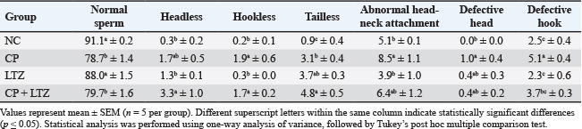
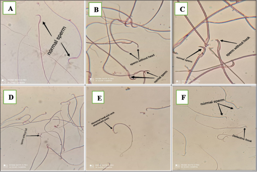
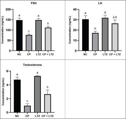
AbstractBackground: Cisplatin (CP) is an effective anticancer agent whose clinical use is limited by organ toxicity, including gonadotoxicity and accompanying male infertility. Letrozole (LTZ) is a nonsteroidal aromatase inhibitor that may improve spermatogenesis by stimulating endogenous testosterone synthesis. Aim: This study assessed the protective effect of LTZ against CP-induced testicular toxicity in a rat model. Methods: Twenty adult male Sprague-Dawley rats were randomly assigned to four groups (n=5): NC, CP (7 mg/kg, single intraperitoneal dose), LTZ (1.5 mg/kg orally for 10 days), and CP + LTZ. The experimental period lasted for 28 days. Body weight, sperm count, sperm motility, sperm morphology, and serum follicle-stimulating hormone (FSH), luteinizing hormone (LH), and testosterone levels were assessed, and testicular tissues were histologically examined. Results: CP administration markedly reduced body weight gain, sperm count, and sperm motility and increased sperm morphological abnormalities. Serum levels of FSH, LH, and testosterone were also reduced in CP-treated rats. Histopathological examination revealed seminiferous tubular degeneration, germ cell necrosis, and interstitial damage. Coadministration of LTZ with CP improved sperm count, motility, and reproductive hormone concentrations and mitigated histopathological alterations. However, the morphological sperm abnormalities remained largely unchanged. Conclusion: CP causes critical structural and functional testicular damage. However, LTZ partially reduces CP-induced reproductive damage by restoring hormonal balance and improving spermatogenic activity. LTZ confers a promising adjunctive therapy to mitigate chemotherapy-induced infertility in males Keywords: Cisplatin, Letrozole, Rat model, Spermatogenesis, Testicular toxicity. IntroductionMale infertility remains a significant health concern globally, accounting for 30%–50% of infertility cases in couples, and impaired spermatogenesis is one of the most common causes of infertility (Eisenberg et al., 2023). Chemotherapy-induced gonadotoxicity is one of the etiological factors of male infertility that has gained increasing attention due to improved cancer survival rates and the emphasis on the quality of life after treatment (Markowska et al., 2024). Cisplatin (CP) is a platinum-based anticancer agent widely used to treat solid tumors (Mariconda et al., 2025). However, its clinical use is significantly limited by serious adverse effects, including nephrotoxicity (Tang et al., 2023), neurotoxicity (Finno et al., 2022), and gonadotoxicity (Abdel-Latif et al., 2022). CP-induced testicular damage poses a significant threat to male fertility, especially in patients of reproductive age, requiring effective strategies to mitigate its detrimental effects on the male reproductive system (Rahimi et al., 2022). Testicular injury from CP is multifactorial and involves oxidative stress (OS), inflammation, DNA damage, and apoptosis (Othman et al., 2023). CP targets rapidly growing cells, leading to the degeneration of seminiferous tubules, depletion of spermatogenic cells, disruption of the blood–testis barrier, and impaired steroidogenesis (Hamam et al., 2022; Mahran et al., 2025). Animal model studies have demonstrated reduced sperm count, motility, and viability following CP administration (Rahimi et al., 2022), accompanied by histopathological changes, including vacuolization of Sertoli cells, tubular atrophy, and Leydig cell dysfunction (Alharbi et al., 2025). Despite advances in fertility preservation techniques, such as sperm cryopreservation, these options are not always feasible or accessible, necessitating pharmacological interventions that can protect or restore spermatogenesis following chemotherapy. Aromatase is a key enzyme that converts androgens (e.g., testosterone) into estrogens (Alemany, 2022). Estrogens play a physiological role in male reproduction, but higher estrogen levels or an altered testosterone-to-estrogen ratio can negatively affect spermatogenesis, gonadotropin secretion, and testicular function (McBride and Coward, 2016). Aromatase inhibitors have emerged as promising agents to manage certain forms of male infertility. Letrozole (LTZ) is a nonsteroidal aromatase inhibitor that is clinically used to treat hormone-dependent breast cancer (Goldrat et al., 2022). It has been investigated for off-label use in male reproductive disorders (Ullur et al., 2025). LTZ reduces estrogen synthesis by inhibiting aromatase activity, thereby increasing endogenous testosterone levels and improving the hormonal milieu necessary for normal spermatogenesis. Previous studies have demonstrated that LTZ can enhance sperm motility and concentration, as well as serum testosterone levels, in males with oligospermia (Guo et al., 2022; Yang et al., 2022). LTZ improves spermatogenic activity in animal models (Nokhbeh Zaeem et al., 2025), which could be useful in treating CP-induced testicular injury. The rat model is widely used in reproductive toxicology studies because its spermatogenic cycle and hormonal regulation are well characterized (Liu et al., 2023). CP-induced testicular injury in rats mimics the pathological changes seen in humans (Hamam et al., 2022), making it an appropriate experimental model for evaluating therapeutic interventions aimed at preserving male fertility. Therefore, this study was designed to investigate the effect of LTZ on spermatogenesis in a rat model of CP-induced testicular tissue injury. This study aimed to elucidate the potential role of LTZ in improving chemotherapy-induced male reproductive toxicity. The results of this study may provide a basis for future translational research and substantiate the development of new strategies to enhance male fertility in oncologic settings. Materials and MethodsAnimals and their treatment groupsTwenty male Sprague-Dawley rats (weight, 210–230 g) were provided by the Animal House at the College of Veterinary Medicine, University of Sulaimani, Iraq. The rats were housed in polypropylene plastic cages with wood chips as bedding and acclimatized to the laboratory conditions at 24 ± 1ºC under a 12-hour dark–light cycle. Commercial rat pellets and water were provided ad libitum. Rats were acclimatized to the environment for two weeks before being used in the experiment. The rats were randomly divided into four groups, each containing five rats. The sample size of five rats per group was selected based on ethical considerations to minimize animal use in accordance with the principles of the 3Rs (Replacement, Reduction, and Refinement), while maintaining sufficient statistical power to detect biologically relevant differences. Group 1 served as a negative control (NC) and received a single intraperitoneal injection of isotonic saline and orally administered corn oil (0.6 mL/kg) for 10 days, starting nine days before CP treatment. Group 2 was injected intraperitoneally with CP (50 mg/100 ml, Code 1876A, Faulding Pharmaceuticals Pic, Warwickshire, UK) at a single dose of 7 mg/kg (Ateşşahin et al., 2006) on day 10 of the experiment. Group 3 was administered LTZ (Swiss Medic, Switzerland) orally at 1.5 mg/kg, suspended in corn oil, for 10 days, starting nine days before CP treatment (Abed et al., 2022). Group 4 received CP (7 mg/kg on day 10) and LTZ (1.5 mg/kg for 10 days). Isotonic saline was used as the CP vehicle. The study duration was 28 days. Rats were weighed on days 0, 7, 14, 21, and 28 using a digital balance. Animal sacrifice and blood and testicular sample collectionOn day 28, the rats were weighed and anesthetized with an intraperitoneal injection of a mixture of ketamine (100 mg/kg) and xylazine (20 mg/kg) (Mohammed-Rashid and Dyary, 2025). They were then sacrificed by exsanguination (blood was drawn from the heart). The collected blood was stored in plain tubes to separate the serum, which was later used to determine the levels of FSH, LH, and testosterone. The testicular specimens were removed immediately after sacrifice and washed thoroughly with isotonic saline. The organs were then cut into pieces, fixed in 10% neutral buffered formalin for at least two days, and embedded in paraffin wax. Histopathological examination of the testicular tissuesTesticular tissue sections of about 4.5×4.5×4.0 µm were cut from the embedded tissues using a microtome. Sections were mounted on glass slides using a hot plate and stained with Harris’s hematoxylin and eosin for light microscopic study. Tissue sections were examined for histopathological alterations, including hypospermatogenesis, germinal epithelium exfoliation, hydropic degeneration, and necrosis. Lesions were scored as 0 (no alterations), 1 (degenerative changes involving 1%–25% of the tissue sections), 2 (degenerative changes involving 26%–55% of the tissue sections), 3 (degenerative changes involving 51%–75% of the tissue sections), and 4 (degenerative changes involving 76%–100% of the tissue sections). Sperm countThe epididymal sperm count procedure was initiated by combining 200 mg of caudal epididymis with 1.8 ml of 0.9% NaCl at 37°C in a 30-mm Petri dish. The caudal epididymis was sliced into four pieces using a pair of scissors, and the tissue was left alone for 30–60 seconds to allow sperm to leak from the tubules. The fluid was collected in a 1.5-ml Eppendorf tube, followed by sperm cell count, morphology, and semen analysis. About 20 µl of semen was mixed with 180 µl of a diluent prepared by adding sodium bicarbonate (5 g), formalin (1 ml), and distilled water (up to 1000 ml) in an enhanced Neubauer hemocytometer with a cover glass, and the sperm concentration was calculated as the average of five distinct fields under the Neubauer chamber multiplied by 10^4 to yield the total sperm count (Mohammed et al., 2025). Sperm abnormalitiesA drop of semen suspension was placed on a warm slide to analyze sperm morphology. The smear was then air-dried, stained with hematoxylin for 15 minutes, washed with tap water, dried at room temperature, and finally stained with 1% eosin for 10 minutes. Smears were inspected at 100× magnification while submerged in oil. Per animal, 400 sperm cells were analyzed to identify morphological anomalies. Any abnormalities in the anatomy and morphology of the head, the tail, or both were regarded as abnormal, and the number of abnormal spermatozoa was counted. Sperm motilitySemen samples (10 µl) were immediately placed straight onto a slide that had been warmed to 37 °C. The slide was then covered with a cover slip and examined under 400× magnification using phase-contrast optics. Ten microscope fields were chosen at random, and each field was used to measure the motility of 10 spermatozoa. As a result, 100 spermatozoa were randomly evaluated for motility, and the results were classified as motile and immotile spermatozoa (Mohammed et al., 2025). Serum biochemistrySerum levels of FSH, LH, and testosterone were assessed using the enzyme-linked immunosorbent assay (ELISA) method as directed by the manufacturer’s kits of each kit in Rat FSH ELISA Kit (BT LAB, Code EA0015Ra, China), Rat LH ELISA Kit (BT LAB, Code EA0012Ra, China), and Rat Testosterone ELISA Kit (BT LAB, Code EA0023Ra, China). Statistical analysisData were statistically evaluated using the statistical package for social sciences (IBM SPSS version 24.0, Chicago, IL) and expressed as means ± standard error of means. A one-way analysis of variance and Tukey’s post hoc test were used to determine differences between groups for parametric data, and the Kruskal–Wallis test was used to compare nonparametric data. A difference of ≤ 0.05 between the groups was considered significant. Ethical approvalThe study protocol was approved by the scientific and ethical committees of the College of Veterinary Medicine, University of Sulaimani (approval number AUP-2025-15). ResultsBody weightsThe mean body weights of the rats in the experimental groups at days 0, 7, 14, 21, and 28 are shown in Fig. 1. On day 0, no statistically significant differences were observed among the groups, confirming successful randomization, comparable initial physiological status, and the absence of baseline weight bias. This outcome validates subsequent intergroup comparisons. CP administration resulted in a significant reduction in body weight from day seven onward compared with the NC group (p < 0.05). LTZ alone did not significantly alter body weight relative to controls. Coadministration of LTZ partially attenuated CP-induced weight loss during the early and mid-experimental periods and restored body weight to levels comparable to those of NCs by day 28. Sperm count and motilityCP administration significantly reduced sperm concentration and motility compared with the NC group (p < 0.05). LTZ alone had no significant effect on either parameter (Fig. 2). The coadministration of CP and LTZ significantly improved CP-induced reductions in sperm count and motility, restoring the values to levels comparable to the NCs. Sperm morphologyTable 1 summarizes the percentage distribution of normal and morphologically abnormal spermatozoa among the experimental groups. Treatment with CP caused a significant deterioration of sperm morphology compared with the NC group. The percentage of normal sperm was significantly reduced (p < 0.05) in the CP-treated group, with a significant increase in total morphological abnormalities. The most common defect observed in the CP-treated group was abnormal head–neck attachment, with the highest frequency among the recorded abnormalities. In addition, the incidence of spermatozoa with a defective or no hook, lacking he tail, lacking the head, or with an abnormal head–neck attachment increased significantly in the CP-only-treated rats compared with the NCs (Fig. 3). Treatment with LTZ alone in group 3 did not cause marked alterations in most morphological parameters compared with the NC group, indicating that LTZ did not adversely affect the spermatozoa’s structural integrity. However, the coadministration of LTZ with CP did not reduce the percentage of abnormal spermatozoa compared with the CP-alone group (79.7% vs 78.7%, respectively). Overall, CP induces significant morphological abnormalities in the spermatozoa, mostly affecting the head–neck junction, whereas coadministration of LTZ with CP confers partial protection against CP-induced spermatogenic damage. FSH, LH, and testosterone levelsCP administration significantly disrupted reproductive hormone homeostasis (Fig. 4). Serum FSH levels were significantly lower in the CP group than in the NC and LTZ groups (p < 0.05). The coadministration of CP and LTZ significantly increased FSH concentrations compared with CP alone, but the values remained significantly lower than those observed in the NC and LTZ groups. Serum LH levels were markedly lower in the CP group than in the NC and LTZ groups. In contrast, LH levels were partially restored in the CP + LTZ group compared with CP alone, and LH concentrations in the LTZ group were comparable to those in the NC group.
Fig. 1. Body weights of the rats in the experimental groups at the indicated time points were recorded. Data are presented as mean ± SEM (n=5 per group). Different superscript letters denote statistically significant differences among groups (p < 0.05). Statistical analysis was performed using one-way analysis of variance, followed by Tukey’s post hoc multiple comparison test. Serum testosterone levels were profoundly lower following CP administration, indicating impaired Leydig cell steroidogenic activity. LTZ alone elevated testosterone levels compared with the NC group, but the difference was not statistically significant. Importantly, coadministration of LTZ with CP significantly attenuated CP-induced testosterone reduction, restoring levels toward NC values, although they remained significantly lower than those in the NC and LTZ groups. Collectively, these findings demonstrate that CP administration significantly suppresses gonadotropins and testosterone, while LTZ exerts a protective role in preserving steroidogenic capacity under CP-induced testicular injury.
Fig. 2. Effects of LTZ on sperm count and motility in rats with CP-induced testicular toxicity. Values are expressed as mean ± SEM. Different superscript letters indicate significant differences among groups at p < 0.05. Statistical analysis was performed using one-way analysis of variance, followed by Tukey’s post hoc multiple comparison test. Table 1. Percentage distribution of normal and morphologically abnormal spermatozoa in the control and treated groups
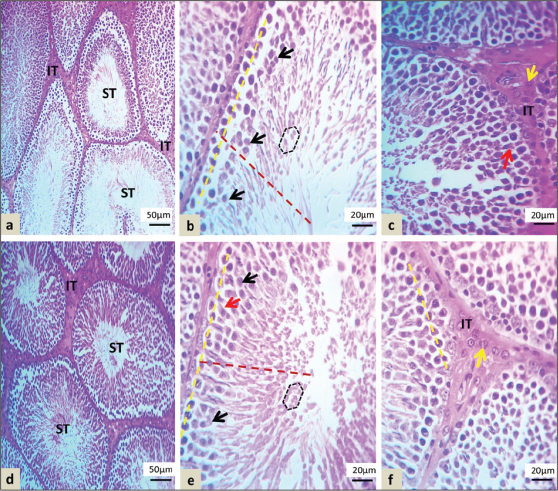
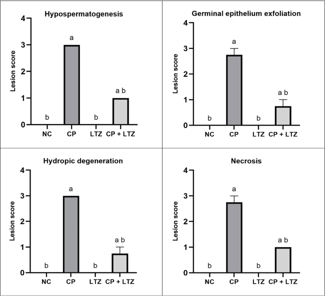
Histopathological findingsThe microscopic features of rats from the NC group showed intact histologic structures, with organized, multiple, rounded seminiferous tubules supplied by normal vasculature and composed of germinal epithelium (4–6 layers) at different stages of spermatogenesis (from spermatogonia to spermatozoa), with high cellularity of spermatids. The lumina of the seminiferous tubules were filled with the mature sperm’s whirling flagella. The interstitial spaces between the tubules contained intact Leydig cells (Fig. 5 a-c). In the CP group, lesion severity was increased and characterized by seminiferous tubule dilation, germ lineage (spermatogonia) sloughing or exfoliation, and necrotic spermatogonia that showed pyknotic nuclear changes. The primary and secondary spermatocytes underwent severe degeneration, marked germ cell depletion, and a reduction in spermatozoa and spermatid numbers, as well as marked interstitial degeneration with pyknotic Leydig cells (Fig. 5 d-f). Rats treated with LTZ showed well organized and nearly normal histological structures of seminiferous tubules with germinal cells, as well as maintaining their associations and cellularity with the intact interstitial cells (Fig. 6 d-f). The semiquantitative scoring of testicular histopathological lesions, including hypospermatogenesis, germinal epithelium exfoliation, hydropic degeneration, and necrosis, in the different experimental groups is shown in Fig. 7. Compared with the NC and LTZ groups, CP administration significantly increased lesion scores for all evaluated parameters. LTZ alone did not induce significant histopathological alterations compared with NC. Coadministration of CP and LTZ reduced CP-induced histopathological damage, as reflected by significantly lower lesion scores compared with CP alone, although values remained higher than those of the NCs.
Fig. 3. Representative photomicrographs of sperm morphological abnormalities in rat epididymal samples. (A) Normal spermatozoon; (B) headless spermatozoon; (C) hookless spermatozoon; (D) tailless spermatozoon; (E) spermatozoon exhibiting abnormal head-neck attachment and defective head morphology; (F) spermatozoon with defective hook attachment. Stained with 1% Y-eosin; magnification ×400.
Fig. 4. Effects of CP and LTZ on serum reproductive hormone levels in male rats. Values are expressed as mean ± SEM. Different letters above the bars indicate significant differences among groups at p < 0.05. Statistical analysis was performed using one-way analysis of variance, followed by Tukey’s post hoc test.
Fig. 5. Light microscopic sections of the seminiferous tubules. a–c: Normal arrangement of seminiferous tubules (ST) at different spermatogenic stages (red dashed lines) and spermatozoa with normal cellularity, intact Sertoli cells (red arrows), and spermatozoa that filled the lumen, well-organized interstitial space (IT) with intact Leydig cells (yellow arrows) in the NC group. d-f: Dilated seminiferous tubules with a disorganized germinal epithelium, hypospermatogenesis (red dashed line), exfoliated spermatogonia with pyknotic features (yellow dashed lines), marked spermatogenic cell degeneration (red dashed lines), and degenerated Leydig cells with moderate interstitial edema (yellow arrows) in the CP group (hematoxylin and eosin stain). LTZ administration with CP resulted in an obvious repair of seminiferous tubule abnormalities. The combination enhanced the mitotic activity of spermatogenic and Leydig cells. It reduced CP-induced seminiferous tubule damage, with only mild enlargement of seminiferous tubules and mild degeneration of germ cells, including primary and secondary spermatocytes, with Sertoli cells and intact interstitial cells within a normal, organized interstitial space (Fig. 6 a-c).
Fig. 6. Light microscopic sections of the seminiferous tubules of rats. a–c: Mild dilation of the seminiferous tubules (ST), mild degeneration of spermatogenic cells (red dashed lines), and Sertoli cells (red arrows), detached and mild degeneration of spermatogonia (yellow dashed lines), normal interstitial space, and intact Leydig cells in the CP + LTZ group. d–f: Normal histological structures of seminiferous tubules (ST), intact germ cells (yellow dashed lines) with spermatogenic cells (red dashed lines), and Sertoli cells (red arrows), normal morphology with the organization of interstitial space (IT) and Leydig cells as indicated by yellow arrows in the LTZ group (hematoxylin and eosin stain). DiscussionThis study demonstrates that CP induces marked reproductive toxicity in male rats, as evidenced by reduced body weight gain, impaired spermatogenesis, altered sperm quality, disruption of reproductive hormone balance, and marked histopathological damage of the testicular parenchyma. Oral administration of LTZ showed a partial protective effect against CP-induced structural and functional changes, particularly in sperm count, motility, reproductive hormones, and histological integrity. The significant reduction in body weight observed in CP-treated rats from day 7 onward is consistent with the well-documented systemic toxicity of CP (Katanić Stanković et al., 2023). Body weight loss is associated with reduced feed intake, gastrointestinal toxicity, metabolic stress, and generalized catabolic effects associated with platinum-based chemotherapeutic agents (Konstantinidis et al., 2025). Reproductive toxicity induced by CP was obvious through a marked reduction in sperm concentration and motility. These findings are consistent with previous studies demonstrating that CP disrupts spermatogenesis, primarily through OS, DNA crosslinking, mitochondrial dysfunction, and apoptosis of rapidly dividing germ cells (Hamam et al., 2022; Peng et al., 2023; Zhang et al., 2025). Due to their high proliferative potential, spermatogonia and primary spermatocytes are particularly susceptible to CP-induced DNA damage (Rahimi et al., 2022). As a result, germinal epithelium depletion and compromised spermatogenesis lead to reduced epididymal sperm output and impaired motility (Hasan et al., 2022). The significant increase in total sperm morphological abnormalities in the CP group further confirms impaired spermatogenic proliferation. The predominance of head–neck attachment defects indicates impairment in spermiogenesis, particularly during chromatin condensation, acrosome formation, and flagellar assembly (Yogo, 2022). Structural disarrangement of the head–neck junction indicates faulty microtubule organization, which is highly susceptible to OS and cytoskeletal disruption (Gunes et al., 2020).
Fig. 7. Histopathological lesion scores in the testicular tissue of the experimental groups. Different letters above the bars indicate statistically significant differences among the groups (p < 0.05). Statistical comparisons were performed using the Kruskal–Wallis test followed by an appropriate post hoc multiple comparison test. CP administration markedly reduced serum concentrations of FSH, LH, and testosterone, indicating disruption of the hypothalamic–pituitary–gonadal axis. Decreased LH concentration directly reduced Leydig cell stimulation, diminishing testosterone synthesis (Lei et al., 2025). The histological manifestations of pyknotic and degenerated Leydig cells detected in the CP group confirm this endocrine inhibition. Decreased FSH concentrations may impair Sertoli cell function, reduce support for germ cell maturation, and contribute to hypospermatogenesis (Li et al., 2024). Low testosterone levels further arrest spermatogenesis, as intratesticular testosterone is crucial for maintaining meiosis and spermiogenesis (Grande et al., 2022). Altogether, the combined suppression of gonadotropin and androgen production provides a mechanistic explanation for the marked impairment of spermatogenesis observed in CP-treated rats. Treatment with LTZ alone did not adversely affect sperm parameters, hormone concentrations, or testicular histology, indicating that under physiological conditions, short-term aromatase inhibition does not inhibit male reproductive function. Excessive estrogen suppression can, in theory, disrupt spermatogenesis (Cooke and Walker, 2022; Dewaele et al., 2022). However, LTZ preserved the testicular structural and functional integrity in this study. Coadministration of LTZ with CP significantly improved sperm count and motility and partially restored the levels of FSH, LH, and testosterone. Mechanistically, LTZ suppresses the aromatase-mediated conversion of testosterone to estradiol, reducing the negative estrogenic feedback at the hypothalamic–pituitary level (Maurya et al., 2026). The result is enhanced gonadotropin release, especially LH, stimulating Leydig cell steroidogenesis (Kucherov et al., 2011). Partial restoration of testosterone in the CP + LTZ group coincides with improved functional capacity of Leydig cells. Although LTZ improved quantitative sperm parameters and endocrine function, it did not reduce the overall percentage of morphologically abnormal spermatozoa. Although LTZ stimulates spermatogenic output and steroidogenic activity, it may not fully inhibit CP-induced structural DNA damage and cytoskeletal alterations during spermiogenesis. Morphological sperm defects reflect irreversible genomic or microtubular injury that may persist despite hormonal restoration. The histopathological findings robustly support the functional data. Seminiferous tubular dilation, germ cell necrosis, exfoliation, reduced spermatogenic layers, and interstitial degeneration were observed in CP-treated testes. These lesions characterize chemotherapeutic gonadotoxicity and reflect oxidative injury, mitochondrial dysfunction, and apoptosis (Markowska et al., 2024; Sriram et al., 2024). Coadministration of CP with LTZ significantly mitigated these changes, resulting in improved tubular organization, preserved Sertoli cell structure, enhanced spermatogenic layers, and preserved interstitial architecture. The reduced Leydig cell degeneration in the CP + LTZ group was correlated with improved testosterone concentrations, highlighting the mutual relationship between structural integrity and endocrine function. Although mild degeneration was observed in the CP + LTZ group, the significant improvement compared with CP alone indicates that LTZ confers cytoprotective effects, possibly through endocrine modulation. Altogether, the present results indicate that CP induces testicular toxicity via multiple mechanisms, including direct germ cell cytotoxicity, Leydig cell dysfunction, gonadotropin secretion inhibition, and distortion of spermatogenic tissue architecture. LTZ partially mitigates these effects by stimulating gonadotropin release, restoring steroidogenic capacity, and preserving seminiferous tubular integrity. However, the partial restoration of sperm morphology and hormone levels indicates that LTZ confers partial protection against CP-induced gonadal injury. Persistent morphological deformities indicate irreversible DNA damage and structural alterations induced during the early stages of germ cell division. ConclusionLTZ partially attenuated CP-induced testicular toxicity in rats, improving sperm count, motility, reproductive hormone levels, and histopathological injury scores. However, the sperm morphological abnormalities were not substantially corrected. These findings support further investigation of LTZ as a potential modulator of chemotherapy-related male reproductive toxicity, although mechanistic and fertility outcome studies are still required. AcknowledgmentsNot applicable. FundingThis research received no funding. Authors’ contributionsThe MOM conducted the research and collected and analyzed the data. HOD conceptualized and conducted the research, analyzed the data, and wrote the manuscript. Conflict of interestThe authors have no conflicts of interest to declare. Data availabilityAll data supporting the findings of this study are available within the manuscript, and no additional data sources are required. ReferencesAbdel-Latif, R., Fathy, M., Anwar, H.A., Naseem, M., Dandekar, T. and Othman, E.M. 2022. Cisplatin-induced reproductive toxicity and oxidative stress: ameliorative effect of kinetin. Antioxidants 11, 863; doi:10.3390/antiox11050863 Abed, A., Judi, H. and Enad, A. 2022. Effect of letrozole administration as a selective aromatase inhibitor on male rat’s reproductive performance. Arch. Razi Inst. 77, 367-373; doi:10.22092/ARI.2021.356539.1864 Alemany, M. 2022. The roles of androgens in humans: biology, metabolic regulation and health. Int. J. Mol. Sci. 23, 11952; doi:10.3390/ijms231911952 Alharbi, F.K., Ali, L.S., Salem, G.A., Elashmouny, N., El-Kholy, S.S., Essawi, W.M., Helal, A.I., Ibrahim, H.S.A., Dahran, N. and El-Shetry, E.S. 2025. Glycyrrhizin alleviated cisplatin-induced testicular injury by inhibiting the oxidative, apoptotic, hormonal, and histological alterations. Am. J. Vet. Res. 86, ajvr.24.10.0288; doi:10.2460/ajvr.24.10.0288 Ateşşahin, A., Karahan, İ., Türk, G., Gür, S., Yılmaz, S. and Çeribaşı, A.O. 2006. Protective role of lycopene on cisplatin-induced changes in sperm characteristics, testicular damage and oxidative stress in rats. Reprod. Toxicol. 21, 42-47; doi:10.1016/j.reprotox.2005.05.003 Cooke, P.S. and Walker, W.H. 2022. Nonclassical androgen and estrogen signaling is essential for normal spermatogenesis. Semin. Cell Dev. Biol. 121, 71-81; doi:10.1016/j.semcdb.2021.05.032 Dewaele, A., Dujardin, E., André, M., Albina, A., Jammes, H., Giton, F., Sellem, E., Jolivet, G., Pailhoux, E. and Pannetier, M. 2022. Absence of testicular estrogen leads to defects in spermatogenesis and increased semen abnormalities in male rabbits. Genes 13, 2070; doi:10.3390/genes13112070 Eisenberg, M.L., Esteves, S.C., Lamb, D.J., Hotaling, J.M., Giwercman, A., Hwang, K. and Cheng, Y.-S. 2023. Male infertility. Nat. Rev. Dis. Primers 9, 49; doi:10.1038/s41572-023-00459-w Finno, C.J., Chen, Y., Park, S., Lee, J.H., Perez-Flores, M.C., Choi, J. and Yamoah, E.N. 2022. Cisplatin neurotoxicity targets specific subpopulations and K+ channels in tyrosine-hydroxylase positive dorsal root ganglia neurons. Front. Cell. Neurosci. 16, 853035; doi:10.3389/fncel.2022.853035 Goldrat, O., De Cooman, M., Mailliez, A., Delbaere, A., D’Orazio, E., Demeestere, I. and Decanter, C. 2022. Efficacy and safety of controlled ovarian hyperstimulation with or without letrozole for fertility preservation in breast cancer patients: a multicenter retrospective study. Europ. J. Cancer 174, 134-141; doi:10.1016/j.ejca.2022.07.017 Grande, G., Barrachina, F., Soler-Ventura, A., Jodar, M., Mancini, F., Marana, R., Chiloiro, S., Pontecorvi, A., Oliva, R. and Milardi, D. 2022. The role of testosterone in spermatogenesis: lessons from proteome profiling of human spermatozoa in testosterone deficiency. Front. Endocrinol. 13, 852661; doi:10.3389/fendo.2022.852661 Gunes, S., Sengupta, P., Henkel, R., Alguraigari, A., Sinigaglia, M.M., Kayal, M., Joumah, A. and Agarwal, A. 2020. Microtubular dysfunction and male infertility. World J. Men Health 38, 9-23; doi:10.5534/wjmh.180066 Guo, B., Li, J.J., Ma, Y.L., Zhao, Y.T. and Liu, J.G. 2022. Efficacy and safety of letrozole or anastrozole in the treatment of male infertility with low testosterone-estradiol ratio: a meta-analysis and systematic review. Andrology 10, 894-909; doi:10.1111/andr.13185 Hamam, E.T., Awadalla, A. and Aboul-Naga, A.M. 2022. Zinc oxide nanoparticles attenuate prepubertal exposure to cisplatin-induced testicular toxicity and spermatogenesis impairment in rats. Toxicology 468, 153102; doi:10.1016/j.tox.2022.153102 Hasan, H., Bhushan, S., Fijak, M. and Meinhardt, A. 2022. Mechanism of inflammatory associated impairment of sperm function, spermatogenesis and steroidogenesis. Front. Endocrinol. 13, 897029; doi:10.3389/fendo.2022.897029 Katanić Stanković, J.S., Selaković, D. and Rosić, G. 2023. Oxidative damage as a fundament of systemic toxicities induced by cisplatin—the crucial limitation or potential therapeutic target? Int. J. Mol. Sci. 24, 14574; doi:10.3390/ijms241914574 Konstantinidis, I., Tsokkou, S., Gavriilaki, E., Delis, G. and Papamitsou, T. 2025. Protective role of key micronutrients in chemotherapy-induced organ toxicity: a comprehensive review of mechanistic insights and clinical implications. Nutrients 17, 2838; doi:10.3390/nu17172838 Kucherov, A., Polotsky, A.J., Menke, M., Isaac, B., McAvey, B., Buyuk, E., Bradford, A.P., Hickmon, C., Babbs, B. and Berga, S. 2011. Aromatase inhibition causes increased amplitude, but not frequency, of hypothalamic-pituitary output in normal women. Fertil. Steril. 95, 2063-2066; doi:10.1016/j.fertnstert.2011.01.158 Lei, T., Yang, Y. and Yang, W.-X. 2025. Luteinizing hormone regulates testosterone production, Leydig cell proliferation, differentiation, and circadian rhythm during spermatogenesis. Int. J. Mol. Sci. 26, 3548; doi:10.3390/ijms26083548 Li, L.; Lin, W., Wang, Z., Huang, R., Xia, H., Li, Z., Deng, J., Ye, T., Huang, Y. and Yang, Y. 2024. Hormone regulation in testicular development and function. Int. J. Mol. Sci. 25, 5805; doi:10.3390/ijms25115805 Liu, W., Zhang, B., Yao, Q., Feng, X., Shen, T., Guo, P., Wang, P., Bai, Y., Li, B. and Wang, P. 2023. Toxicological effects of micro/nano-plastics on mouse/rat models: a systematic review and meta-analysis. Front. Public Health. 11, 1103289; doi:10.3389/fpubh.2023.1103289 Mahran, Y., Badr, A.M., Aloyouni, S., Alkahtani, M.M., Sarawi, W.S., Ali, R., Alsultan, D., Almufadhili, S., Almasud, D.H. and Hasan, I.H. 2025. Morin hydrate protects against cisplatin-induced testicular toxicity by modulating ferroptosis and steroidogenesis genes’ expression and upregulating Nrf2/Heme oxygenase-1. Sci. Rep. 15, 22720; doi:10.1038/s41598-025-08235-4 Mariconda, A., Ceramella, J., Catalano, A., Saturnino, C., Sinicropi, M.S. and Longo, P. 2025. Cisplatin, the timeless molecule. Inorganics 13, 246; doi:10.3390/inorganics13070246 Markowska, A., Antoszczak, M., Markowska, J. and Huczyński, A. 2024. Gynotoxic effects of chemotherapy and potential protective mechanisms. Cancers 16, 2288; doi:10.3390/cancers16122288 Maurya, V.P., Tandon, A., Singh, R., Chouksey, P., Jain, V., Moscote-Salazar, L.R. and Agrawal, A. 2026. Expression, role, and clinical implications of P450 aromatase enzymes in the pituitary gland: a scoping review. Neurologico Spinale Medico Chirurgico 9. 7-13; doi:10.4103/nsmc.nsmc_14_25 McBride, J.A. and Coward, R.M. 2016. Recovery of spermatogenesis following testosterone replacement therapy or anabolic-androgenic steroid use. Asian J. Androl. 18, 373-380; doi:10.4103/1008-682X.173938 Mohammed-Rashid, S.S. and Dyary, H.O. 2025. Effect of catechin on hepatotoxicity induced by combined doxorubicin and paclitaxel treatment. J. Toxicol. 6775839; doi:10.1155/jt/6775839 Mohammed, M.O., Muhamad, F.A. and Dyary, H.O. 2025. Allicin attenuates doxorubicin-induced testicular and systemic toxicity in rats via antioxidant and anti-apoptotic mechanisms. Reprod. Toxicol. 109082; doi:10.1016/j.reprotox.2025.109082 Nokhbeh Zaeem, S., Heydari Nasrabadi, M., Salehipour, M. and Ehtesham, S. 2025. Letrozole and crocin: protecting Leydig cells and modulating androgen receptor and CYP19 gene expression in busulfan-induced azoospermia. Animals 15, 697; doi:10.3390/ani15050697 Othman, E.M., Habib, H.A., Zahran, M.E., Amin, A. and Heeba, G.H. 2023. Mechanistic protective effect of cilostazol in cisplatin-induced testicular damage via regulation of oxidative stress and TNF-α/NF-κB/caspase-3 pathways. Int. J. Mol. Sci. 24, 12651; doi:10.3390/ijms241612651 Peng, Z., Xin, W.Z., Sheng, Z.Y., Zi, T., Nan, L.Y., Lin, W.Z., Jun, L.F. and Xia, L.X. 2023. Melatonin alleviates cisplatin-induced mice spermatogenesis defects. Reprod. Toxicol. 119, 108391; doi:10.1016/j.reprotox.2023.108391 Rahimi, A., Asadi, F., Rezghi, M., Kazemi, S., Soorani, F. and Memariani, Z. 2022. Natural products against cisplatin-induced male reproductive toxicity: a comprehensive review. J. Biochem. Mol. Toxicol. 36, e22970; doi:10.1002/jbt.22970 Sriram, S., Macedo, T., Mavinkurve-Groothuis, A., van de Wetering, M. and Looijenga, L.H. 2024. Alkylating agents-induced gonadotoxicity in prepubertal males: Insights on the clinical and preclinical front. Clin. Transl. Sci. 17, e13866; doi:10.1111/cts.13866 Tang, C., Livingston, M.J., Safirstein, R. and Dong, Z. 2023. Cisplatin nephrotoxicity: new insights and therapeutic implications. Nat. Rev. Nephrol. 19, 53-72; doi:10.1038/s41581-022-00631-7 Ullur, P., Kubera, N., Manikandan, S., Nanda, N., Venkatraman, S. and Ram, A. 2025. Efficacy and safety of letrozole in improving semen parameters of subfertile men with moderate-to-severe oligoasthenoteratozoospermia: A Placebo-controlled randomized trial. J. Hum. Reprod. Sci. 18, 2-8; doi:10.4103/jhrs.jhrs_160_24 Yang, C., Li, P. and Li, Z. 2022. Clinical application of aromatase inhibitors to treat male infertility. Hum. Rep. 28, 30-50; doi:10.1093/humupd/dmab036 Yogo, K. 2022. Molecular basis of the morphogenesis of sperm head and tail in mice. Reprod. Med. Biol. 21, e12466; doi:10.1002/rmb2.12466 Zhang, H., Zhang, Y.-z., Liu, W., Tang, S., Ren, S., Wang, Z., Zhu, H.-y., Li, X.-d., Zhang, J. and Li, W. 2025. Effect of stem extract of Schisandra chinensis on improving spermatogenesis disorder induced by cisplatin in vivo and in vitro. Phytomedicine 136, 156274; doi:10.1016/j.phymed.2024.156274 | ||



| How to Cite this Article |
| Pubmed Style Mohammed MO, Dyary HO. Effect of letrozole on cisplatin-induced testicular tissue damage in a rat model. Open Vet. J.. 2026; 16(8): 5309-5320. doi:10.5455/OVJ.2026.v16.i8.25 Web Style Mohammed MO, Dyary HO. Effect of letrozole on cisplatin-induced testicular tissue damage in a rat model. https://www.openveterinaryjournal.com/?mno=312116 [Access: August 08, 2026]. doi:10.5455/OVJ.2026.v16.i8.25 AMA (American Medical Association) Style Mohammed MO, Dyary HO. Effect of letrozole on cisplatin-induced testicular tissue damage in a rat model. Open Vet. J.. 2026; 16(8): 5309-5320. doi:10.5455/OVJ.2026.v16.i8.25 Vancouver/ICMJE Style Mohammed MO, Dyary HO. Effect of letrozole on cisplatin-induced testicular tissue damage in a rat model. Open Vet. J.. (2026), [cited August 08, 2026]; 16(8): 5309-5320. doi:10.5455/OVJ.2026.v16.i8.25 Harvard Style Mohammed, M. O. & Dyary, . H. O. (2026) Effect of letrozole on cisplatin-induced testicular tissue damage in a rat model. Open Vet. J., 16 (8), 5309-5320. doi:10.5455/OVJ.2026.v16.i8.25 Turabian Style Mohammed, Mardin Omer, and Hiewa Othman Dyary. 2026. Effect of letrozole on cisplatin-induced testicular tissue damage in a rat model. Open Veterinary Journal, 16 (8), 5309-5320. doi:10.5455/OVJ.2026.v16.i8.25 Chicago Style Mohammed, Mardin Omer, and Hiewa Othman Dyary. "Effect of letrozole on cisplatin-induced testicular tissue damage in a rat model." Open Veterinary Journal 16 (2026), 5309-5320. doi:10.5455/OVJ.2026.v16.i8.25 MLA (The Modern Language Association) Style Mohammed, Mardin Omer, and Hiewa Othman Dyary. "Effect of letrozole on cisplatin-induced testicular tissue damage in a rat model." Open Veterinary Journal 16.8 (2026), 5309-5320. Print. doi:10.5455/OVJ.2026.v16.i8.25 APA (American Psychological Association) Style Mohammed, M. O. & Dyary, . H. O. (2026) Effect of letrozole on cisplatin-induced testicular tissue damage in a rat model. Open Veterinary Journal, 16 (8), 5309-5320. doi:10.5455/OVJ.2026.v16.i8.25 |


