





| Research Article | ||
Open Vet. J.. 2026; 16(8): 5285-5293 !
Open Veterinary Journal, (2026), Vol. 16(8): 5285–5293 Research Article Gallic acid enhances metformin-mediated improvement of ovarian architecture and estrogen receptor alpha expression in an estradiol benzoate-induced rat model of PCOS-like ovarian dysfunctionYudit Oktanella1*, Viski Fitri Hendrawan1, Gretania Residiwati1, Aulia Firmawati1, Andre Giovanni2, Widjiati Widjiati3, Ghina Kamilah4, Nur Fitrah Ramadhani Putri4, Jamilaturrosyidah Jamilaturrosyidah4, Nabilla Rizky Mahalita4, Tiara Arum Setyanti4 and Edo Brendo Stevano51Department of Veterinary Reproduction, Faculty of Veterinary Medicine, Universitas Brawijaya, Malang, Indonesia 2International Program in Ornamental Fish Technology and Aquatic Animal Health, International College, National Pingtung University of Science and Technology, Neipu, Taiwan 3Department of Veterinary Embryology, Faculty of Veterinary Medicine, Universitas Airlangga, Surabaya, Indonesia 4Professional Program of Veterinary Medicine, Faculty of Veterinary Medicine, Universitas Brawijaya, Malang, Indonesia 5Faculty of Medicine, Universitas Sriwijaya, Palembang, Indonesia *Corresponding Author: Yudit Oktanella. Department of Veterinary Reproduction, Faculty of Veterinary Medicine, Universitas Brawijaya, Malang, Indonesia. Email: yudito [at] ub.ac.id Submitted: 25/02/2026 Revised: 02/06/2026 Accepted: 17/06/2026 Published: 08/08/2026 © 2025 Open Veterinary Journal
AbstractBackground: Polycystic ovary syndrome (PCOS) is associated with anovulation, cystic ovarian morphology, and altered ovarian estrogen signaling. Estradiol benzoate-induced rodent models are useful for reproducing persistent estrus and cystic ovarian morphology, although they do not fully represent the endocrine and metabolic complexity of human PCOS. Gallic acid has antioxidant, anti-inflammatory, and cytoprotective properties that may complement metformin therapy. Aim: This study evaluated the adjunctive effect of gallic acid with metformin on ovarian histomorphology, follicular dynamics, and estrogen receptor alpha (ERα) immunoexpression in an estradiol benzoate-induced rat model of PCOS-like ovarian dysfunction. Methods: Eighteen female Wistar rats were induced with estradiol benzoate [2 mg/kg body weight (BW), intraperitoneally] for four consecutive days and then randomized into three PCOS-induced groups (n=6/group): [PCOS control (CP); 1% carboxymethylcellulose sodium], metformin monotherapy (T1; 10 mg/rat/day, approximately 67–77 mg/kg BW/day), and combination therapy (T2; metformin 10 mg/rat/day plus gallic acid 50 mg/kg BW/day). Treatments were administered orally for 14 days. Ovaries were collected for hematoxylin–eosin staining and ERα immunohistochemistry. Results: Compared with CP, metformin was associated with improved follicular morphology, and the metformin plus gallic acid group showed the most favorable histological profile. Follicular cyst counts decreased from 2.80 ± 0.54 (CP) to 0.50 ± 0.55 (T1) and 0.16 ± 0.41 (T2). Atretic follicles decreased from 7.50 ± 1.21 (CP) to 5.33 ± 0.20 (T1) and 2.67 ± 0.75 (T2), whereas corpora lutea counts increased from 4.50 ± 0.83 (CP) to 6.33 ± 0.21 (T1) and 11.83 ± 1.45 (T2). ERα-positive area was highest in CP (41.88% ± 4.12%) and was significantly lower in T2 (28.75% ± 2.15%; p < 0.05), whereas T1 showed an intermediate, non-significant reduction (35.07% ± 2.29%). Conclusion: Within the limitations of this exploratory design, adjunctive gallic acid with metformin improved ovarian histomorphology and reduced ERα immunopositivity relative to the PCOS control group. Further studies including healthy controls, gallic acid monotherapy, endocrine-metabolic profiling, oxidative stress markers, and molecular validation are required to confirm mechanistic effects. Keywords: Estrogen Receptor-α (ERα), Folliculogenesis, Gallic Acid, Metformin, Polycystic Ovary Syndrome (PCOS), Rats. IntroductionReproductive disorders can disrupt ovarian cyclicity and function, thereby reducing fertility and productivity in animals (Chen et al., 2022). Ovarian dysfunction affects folliculogenesis, ovulation, and corpus luteum formation, and cystic follicular conditions are characterized by persistent anovulatory follicles and altered ovarian architecture (Mason et al., 2014; Ibáñez et al., 2017; Fiorentino et al., 2023). Experimental rodent models allow controlled evaluation of ovarian morphological responses to candidate treatments (Oktanella et al., 2023; Aaly-Gharibeh et al., 2024), but each model reflects only selected aspects of the complex PCOS phenotype. Polycystic ovary syndrome (PCOS) is a heterogeneous endocrine-metabolic disorder characterized by ovulatory dysfunction, hyperandrogenism, and polycystic ovarian morphology in humans (Shah et al., 2022; Chaudhuri, 2023; Akinola et al., 2024; Pala et al., 2025). Estradiol benzoate-induced rodent models primarily reproduce persistent estrus, ovarian cystic morphology, follicular arrest, and altered estrogen-responsiveness. Therefore, this model is best interpreted as a PCOS-like ovarian dysfunction model rather than a complete representation of human PCOS, particularly because metabolic abnormalities such as insulin resistance and hyperandrogenism may not be fully reproduced. Estrogen signaling is central to follicular growth, granulosa cell function, and ovarian tissue remodeling. These effects are mediated mainly through estrogen receptor alpha (ERα), estrogen receptor beta, and G-protein-coupled estrogen receptor in the ovary. Altered ERα expression may indicate disrupted ovarian estrogen responsiveness in cystic ovarian conditions and may be useful for evaluating treatment-associated changes in ovarian tissue (Chauvin et al., 2022; Lakshmi et al., 2023). In estradiol-induced models, persistent estrogenic stimulation may be associated with increased ERα immunoreactivity in ovarian compartments, although interpretation requires caution without a healthy non-induced control group. Metformin is widely used in PCOS management because of its insulin-sensitizing and endocrine-modulating effects (Attia et al., 2023). It can support ovulatory function by improving metabolic homeostasis and reducing endocrine disturbances that impair follicular maturation (Pasquali, 2015; Rotimi et al., 2021). However, metformin alone may not fully address ovarian oxidative stress and inflammatory injury, which are frequently implicated in follicular arrest and atresia. Gallic acid (3,4,5-trihydroxybenzoic acid) is a polyphenolic compound found in many plants and is recognized for antioxidant, anti-inflammatory, and cytoprotective activities (Tohma et al., 2019; Gezer et al., 2024; Kumar et al., 2025). Previous animal studies suggest that gallic acid may reduce oxidative damage and improve ovarian histological features in experimentally induced ovarian dysfunction (Mazloom et al., 2019; Shah et al., 2022). Gallic acid has also been reported to support in vitro follicular development (Silva et al., 2021). These properties provide a rationale for evaluating gallic acid as an adjunct to metformin in PCOS-like ovarian dysfunction. Therefore, the present study was designed to evaluate whether gallic acid as an adjunct to metformin improves ovarian histopathology and ERα immunoexpression in an estradiol benzoate-induced rat model of PCOS-like ovarian dysfunction. We hypothesized that the combination treatment would be associated with fewer cystic and atretic follicles, more luteal structures, and lower ERα immunopositivity compared with metformin monotherapy and the PCOS control group. Materials and MethodsStudy site and animalsThe study was conducted from April to October 2024 at the Animal Laboratory and Animal Disease Diagnostic Laboratory, Faculty of Veterinary Medicine, and the Anatomical Pathology Laboratory, Faculty of Medicine, Universitas Brawijaya, Indonesia. Eighteen female Wistar rats (Rattus norvegicus), 8 weeks old and weighing 130–150 g, were used. After a 7-day acclimatization period, animals were housed in plastic cages with sterile pine shavings under controlled conditions (22°C ± 2°C; 14 hours light/10 hour dark cycle) and were provided standard feed and water ad libitum. Estrous cycle monitoring and synchronizationEstrous cyclicity was monitored twice daily (morning and evening) during acclimatization using vaginal cytology. A cotton swab moistened with 0.9% NaCl was gently inserted into the vagina and rotated to collect epithelial cells. The collected cells were transferred onto a glass slide, stained with methylene blue, and examined under a light microscope (400 × to 1,000 × magnification). Estrous stages were identified based on the relative proportions of nucleated epithelial cells, cornified epithelial cells, and leukocytes. Only rats showing regular cyclic patterns were included. Estrus synchronization was performed using two injections of prostaglandin F2α [PGF2α; 25 mg/kg body weight (BW), Capriglandin, manufactured by Sanbe Farma, Bandung, Indonesia administered 17 hours apart. PCOS inductionTwenty hours after the second PGF2α injection, each rat received estradiol benzoate (Java Animal Care, Bogor, Indonesia) at 2 mg/kg BW by intraperitoneal injection once daily for four consecutive days. The estradiol benzoate preparation was administered as an oil-based formulation with a final injection volume of 0.13 ml per rat. Model induction was considered successful when vaginal cytology showed persistent cornification, defined as predominance of cornified epithelial cells in repeated smears for at least two consecutive days after induction. This protocol was used to generate a PCOS-like ovarian phenotype characterized by persistent estrus and cystic ovarian morphology; hormonal and metabolic confirmation was not performed. Experimental design and drug administrationTwo days after confirmation of the PCOS-like model, rats were randomly allocated into three groups (n=6/group). The PCOS control (CP) group received 1% carboxymethylcellulose sodium (CMC-Na) orally at 1 ml/day. The metformin group (T1) received metformin monotherapy at 10 mg/rat/day, equivalent to approximately 67–77 mg/kg BW/day based on the body-weight range used in this study. The combination group (T2) received metformin (10 mg/rat/day) plus gallic acid (50 mg/kg BW/day). All treatments were administered orally by gavage for 14 consecutive days, a period selected to cover approximately three rat estrous cycles and to follow previous rodent PCOS treatment protocols (Tohma et al., 2019; Moazzam et al., 2024). Metformin (Dexa Medica, Tangerang, Indonesia) and gallic acid (>98%, Sigma-Aldrich, St. Louis, MO, USA) were dissolved or suspended in 1% CMC-Na and freshly prepared daily. No formal a priori power analysis was performed; the sample size was based on comparable exploratory animal studies and ethical considerations to minimize animal use. Sample collection and euthanasiaOn day 15, immediately after completion of the 14-day treatment period, rats were anesthetized by intramuscular injection of ketamine (100 mg/kg BW; Ket-A-100, Agrovet Market S.A., Lima, Peru) and xylazine (20 mg/kg BW; Xylazil-20, Troy Laboratories, Glendenning, NSW, Australia). Euthanasia was then performed by cervical dislocation. The ovaries were surgically removed, rinsed with physiological saline (0.9% NaCl) to remove blood, and fixed immediately in 10% neutral-buffered formalin for histopathological and immunohistochemical analyses. Histopathology and follicle assessmentFixed ovarian tissues were dehydrated through graded ethanol, cleared in xylene, embedded in paraffin, and sectioned at 4–5 µm. Sections were stained with hematoxylin and eosin and examined using a light microscope (Olympus CX-23, Olympus Corp., Tokyo, Japan). Follicular structures were identified according to established ovarian histomorphological criteria (Abd El-Galil and Mohammed, 2021). Follicular cysts were defined as enlarged antral follicles (largest visible diameter >400 µm in the examined section) with a thin granulosa cell layer and no visible oocyte or cumulus–oocyte complex in the examined section. Because the analysis was based on two-dimensional histological sections, absence of an oocyte was interpreted cautiously and in combination with follicle size and granulosa-layer morphology. Atretic follicles were identified by degenerating granulosa cells, pyknotic nuclei, and collapse of follicular architecture. Corpora lutea were identified as luteinized structures with eosinophilic cytoplasm and were interpreted as histological indicators of luteal activity rather than definitive proof of functional ovulation. Immunohistochemistry (ERα) and quantification
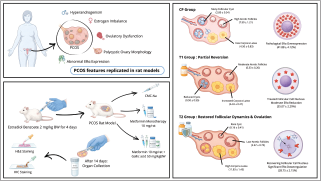
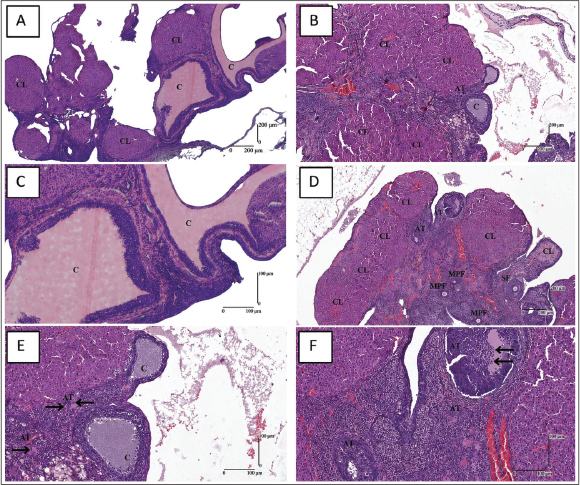
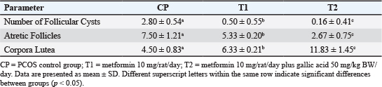
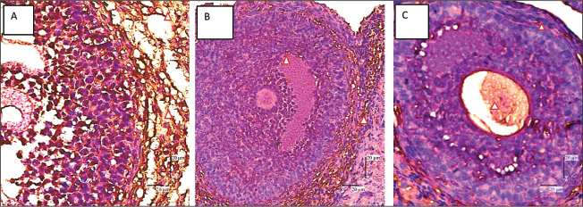
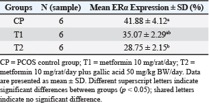
Fig. 1. Experimental workflow showing estradiol benzoate induction of PCOS-like ovarian dysfunction, treatment allocation into CP, T1, and T2 groups, treatment duration, and analytical endpoints. Paraffin-embedded ovarian sections (4–5 µm) were deparaffinized, rehydrated, and subjected to heat-induced antigen retrieval using Tris-EDTA buffer (pH 9.0). Sections were heated in a microwave/steamer until boiling and maintained slightly below boiling for 15–20 minutes, followed by cooling to room temperature. Endogenous peroxidase activity was blocked using 3% H2O2 for 10 minutes, and non-specific binding was blocked using 5% normal goat serum in PBS for 30 minutes at room temperature. Sections were incubated overnight at 4°C with a primary antibody against ERα (rabbit monoclonal anti-estrogen receptor-α, 1:200 dilution; Abcam, Cambridge, UK). They were then incubated with a horseradish peroxidase (EnVision+ System-HRP Labelled Polymer Anti-Rabbit; Dako, Agilent Technologies, Santa Clara, CA, USA). Immunoreactivity was visualized using the Dako Liquid DAB+ Substrate Chromogen System. A light hematoxylin counterstain was used for tissue orientation during microscopic evaluation. Digital images were captured from five comparable non-overlapping fields per ovary at 400× magnification using identical illumination and exposure settings. ERα immunoexpression was quantified using ImageJ software (National Institutes of Health, Bethesda, MD, USA). Images were processed using color separation to identify DAB-positive staining, and background was corrected using unstained tissue areas from the same image. A consistent threshold setting was then applied to all images within the same staining batch. ERα expression was calculated as the percentage of DAB-positive area relative to total tissue area in each field, and the mean value per animal was used for statistical analysis. Follicle counting and immunohistochemistry (IHC) quantification were performed using predefined criteria and standardized image settings; however, the absence of formal blinded assessment is acknowledged as a limitation. The overall experimental workflow is summarized in Figure 1. Statistical analysisQuantitative data are presented as mean ± standard deviation (SD). Statistical analyses were performed using International Business Machines Statistical Package for the Social Sciences Statistics version 25 (IBM Corp., Armonk, NY, USA). The analyzed variables were follicular cyst counts, atretic follicle counts, corpora lutea counts, and percentage area of ERα-positive staining. Normality was assessed using the Shapiro–Wilk test, and homogeneity of variance was assessed using Levene’s test. All variables met the assumptions for parametric analysis and were therefore analyzed using one-way analysis of variance (ANOVA), followed by Tukey’s honestly significant difference post hoc test. A p-value < 0.05 was considered statistically significant. Different superscript letters in the tables indicate significant post hoc differences among groups. Ethical approvalThe Research Ethics Committee of Brawijaya University granted this study a certificate of ethical clearance with the approval number 079-KEP-UB-2024. All experiments were carried out in strict compliance with the Guide for the Care and Use of Laboratory Animals and also followed the institutional guidelines for the humane treatment of animals. ResultsOvarian histopathology and follicle dynamicsRepresentative ovarian sections are shown in Figure 2. The PCOS control group (CP) showed cystic ovarian morphology characterized by enlarged antral follicles, thin granulosa cell layers, and follicular degeneration. Metformin-treated rats (T1) and rats receiving metformin plus gallic acid (T2) showed fewer cystic and atretic follicles. The T2 group also showed a higher number of luteal structures than the other groups, although these structures were interpreted as histological indicators and not as definitive functional confirmation of ovulation. Table 1 summarizes the quantitative analysis of follicular dynamics. Follicular cyst counts differed significantly among groups (p < 0.05). The number of follicular cysts decreased progressively from CP to T1 and T2, with significant post hoc differences among the three groups (p < 0.05). Atretic follicle counts also differed significantly among groups (p < 0.05). The T2 group showed the lowest number of atretic follicles and differed significantly from both CP and T1, suggesting reduced follicular degeneration in the combination treatment group. The number of corpora lutea differed significantly among groups (p < 0.05), with the highest count observed in T2. This finding suggests increased luteal structure formation after combination therapy. However, because progesterone levels, oocyte recovery, and continuous post-treatment estrous monitoring were not performed, the corpora lutea count should be interpreted as an indirect histological marker of ovulatory activity. ERα expression in ovarian tissueImmunohistochemical staining revealed ERα immunoreactivity as brown nuclear staining localized mainly in granulosa and theca cell compartments (Fig. 3). The CP group showed the highest ERα-positive staining area, consistent with increased ovarian estrogen receptor immunoreactivity in this estradiol benzoate-induced model. Quantitative analysis confirmed a significant group effect for ERα-positive area (Table 2; one-way ANOVA, p < 0.05). The CP group exhibited the highest mean ERα expression (41.88% ± 4.12%). The combination treatment group (T2) showed significantly lower ERα expression (28.75% ± 2.15%) than CP (p < 0.05). The metformin monotherapy group (T1) showed an intermediate ERα-positive area (35.07% ± 2.29%) and did not differ significantly from either CP or T2, as indicated by the shared superscript letters. Therefore, the ERα result supports a significant reduction only for the combination group relative to the PCOS control group. DiscussionThe results showed that metformin plus gallic acid produced more favorable ovarian histomorphological outcomes than the PCOS control treatment in an estradiol benzoate-induced rat model of PCOS-like ovarian dysfunction. The combination group had fewer follicular cysts and atretic follicles and more luteal structures than the PCOS control group. The ERα immunopositivity was also significantly reduced in the combination group, but an intermediate and statistically non-significant reduction of ERα positive area from metformin monotherapy was observed. This evidence may indicate a possible adjunct effect of gallic acid, but it has been considered as an exploratory study with limited endpoints.
Fig. 2. Representative hematoxylin–eosin-stained ovarian sections (4–5 µm) showing ovarian morphology across groups. Figure 2a,b: PCOS control group (CP); Figure 2c,d: metformin group (T1; metformin 10 mg/rat/day); Figure 2e,f: combination group (T2; metformin 10 mg/rat/day plus gallic acid 50 mg/kg BW/day). C: cystic follicle; CL: corpus luteum/luteal structure; AT: atretic follicle; SF: secondary follicle; MPF: multilaminar primary follicle; black arrow: apoptotic cell. Magnification: 40× and 100× Table 1. Number of follicular cysts, atretic follicles, and corpora lutea in rat ovaries.
The estradiol benzoate-induced model in this study is useful for generating persistent estrus and cystic ovarian morphology that also pertains to ovarian features of PCOS-based dysfunction. However, it is not able to fully replicate the endocrine-metabolic phenotype of human PCOS, particularly hyperandrogenism and insulin resistance. Therefore, the present findings indicate improvement relative to the PCOS control condition and cannot demonstrate complete restoration to a normal physiological state because a healthy non-induced control group was not included.
Fig. 3. Immunohistochemical analysis of ERα expression in ovarian tissue at 400× magnification. A: PCOS control group (CP); B: metformin group (T1; metformin 10 mg/rat/day); C: combination group (T2; metformin 10 mg/rat/day plus gallic acid 50 mg/kg BW/day). Red arrowhead: ERα-positive staining. Table 2. ERα expression in rat ovaries from all groups.
Compared to the PCOS control group, metformin improved numerous ovarian histological parameters. This finding corroborates previous reports that metformin may improve ovarian function by a combination of metabolic and endocrine-modulating effects (Pasquali, 2015; Gao et al., 2024). In the current study, the metformin treatment group demonstrated decreased cystic follicle count and increased luteal structure count compared with the PCOS control group, but ERα-positive area did not vary significantly between the metformin group and CP or T2. As a result, the metformin response should be characterized as partial histological improvement rather than complete normalization. Gallic acid likely offered further benefits by presenting antioxidant and anti-inflammatory activities in other studies (Mazloom et al., 2019; Rotimi et al., 2021; Shah et al., 2022; Gezer et al., 2024). In PCOS-like ovarian dysfunction, oxidative stress and low-grade inflammation are implicated in follicular arrest and atresia (Banerjee et al., 2023; Barcińska et al., 2024). However, oxidative stress markers, inflammatory cytokines, and gene expression were not quantified in the current study. Thus, gallic acid's antioxidant and anti-inflammatory activities are presented as literature-driven hypotheses and not as mechanistic results from this experiment. ERα contributes to ovarian reactivity to estrogenic stimuli and follicular remodeling (Camaioni et al., 2022; Chauvin et al., 2022). Greater ERα immunopositivity in CP may reflect altered receptor expression associated with persistent estrogenic stimulation in the estradiol benzoate-induced model. The lower ERα-positive area in T2 is linked to fewer cystic and atretic follicles, thereby indicating an association with improvement of ovarian morphology and decreased ERα immunoreactivity. However, because ERα was assessed only by immunohistochemistry, without RT-qPCR or protein quantification by other methods, this finding should be seen as a tissue-level observation rather than evidence of transcriptional regulation. More corpora lutea in T2 suggests increased luteal structure formation. However, corpora lutea counted in a single histological time point cannot definitively confirm active ovulation, because they may include residual luteal structures or luteinized follicles. Functional confirmation would require additional indicators such as progesterone measurement, oocyte recovery, fertility outcomes, or longitudinal estrous-cycle monitoring after treatment. Several molecular pathways have been linked to PCOS pathophysiology and polyphenol activity, including adenosine monophosphate-activated protein kinase, PI3K/AKT, mitogen-activated protein kinase, nuclear factor kappa-light-chain-enhancer of activated B cells, nuclear factor erythroid 2-related factor 2, and inflammasome-related signaling (Candelaria et al., 2013; Lin et al., 2022; Salahi et al., 2022; Zhou et al., 2022; Wang et al., 2023; Wang et al., 2024; Wei et al., 2026). In this study, these pathways were not experimentally evaluated. Accordingly, they are presented only as possible mechanisms that may explain the histological and IHC patterns and should be tested in future studies using oxidative stress assays, hormonal profiling, RT-qPCR, and protein validation. There are some significant limitations to this study. First, the absence of any healthy non-PCOS control group precluded any interpretation of whether the treatments restored parameters to their normal baseline levels. Second, there was no gallic acid-only group and therefore the independent effect of gallic acid and the potential for additive or synergistic interactions with metformin cannot be determined. Third, PCOS induction was confirmed through vaginal cytology and ovarian morphology without androgen, insulin, glucose, LH, FSH, estradiol, or progesterone measurements. Fourth, the sample size is small (n=6/group), no formal a priori power analysis was completed, and only one dose and one treatment duration were evaluated. Additional limitations include reliance on histology and IHC as primary endpoints, lack of reverse transcription quantitative polymerase chain reaction or other quantitative molecular validation for ERα, and absence of formal blinded evaluation. These limitations restrict the mechanistic strength of the conclusions. Future studies should include healthy and disease controls, gallic acid monotherapy, multiple doses, larger sample sizes with a priori power analysis, hormonal and metabolic profiling, oxidative stress markers, and molecular confirmation of ERα and related pathways. Overall, the results indicate that gallic acid combined with metformin may improve ovarian histomorphology and reduce ERα immunopositivity relative to untreated PCOS-like control rats. The findings support further investigation of gallic acid as an adjunctive therapy, but they do not establish complete physiological restoration or a definitive molecular mechanism. ConclusionIn this estradiol benzoate-induced PCOS-like ovarian dysfunction model, adjunctive gallic acid with metformin improved ovarian histomorphological features compared to the PCOS control group, as demonstrated by fewer cystic and atretic follicles and more luteal structures. The ERα immunopositivity of combination treatment also decreased compared to the PCOS control group, while ERα immunopositivity of metformin monotherapy was intermediate and non-significant. Our findings support the potential of gallic acid as an adjunct to metformin; however, confirmation must be carried out using studies with healthy controls, gallic acid monotherapy, endocrine-metabolic measurements, oxidative stress markers, and molecular validation. AcknowledgmentsThe authors gratefully appreciate the Directorate of Research and Community Services, Universitas Brawijaya, for the institutional support. We also appreciate the technical support from the staff of the Animal Laboratory and Animal Disease Diagnostic Laboratory, Faculty of Veterinary Medicine, and the Anatomical Pathology Laboratory, Faculty of Medicine, Universitas Brawijaya. Conflict of interestThe authors declare that there is no conflict of interest. No financial support, personal relationships, or affiliations with any commercial entities influenced the research, authorship, or publication of this manuscript. FundingThe Directorate of Research and Community Services at Universitas Brawijaya funded this research under the Hibah Penelitian Madya scheme (Grant No. 00145.13/UN10.A0501/B/PT.01.03.2/2024). Authors' contributionsAnalysis and interpretation: A.F., G.R., A.G., and E.B.S. Data collection: V.F.H., N.F.R.P., J., N.R.M., and T.A.S. Writing the article: Y.O., G.K., N.F.R.P., and N.R.M. Critical revision of the article: W.W. Statistical analysis: G.K. and N.F.R.P. Obtained funding: Y.O. and V.F.H. Overall responsibility: Y.O. All authors have read and agreed to the final version of the manuscript. Data availabilityThe data underlying the results of this study can be obtained from the corresponding author, if there is a reasonable request. ReferencesAaly-Gharibeh, Z., Hosseinchi, M. and Shalizar-Jalali, A. 2024. Effect of nanocurcumin on fertility in murine model of polycystic ovary syndrome. Vet. Res. Forum 15, 113–117. Abd El-Galil, M.M. and Mohammed, S.F. 2021. The possible effect of flaxseed extract on letrozole-induced polycystic ovary rat model: correlative histological and functional study. Al-Azhar. Med. J. 50, 3051–3096. Akinola, L.A., Inyangudo, G.N., Ottun, A.T., Olumodeji, A.M., Adewunmi, A.A., Olalere, F.H., Omololu, O.M. and Ogungbemile, B.D. 2024. Exploring serum vitamin D, sex hormones, and lipid profile disparities in women with and without polycystic ovarian syndrome: a case-control study. Cureus 16, e60975. Attia, G.M., Almouteri, M.M. and Alnakhli, F.T. 2023. Role of metformin in polycystic ovary syndrome (PCOS)-related infertility. Cureus 15, e44493. Banerjee, S., Cooney, L.G. and Stanic, A.K. 2023. Immune dysfunction in polycystic ovary syndrome. ImmunoHorizons 7, 323–332. Barcińska, J.M., Ostrowska-Czyżewska, A. and Szwed, D. 2024. Immunological abnormalities In PCOS. Qual. Sport. 36, 56517. Camaioni, A., Ucci, M.A., Campagnolo, L., De Felici, M. and Klinger, F.G. 2022. The process of ovarian aging: it is not just about oocytes and granulosa cells. J. Assist. Reprod. Genet. 39, 783–792. Candelaria, N.R., Liu, K. and Lin, C.Y. 2013. Estrogen receptor alpha: molecular mechanisms and emerging insights. J. Cell. Biochem. 114, 2203–2208. Chaudhuri, A. 2023. Polycystic ovary syndrome: causes, symptoms, pathophysiology, and remedies. Obes. Med. 39, 100480. Chauvin, S., Cohen-Tannoudji, J. and Guigon, C.J. 2022. Estradiol signaling at the heart of folliculogenesis: its potential deregulation in human ovarian pathologies. Int. J. Mol. Sci. 23, 512. Chen, P., Jia, R., Liu, Y., Cao, M., Zhou, L. and Zhao, Z. 2022. Progress of adipokines in the female reproductive system: a focus on polycystic ovary syndrome. Front. Endocrinol. 13, 881684. Fiorentino, G., Cimadomo, D., Innocenti, F., Soscia, D., Vaiarelli, A., Ubaldi, F.M., Gennarelli, G., Garagna, S., Rienzi, L. and Zuccotti, M. 2023. Biomechanical forces and signals operating in the ovary during folliculogenesis and their dysregulation: implications for fertility. Hum. Reprod. Update 29, 1–23. Gao, J., Huang, Y., Song, Y., Huang, X., Zhang, D. and Hou, Z. 2024. Therapeutic effects of Althaea officinalis L. and metformin on estradiol-induced polycystic ovary syndrome in rats: insights into The PI3K/AKT pathway, inflammation, and oxidative stress. Cell J. 26, 473–486. Gezer, A., Üstündağ, H., Kılıç Baygutalp, N., Erbaş, E. and Özkaraca, M. 2024. The protective effect of gallic acid against bisphenol a-induced ovarian toxicity and endocrine disruption in female rats. J. Med. Food. 27, 651–660. Ibáñez, L., Oberfield, S.E., Witchel, S., Auchus, R.J., Chang, R.J., Codner, E., Dabadghao, P., Darendeliler, F., Elbarbary, N.S., Gambineri, A., Garcia Rudaz, C., Hoeger, K.M., López-Bermejo, A., Ong, K., Peña, A.S., Reinehr, T., Santoro, N., Tena-Sempere, M., Tao, R., Yildiz, B.O., Alkhayyat, H., Deeb, A., Joel, D., Horikawa, R., De Zegher, F. and Lee, P.A. 2017. An International consortium update: pathophysiology, diagnosis, and treatment of polycystic ovarian syndrome in adolescence. Horm. Res. Paediatr. 88, 371–395. Kumar, S., Saharan, R., Saini, V. and Saini, A. 2025. Gallic acid: a wonderful remedy in medicinal field. Curr. Tradit. Med. 11(2), e111023222052. Lakshmi, J.N., Babu, A.N., Kiran, S.S.M., Nori, L.P., Hassan, N., Ashames, A., Bhandare, R.R. and Shaik, A.B. 2023. Herbs as a source for the treatment of polycystic ovarian syndrome: a systematic review. BioTech 12(1), 4. Lin, N., Lin, J., Plosch, T., Sun, P. and Zhou, X. 2022. An oxidative stress-related gene signature in granulosa cells is associated with ovarian aging. Oxidative Med. Cellular Longevity 2022, 1070968. Mason, H.D., Dilaver, N. and Rice, S. 2014. Ovarian dysfunction in polycystic ovary syndrome. In Polycystic Ovary Syndrome: Current and Emerging Concepts. Ed. Pal, L. New York, NY: Springer, pp: 53–70. Mazloom, B.F., Edalatmanesh, M.A. and Hosseini, S.E. 2019. Gallic acid reduces inflammatory cytokines and markers of oxidative damage in a rat model of estradiol-induced polycystic ovary. Comp. Clin. Pathol. 28, 1281–1286. Moazzam, A., Saleem, A., Shah, S., Hussain, L., Baig, M.M.F.A., Alshammari, A., Albekairi, N.A. and Akhtar, M.F. 2024. A combination of gliclazide and metformin attenuates obesity-induced polycystic ovary syndrome in female Wistar rats. Heliyon 10, e29015. Oktanella, Y., Untari, H., Wuragil, D.K., Ismiawati, H., Hasanah, N.A., Agustina, G.C. and Pratama, D.A.O. 2023. Evaluation of renal disturbance in animal models of polycystic ovary syndrome. Open Vet. J. 13(8), 1003–1011. Pala, K.M., Tomaszewska, W., Siudziński, P., Łyko, M., Skoczylas, A., Golińska, M., Nowak, A., Kurasz, J., Podlasiewicz, W., Dudziak, P. and Maj, W. 2025. The Hormonal dysregulation and its consequences in Obese Women—overview. J. Educ. Health. Sport. 77, 56907. Pasquali, R. 2015. Metformin in women with PCOS, Pros. Endocrine 48, 422–426. Rotimi, D., Ojo, O.A., Emmanuel, B.A., Ojo, A.B., Elebiyo, T.C., Nwonuma, C.O. and Oluba, O.M. 2021. Protective impacts of gallic acid against cadmium-induced oxidative toxicity in the ovary of rats. Comp. Clin. Pathol. 30, 453–460. Salahi, E., Amidi, F., Zahiri, Z., Aghahosseini, M., Mashayekhi, F., Amani Abkenari, S., Hosseinishenatal, S. and Sobhani, A. 2022. The effect of mitochondria-targeted antioxidant MitoQ10 on redox signaling pathway components in PCOS mouse model. Arch. Gynecol. Obstet. 305, 985–994. Silva, G.A.L., Araújo, L.B., Silva, L.C.R., Gouveia, B.B., Barberino, R.S., Lins, T.L.B.G., Monte, A.P.O., Macedo, T.J.S., Santos, J.M.S., Menezes, V.G., Silva, R.L.S. and Matos, M.H.T. 2021. Gallic acid promotes the in vitro development of sheep secondary isolated follicles involving the phosphatidylinositol 3-kinase pathway. Anim. Reprod. Sci. 230, 106767. Tohma, Y., Onalan, G., Tepeoglu, M., Bayraktar, N., Colak, E., Ozcimen, E. and Zeyneloglu, H. 2019. Phosphodiesterase 4 inhibitor plus metformin is superior to metformin alone for the treatment of polycystic ovary syndrome: a rat model study. Exp. Ther. Med. 17, 4013–4022. Ul Haq Shah, M.Z., Soni, M., Shrivastava, V.K., Mir, M.A. and Muzamil, S. 2022. Gallic acid reverses ovarian disturbances in mice with letrozole-induced PCOS via modulating Adipo R1 expression. Toxicol. Rep. 9, 1938–1949. Wang, F., Yin, Y., Nie, X., Zou, Y., Tong, X., Tong, Y., Zhou, J. and Tan, Y. 2023. Protocatechuic acid alleviates polycystic ovary syndrome symptoms in mice by PI3K signaling in granulosa cells to relieve ROS pressure and apoptosis. Gynecol. Endocrinol. 39, 2228917. Wang, Y.Q., Dong, Y.W., Qu, H.X., Qi, J.J., Yan, C.X., Wei, H.K., Sun, H., Sun, B.X. and Liang, S. 2024. Oleanolic acid promotes porcine oocyte maturation by activating the Nrf2/HO-1 signalling pathway. Theriogenology 230, 203–211. Wei, H., Zhang, Z., Zhang, S., Wang, J., Cui, X., Zhang, Z., Yu, J., Lei, X., Zhuge, X. and Huo, P. 2026. Resveratrol improves follicular development in PCOS rats by inhibiting the inflammatory response and pyroptosis of granulosa cells. Biol. Reprod. 114(4), 1227–1240. Zhou, S., Zhao, A., Wu, Y., Mi, Y. and Zhang, C. 2022. Protective effect of grape seed proanthocyanidins on oxidative damage of chicken follicular granulosa cells by inhibiting FoxO1-mediated autophagy. Front. Cell. Dev. Biol. 10, 762228. | ||
| How to Cite this Article |
| Pubmed Style Oktanella Y, Hendrawan VF, Residiwati G, Firmawati A, Giovanni A, Widjiati W, Kamilah G, Putri NFR, Jamilaturrosyidah J, Mahalita NR, Setyanti TA, Stevano EB. Gallic acid enhances metformin-mediated improvement of ovarian architecture and estrogen receptor alpha expression in an estradiol benzoate-induced rat model of PCOS-like ovarian dysfunction. Open Vet. J.. 2026; 16(8): 5285-5293. doi:10.5455/OVJ.2026.v16.i8.23 Web Style Oktanella Y, Hendrawan VF, Residiwati G, Firmawati A, Giovanni A, Widjiati W, Kamilah G, Putri NFR, Jamilaturrosyidah J, Mahalita NR, Setyanti TA, Stevano EB. Gallic acid enhances metformin-mediated improvement of ovarian architecture and estrogen receptor alpha expression in an estradiol benzoate-induced rat model of PCOS-like ovarian dysfunction. https://www.openveterinaryjournal.com/?mno=311909 [Access: August 08, 2026]. doi:10.5455/OVJ.2026.v16.i8.23 AMA (American Medical Association) Style Oktanella Y, Hendrawan VF, Residiwati G, Firmawati A, Giovanni A, Widjiati W, Kamilah G, Putri NFR, Jamilaturrosyidah J, Mahalita NR, Setyanti TA, Stevano EB. Gallic acid enhances metformin-mediated improvement of ovarian architecture and estrogen receptor alpha expression in an estradiol benzoate-induced rat model of PCOS-like ovarian dysfunction. Open Vet. J.. 2026; 16(8): 5285-5293. doi:10.5455/OVJ.2026.v16.i8.23 Vancouver/ICMJE Style Oktanella Y, Hendrawan VF, Residiwati G, Firmawati A, Giovanni A, Widjiati W, Kamilah G, Putri NFR, Jamilaturrosyidah J, Mahalita NR, Setyanti TA, Stevano EB. Gallic acid enhances metformin-mediated improvement of ovarian architecture and estrogen receptor alpha expression in an estradiol benzoate-induced rat model of PCOS-like ovarian dysfunction. Open Vet. J.. (2026), [cited August 08, 2026]; 16(8): 5285-5293. doi:10.5455/OVJ.2026.v16.i8.23 Harvard Style Oktanella, Y., Hendrawan, . V. F., Residiwati, . G., Firmawati, . A., Giovanni, . A., Widjiati, . W., Kamilah, . G., Putri, . N. F. R., Jamilaturrosyidah, . J., Mahalita, . N. R., Setyanti, . T. A. & Stevano, . E. B. (2026) Gallic acid enhances metformin-mediated improvement of ovarian architecture and estrogen receptor alpha expression in an estradiol benzoate-induced rat model of PCOS-like ovarian dysfunction. Open Vet. J., 16 (8), 5285-5293. doi:10.5455/OVJ.2026.v16.i8.23 Turabian Style Oktanella, Yudit, Viski Fitri Hendrawan, Gretania Residiwati, Aulia Firmawati, Andre Giovanni, Widjiati Widjiati, Ghina Kamilah, Nur Fitrah Ramadhani Putri, Jamilaturrosyidah Jamilaturrosyidah, Nabilla Rizky Mahalita, Tiara Arum Setyanti, and Edo Brendo Stevano. 2026. Gallic acid enhances metformin-mediated improvement of ovarian architecture and estrogen receptor alpha expression in an estradiol benzoate-induced rat model of PCOS-like ovarian dysfunction. Open Veterinary Journal, 16 (8), 5285-5293. doi:10.5455/OVJ.2026.v16.i8.23 Chicago Style Oktanella, Yudit, Viski Fitri Hendrawan, Gretania Residiwati, Aulia Firmawati, Andre Giovanni, Widjiati Widjiati, Ghina Kamilah, Nur Fitrah Ramadhani Putri, Jamilaturrosyidah Jamilaturrosyidah, Nabilla Rizky Mahalita, Tiara Arum Setyanti, and Edo Brendo Stevano. "Gallic acid enhances metformin-mediated improvement of ovarian architecture and estrogen receptor alpha expression in an estradiol benzoate-induced rat model of PCOS-like ovarian dysfunction." Open Veterinary Journal 16 (2026), 5285-5293. doi:10.5455/OVJ.2026.v16.i8.23 MLA (The Modern Language Association) Style Oktanella, Yudit, Viski Fitri Hendrawan, Gretania Residiwati, Aulia Firmawati, Andre Giovanni, Widjiati Widjiati, Ghina Kamilah, Nur Fitrah Ramadhani Putri, Jamilaturrosyidah Jamilaturrosyidah, Nabilla Rizky Mahalita, Tiara Arum Setyanti, and Edo Brendo Stevano. "Gallic acid enhances metformin-mediated improvement of ovarian architecture and estrogen receptor alpha expression in an estradiol benzoate-induced rat model of PCOS-like ovarian dysfunction." Open Veterinary Journal 16.8 (2026), 5285-5293. Print. doi:10.5455/OVJ.2026.v16.i8.23 APA (American Psychological Association) Style Oktanella, Y., Hendrawan, . V. F., Residiwati, . G., Firmawati, . A., Giovanni, . A., Widjiati, . W., Kamilah, . G., Putri, . N. F. R., Jamilaturrosyidah, . J., Mahalita, . N. R., Setyanti, . T. A. & Stevano, . E. B. (2026) Gallic acid enhances metformin-mediated improvement of ovarian architecture and estrogen receptor alpha expression in an estradiol benzoate-induced rat model of PCOS-like ovarian dysfunction. Open Veterinary Journal, 16 (8), 5285-5293. doi:10.5455/OVJ.2026.v16.i8.23 |


