





| Research Article | ||
Open Vet. J.. 2026; 16(8): 5340-5351 !
Open Veterinary Journal, (2026), Vol. 16(8): 5340–5351 Research Article Animal model-based generation of strong reactivity polyclonal antibodies against Toxoplasma gondii GRA7Siti Fadilla1,2, Loeki Enggar Fitri3,4*, Nuning Winaris3,4, Aulia Rahmi Pawestri3,4 Dearikha Karina Mayashinta3,4, Hanifa Rizky Rahmawati4, Ovi Sofia5, Dodit Suprianto6,7 and Suci Megasari81Master Program in Biomedical Science, Faculty of Medicine, Universitas Brawijaya, Malang, East Java, Indonesia 2Medical Doctor Profession Education, Faculty of Medical and Health Science, Maulana Malik Ibrahim State, Islamic University, Malang, Indonesia 3Department of Clinical Parasitology, Faculty of Medicine, Universitas Brawijaya, Malang, East Java, Indonesia 4AIDS, Toxoplasmosis, Opportunistic Disease, and Malaria Research Group, Faculty of Medicine, Universitas Brawijaya, Malang, East Java, Indonesia 5Department of Ophthalmology, Faculty of Medicine, Universitas Brawijaya/Dr. Saiful Anwar General Hospital, Malang, Indonesia 6Doctoral Program in Medical Science, Faculty of Medicine, Universitas Brawijaya, Malang, Indonesia 7Department of Electrical Engineering, Politeknik Negeri Malang, Malang, Indonesia 8Central Biomedical Laboratory, Faculty of Medicine, Universitas Brawijaya *Corresponding Author: Loeki Enggar Fitri. Department of Clinical Parasitology, Faculty of Medicine, Universitas Brawijaya, Malang, East Java, Indonesia. Email: lukief [at] ub.ac.id Submitted: 10/02/2026 Revised: 25/06/2026 Accepted: 10/07/2026 Published: 08/08/2026 © 2025 Open Veterinary Journal
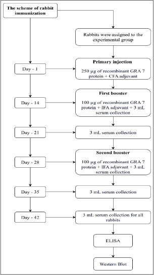
AbstractBackground: Toxoplasmosis is a zoonotic disease caused by the parasite Toxoplasma gondii (T. gondii), which can infect any warm-blooded intermediate host, including humans. Dense Granule Protein 7 (GRA7), which possesses high immunogenicity, is one of the potential T. gondii antigen candidates for diagnostic development. Aim: This study aimed to develop polyclonal antibodies specific to the recombinant GRA7 protein and evaluate their detection capability. Methods: Two male New Zealand White rabbits were immunized with recombinant GRA7 protein using a primary immunization protocol, followed by two booster doses on days 14 and 28. Serum samples were collected on days 14, 21, 28, 35, and 42 to analyze antibody levels using enzyme-linked immunosorbent assay (ELISA), while antibody specificity was tested using Western blot. Results: ELISA analysis demonstrated a significant increase in optical density (OD) values by day 28 (0.519, 1.050, and 1.067), followed by a pronounced peak at day 42 (1.480, 1.511, and 1.547) across all tested antigen concentrations (1, 5, and 10 µg/ml). These findings indicate the development of a robust and sustained humoral immune response. Western blot analysis confirmed specific recognition of the recombinant GRA7 protein at approximately 63 kDa, with protein bands appearing thicker and more distinct in the 10–6 dilution. This indicated that antibodies at the 10–6 dilution have a higher concentration, enabling a stronger signal in Western blot visualization, reflecting a high-affinity antibody response. Conclusion: The polyclonal antibodies generated against the recombinant GRA7 protein demonstrated strong preliminary immunoreactivity and high binding capacity, supporting their potential application as key components in the development of immunological diagnostic tools for toxoplasmosis. Further specificity assays are required to improve the performance of these newly produced antibodies. Keywords: GRA7, Polyclonal antibodies, Toxoplasma gondii, Western Blot, Zoonoses. IntroductionToxoplasmosis is a zoonotic disease caused by Toxoplasma gondii (T. gondii), an obligate intracellular protozoan parasite. Toxoplasma gondii can infect many other warm-blooded animal species, including humans (Abdallah et al., 2019; Akins et al., 2024). This infection is one of the most common chronic diseases worldwide and is estimated to affect approximately one-third of the global population, although its prevalence varies across geographical regions (Farhab et al., 2025). The prevalence of human toxoplasmosis infection in Indonesia was relatively high, ranging from 2% to 51%, as reported in 2007 (Soedarto, 2017). In Brazil, a study reported that the incidence of acute toxoplasmosis among pregnant women was 4.8 per 1,000 individuals, while congenital toxoplasmosis in newborns was recorded at 0.6 per 1,000 live births in 2009 (Mushlih et al., 2020). Toxoplasmosis is asymptomatic in most cases and poses no serious risk to immunocompetent individuals. However, severe consequences may arise, particularly in pregnant women and immunocompromised individuals (Rohmayani et al., 2022). Toxoplasma gondii has a complex life cycle that includes a sexual phase in the definitive host, namely cats, and an asexual phase in intermediate hosts, including humans, birds, and other mammals. In cats, the ingestion of bradyzoite-containing tissue cysts initiates the sexual phase in the small intestine, where bradyzoites invade intestinal epithelial cells, differentiate into microgametes and macrogametes, and subsequently fuse to form zygotes that develop into oocysts and are excreted in the feces in an unsporulated form. Within a few days, oocysts in the environment undergo sporulation and become infective, thereby enabling infection of intermediate hosts through contaminated food, water, or soil, as well as through the consumption of undercooked meat containing tissue cysts. Once ingested by an intermediate host, the parasite releases sporozoites or bradyzoites, which invade the intestinal epithelial cells and differentiate into tachyzoites, which disseminate systemically through the blood and lymph to various tissues. Subsequently, tachyzoites form tissue cysts containing bradyzoites as a persistent, slowly developing stage that can remain in the host throughout life and plays an important role in chronic infection and disease reactivation. This bradyzoite stage is also relevant in the context of antigen expression and diagnostic development, as the antigen profile of T. gondii may vary according to the parasite stage, including differences between tachyzoites and bradyzoites (Attias et al., 2020). The commonly used diagnostic method of toxoplasmosis involves antibody detection through several immunodiagnostic platforms. Various proteins have been used in the diagnosis of toxoplasmosis, which can be achieved by detecting immune responses to different T. gondii antigens. Dense granule protein (GRA) antigen is one of the most significant proteins. GRA is a major protein within the excretory–secretory antigen group, stored in the dense granules of T. gondii. This antigen is also known for its strong ability to stimulate the immune system, as it can induce both cellular and humoral immune responses, including T- and B-cell activation against T. gondii infection (Rezaei et al., 2019). GRA proteins, particularly GRA7, are available in limited commercial diagnostic kits for toxoplasmosis, although this protein is known to accumulate within the parasitophorous vacuole when tachyzoites infect the host cell. Conversely, GRA7 is found in the cytoplasm of infected host cells during the bradyzoite stage. This characteristic is crucial for the recognition of cytotoxic T cells (Tc/CD8+) because GRA7 undergoes processing and presentation through the major histocompatibility complex class I pathway (Kusumaningsih, 2018). GRA proteins are abundantly secreted during infection, enabling their detection in the bloodstream during the early stages of the disease. Their high expression levels, prolonged stability, and strong immunogenic properties make them attractive candidates for recombinant protein production. Moreover, multiple studies have shown that enzyme-linked immunosorbent (ELISA) assays based on recombinant T. gondii GRA (rGRA) proteins are effective in discriminating between acute and chronic infections (Arab-Mazar et al., 2021). The tachyzoite stage of T. gondii is known to induce a predominantly IgG2a antibody response against GRA7 compared with other parasite antigens, underscoring this protein’s strong intrinsic antigenicity. This biased IgG2a response reflects a robust Th1-associated immune activation, which is characteristic of host defense against T. gondii infection. Consequently, GRA7 represents a highly promising candidate for incorporation into sensitive and specific diagnostic platforms for toxoplasmosis, particularly for applications requiring reliable detection of parasite-specific humoral immune responses (Arab-Mazar et al., 2021). Research on T. gondii antigenic epitopes not only expands knowledge on antigen–antibody interactions, antigen structure and function, and its immunological aspects but also plays a crucial role in the development of polyclonal antibodies and reagents for diagnostic purposes. Polyclonal antibodies can be used to detect antigens present in clinical samples and have significantly higher sensitivity than monoclonal antibodies/monoclonal antibody (mAb) (Arab-Mazar et al., 2021; Wang et al., 2023). There are currently no commercially available antibodies against GRA7 as part of a toxoplasmosis detection assay. The local production and characterization of anti-GRA7 antibodies in regions where commercial antibodies are not available is necessary and has emerged as a novel approach. Given the high global prevalence of toxoplasmosis infections and the potential health risks posed by the disease, there is a pressing need to develop more feasible and accurate diagnostic tools. Therefore, it is necessary to develop an antibody-based diagnostic approach to detect antigens through the development of polyclonal antibodies as biomarkers of T. gondii for toxoplasmosis diagnostic devices. Materials and MethodsMaterialsThe in vivo study was conducted using two male New Zealand White rabbits obtained from Perseroan Terbatas (PT). Biomedical Technology, Indonesia. Male New Zealand White rabbits aged 20–24 weeks, weighing 2,500–3,000 g, healthy, with no anatomical deformities, and free from illness during the adaptation period, were included in this study. The exclusion criteria were rabbits exhibiting deformities, illness, or death during the adaptation or treatment period. MethodsFigure 1 summarizes the overall experimental timeline, including immunization, booster administration, serum collection, and downstream analyses. Rabbit immunizationTwo male New Zealand white rabbits, aged 20–24 weeks, were obtained from PT. Biomedical Technology Indonesia. This particular breed of rabbit was selected due to its large size, which guarantees the production of sufficient antiserum, and its longevity (Adiningsih et al., 2019). The sample size was determined in accordance with the 3Rs principle, aiming to minimize animal usage while still obtaining sufficient serum for analysis, as the objective of this study was antibody production and preliminary immunoreactivity assessment rather than statistical comparison. The antigen used was recombinant T. gondii GRA7 protein (Cat. number: abx260168 manufactured by Abbexa Ltd., United Kingdom) at a concentration of 1.56 mg/ml. The recombinant GRA7 protein used contained a Glutathione-S-Transferase (GST) tag with a molecular weight of around 26 kDa. Two of the rabbits were each immunized intramuscularly (I/M) with 250 µg in 500 µl and the addition of Complete Freund’s Adjuvant (CFA) (Sigma-Aldrich, St. Louis, MO, USA) for primary injections, followed by the first booster immunization with 100 µg in 500 µl on day 14, and a second booster with 100 µg in 500 µl on day 28 for each rabbit. For the booster administration, Incomplete Freund’s Adjuvant (IFA) (Sigma-Aldrich, St. Louis, MO, USA) was added. The utilization of diverse adjuvants during immunization was driven by the objective of minimizing toxicity in animal models and formulating a procedure for antibody production. CFA contains heat-killed Mycobacterium tuberculosis, which has been demonstrated to elicit a robust immune response, accompanied by symptoms such as discomfort, inflammation, tissue sloughing, and granulomas. The utilization of IFA, devoid of mycobacteria, in booster doses significantly mitigates these deleterious effects while concurrently imparting a robust adjuvant effect (Care, 2002). Retrieval of polyclonal antibodiesBlood collection for serum sampling was carried out on days 14, 21, 28, and 35 via the auricular vein. The total volume of blood collected was approximately 6 ml to obtain ± 3ml serum. On day 42 of surgery, blood was collected directly from the heart. For anesthesia, rabbits were injected with a mixture of ketamine (35 mg/kgBW) and xylazine (5 mg/kgBW) administered intramuscularly/IM). Once unconscious, a thoracotomy was performed, and 10 ml of blood was aspirated from the heart puncture. Serum was collected by centrifuging the blood samples from both the ear and heart for 15 minutes at 1000 × g, discarding the pellet, and collecting the supernatant (serum). Serum samples were stored at 20°C until further analysis (Louis et al., 2023).
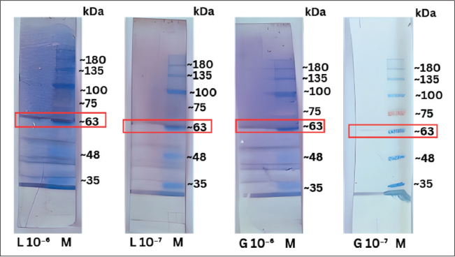
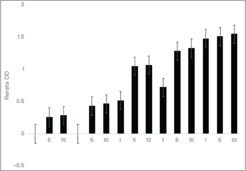
Fig. 1. Research flowchart. Rabbit immunization scheme for the production of polyclonal antibodies against recombinant GRA7. Antibody purification was performed using the saturated ammonium sulfate method. A 5% (w/v) ammonium sulfate solution was prepared in sterile distilled water and mixed using a magnetic stirrer until completely dissolved. Serum samples were mixed with ammonium sulfate solution at a 1:1 (v/v) ratio and thoroughly homogenized. The mixture was incubated at 4°C overnight to facilitate protein precipitation. After incubation, the samples were centrifuged at 7,500 rpm for 10 minutes at 4°C. The supernatant was discarded, and the resulting pellet was resuspended in phosphate-buffered saline (PBS) until homogeneous. The protein suspension was transferred into a pretreated cellulose dialysis membrane and securely sealed using dialysis tubing clips. Dialysis was performed against 1 L of PBS at 4°C for 24 hours under continuous stirring, and the dialysis buffer was replaced 3 times during the process. After dialysis, the samples were centrifuged at 4°C for 10 minutes at 12,000 rpm. The supernatant and pellet were separated, and the pellet was resuspended in 1 × PBS. Subsequently, the purified protein preparation was used for downstream analyses. Immunoblotting was used to characterize the antibodies, while the produced antibody titers were determined using the ELISA checkerboard method. IgG was the immunoglobulin analyzed (Eivazi et al., 2015). Enzyme-linked immunosorbent assayELISA checkerboard titration was performed to determine the ideal concentration of IgG. Antigen recombinant Abbexa GRA7 T. gondii (100 µl) was coated onto microwell plates, incubated overnight (16–18 hours, 4°C), and then washed three times using a wash buffer (0.01 M PBS-Tween 20, pH 7.4). Plates were blocked with blocking buffer (wash buffer with 1% bovine serum albumin, Sigma-Aldrich, St. Louis, MO, USA), 100 µl, overnight at 4°C, and washed three times. The primary antibody obtained from rabbit serum (100 µl) was added according to the sample layout, incubated overnight at 4°C, and washed three times with wash buffer. The goat anti-rabbit IgG (H+L) secondary antibody (31820, Thermo Fisher Scientific Inc., Waltham, MA, USA) conjugated with biotin or peroxidase was added and incubated for 1 hour at room temperature, followed by three washes using washing buffer. For biotin-conjugated secondary antibodies, 100 µl of Merck Millipore Streptavidin, Horseradish peroxidase (HRP) OR03L-200UG (EMD Millipore Corp, USA) was added, incubated for 1 hour at room temperature, and washed three times using wash buffer. In addition, 100 µl of substrate solution (Elabscience, Houston, US) was added, incubated for 10–20 minutes at room temperature in the dark, and the reaction was stopped by adding 50 µl of stop solution (Elabscience, Houston, US). The absorbance was immediately measured at 450 nm with a reference wavelength of 630 nm using a microplate reader (Zenix, DIAFIN DF-320, PT). Sumifin Citra Abadi, Tangerang, Indonesia) (Gao et al., 2023; Sthitmatee et al., 2023). Sodium dodecyl sulfate-polyacrylamide gel electrophoresisProfiling of recombinant T. gondii GRA7 protein samples was performed by diluting the recombinant T. gondii GRA7 protein with PBS (1:200), followed by denaturation of the samples through the addition of reducing sample buffer (RSB) at a 1:1 ratio. RSB stock consisting of 1 ml (pH 6.8) Tris-Cl (BioWorld Science cat #42020224-1, Dublin, Ohio, USA), 0.8 ml glycerol (Merck, Darmstadt, Germany), 1.6 ml 10% (m/v) SDS (A3678-100G, Sigma-Aldrich, St. Louis, MO, USA), 0.4 ml beta-mercaptoethanol (BME; M-7154, Sigma-Aldrich, St. Louis, MO, USA), 0.2 ml 1% (m/v) bromophenol blue (BO149-56, Sigma-Aldrich, St. Louis, MO, USA), and 8 ml ddH2O. Sodium dodecyl sulfate-polyacrylamide gel electrophoresis (SDS-PAGE) gel 12% (3.4 ml ddH2O, 4 ml acrylamide (Merck 1.10784.1000, Darmstadt, Germany). A 2.5 ml gel buffer containing 10 µl ammonium persulfate (APS (A3678-100G, Sigma-Aldrich, St. Louis, MO, USA), 10 µl TEMED (CAS: 110-18-9, Merck, Darmstadt, Germany), and 0.1 ml SDS (A3678-100G, Sigma-Aldrich, St. Louis, MO, USA) was prepared and placed into the electrophoresis chamber and immersed in a running buffer (10x conc.); 30.3 g Tris base (93362, Sigma-Aldrich, St. Louis, MO, USA), 144.4 g glycine (Promega. H5071, Madison, Wisconsin, US), 10 g sodium dodecyl sulfate (A3678-100G, Sigma-Aldrich, St. Louis, MO, USA) up to the marked level. The samples were then loaded into each well of the gel according to the predetermined order, and electrophoresis was performed using a Mini-PROTEAN Tetra Vertical Electrophoresis Cell (Bio-Rad Laboratories, Inc., Hercules, California, US) at a constant voltage of 100 V for 100 minutes. Visualization of the separated protein bands on the PAGE gel was performed by staining the gel with a 0.1% (w/v) Coomassie Brilliant Blue solution [6104-58-1, Merck, Darmstadt, Germany], 40 ml 40% methanol [1.06009.2500, Merck, Darmstadt, Germany], 10 ml 10% glacial acetic acid [1.00063.2500, Merck, Darmstadt, Germany], and ddH2O up 100 ml overnight, followed by background destaining through repeated replacement of the destaining solution (20 ml 20% methanol [1.06009.2500, Merck, Darmstadt, Germany], 10 ml 10% glacial acetic acid [1.00063.2500, Merck, Darmstadt, Germany], and ddH2O up to 100 ml until the protein bands were clearly visible on the PAGE gel (Qi et al., 2022). Western blotWestern blotting was performed to detect the specific interaction between the T. gondii GRA7 antigen and antibodies. Nitrocellulose (NC) membranes (CAS: 9004-70-0, Merck, Darmstadt, Germany) and SDS-PAGE gels were immersed in transfer buffer, and proteins were transferred using a Trans-Blot Semi-Dry system (Trans-Blot® SD Semi-Dry Transfer Cell, Bio-Rad Laboratories, Inc., Hercules, California, US) using 300 mA, 20 V, for 2 hours. The transfer efficiency was confirmed by Ponceau staining. Membranes were blocked with Tris-buffered saline (TBS; Sigma-Aldrich 93362, St. Louis, Missouri, United States) containing 5% skim milk overnight at 4°C, washed, and incubated with primary antibodies for 2 hours at room temperature, followed by washing and incubation with secondary antibodies for 1 hour at room temperature. The membranes were then incubated with streptavidin-HRP for 40 minutes, followed by visual detection using 3,3′,5,5′-tetramethylbenzidine (TMB) chromogenic substrate (TMB; Thermo Fisher Scientific, Waltham, MA, USA) until a protein band (~60 kDa) appeared. The reaction was stopped using sterile distilled water. Membranes were dried and scanned for documentation (Jirapattharasate et al., 2021; Kim et al., 2024). Analysis of dataELISA data were analyzed using one-way analysis of variance with post hoc Tukey using IBM SPSS Statistics 26. The analysis of variance (ANOVA) test was used to determine statistically significant differences in optical density (OD) values among various treatment groups based on differences in antibody concentrations and immunization times. For the Western blot method, the presence and intensity of protein bands following the antigen–antibody reaction were analyzed descriptively. This analysis evaluated whether the antibodies used could recognize the target antigen (e.g., GRA7) based on the appearance of specific size bands (Kpordze et al., 2024). This figure shows the stages and schedule of rabbit immunization using the recombinant GRA7 antigen, which begins with a primary injection on day 1, followed by two boosters on days 14 and 28. Serum samples were collected periodically until day 42, and the resulting serum was analyzed using ELISA and Western blot to evaluate the antibody response to the recombinant GRA7 protein. ResultsPolyclonal antibody concentration in response to recombinant Toxoplasma gondii GRA7 proteinAn ELISA test of polyclonal antibodies from the serum of two rabbits (P1, P2) against the recombinant GRA7 protein was performed on days 14, 21, 28, 35, and 42. This study aimed to assess the successful formation of antibodies in rabbits injected with GRA7 and to evaluate the increase in antibody production following booster administrations. The OD values can serve as a quantitative indicator for determining the strength or concentration of the immune response to the target antigen. Table 1 and Figure 2 present the mean OD values of polyclonal antibodies for treatment groups 1 and 2. On day 14, the antibody levels (OD) remained low or negative, indicating that the initial immune response was not yet optimal. By day 21, an increase was observed at antigen concentrations of 5 and 10 µg/mL, although the antibody levels were still not maximal. On day 28, a significant increase occurred following the first booster, continuing to increase until day 35, albeit less markedly. By day 42, antibody levels were stable across all concentrations, indicating that the immune response had reached its peak. Figure 2 depicts the difference in the mean OD of IgG antibody measurement results on days 14, 21, 28, 35, and 42. Statistical analysis was performed using one-way ANOVA followed by Tukey’s post hoc test. Different notations indicate significant differences in mean OD (p < 0.05); bars indicate standard error (n=2). Statistical analysis showed no significant differences among antigen concentrations (1, 5, and 10 µg/ml); however, immunization time significantly affected antibody levels (p < 0.05). A significant increase was observed on day 28, with antibody levels plateauing after day 35. Western blot analysis of polyclonal antibody–recombinant Toxoplasma gondii GRA7 protein interactionWestern blot analysis revealed the presence and molecular size of the GRA7 protein recognized by polyclonal antibodies in immunized experimental rabbits. As shown in Figure 3, a distinct protein band appears at approximately 63 kDa in both samples (polyclonal antibodies at dilutions of 10–6 and 10–7), differing from the expected molecular weight of the native GRA7, which is around 29–30 kDa. The protein bands appear thicker and more distinct in the 10–6dilution compared to the 10–7 dilution. This indicates that antibodies at the 10–6 dilution have a higher concentration, enabling a stronger signal in Western blot visualization. Although the protein band in the 10–7 dilution appears fainter, its presence indicates that the antibodies can recognize the GRA7 antigen even at lower concentrations. Western blot analysis of the polyclonal antibodies obtained from rabbit serum showed that the GRA7 protein was specifically recognized, as evidenced by the appearance of a protein band at approximately 63 kDa at both 10–6 and 10–7 dilutions. This finding is consistent with the results of the previous ELISA test, in which the OD values increased significantly from day 14 to day 42, with the highest OD on day 42 exceeding 1.5 at antigen concentrations of 5 and 10 µg/ml and at an antibody dilution of 1:1,000,000. These concentrations yielded high OD values, indicating elevated serum antibody levels. However, another band at 35 kDa was also observed in the Western blot result, mainly at 10–6 dilution. These bands were likely due to the cross-reactivity of the polyclonal antibody (pAb). Table 1. Mean OD values of IgG antibody measurement using the enzyme-linked immunosorbent assay method at antigen concentrations of 1, 5, and 10 µg/ml on days 14, 21, 28, 35, and 42.
Fig. 3. Western blot results of polyclonal antibodies at dilutions of 10–6 and 10–7 with lysates from T. gondii culture and recombinant GRA7 protein indicated a molecular weight of approximately 63 kDa (M: marker; L: antigen lysate, refers to a mixture of antigens (proteins) obtained from the lysate of T. gondii culture (including GRA7); G: recombinant GRA7 T. gondii protein. Source: Authors’ documentation.
Fig. 2. Graph of mean OD of IgG antibody measurement using ELISA. DiscussionThe findings of the present study demonstrate that anti-GRA7 polyclonal antibodies possess strong potential as a crucial component in the development of immunological diagnostic tools for the detection of toxoplasmosis, especially during the initial phase of infection. The findings also lay the scientific groundwork for employing polyclonal antibodies as antigen-based biomarkers to achieve more reliable and efficient diagnostic outcomes. ELISA tests on rabbit serum (P1 and P2) induced with the recombinant GRA7 protein showed a consistent pattern of increased humoral immune responses over time, especially after booster administration. On day 14, the average OD values remained low and were negative at an antigen concentration of 1 µg/ml. These findings indicate that antibody production was not yet optimal, as the rabbits’ immune systems had not developed sufficient antibody levels for reliable detection. The negative OD values may also be attributable to the ELISA assay’s detection limits, which exhibit reduced sensitivity at low antigen concentrations. During the early phase of the immune response, antibody titers are often low and may be difficult to detect, particularly when the antigen concentration used in the assay is insufficient (Luo et al., 2019). The GRA7 antigen from T. gondii is a protein antigen recognized by the immune system and is widely used in the development of immunodiagnostic tools. However, antibody production against this antigen following immunization is time-dependent, and a single primary dose in the absence of a booster is insufficient to induce high antibody titers (Arab-Mazar et al., 2021; Wang et al., 2023). However, we acknowledge that other factors may also have contributed to these findings. Potential technical issues include high background signals in the blank wells, insufficient washing steps, pipetting inaccuracies, and general assay variability. In addition, antibody production may not have reached detectable levels at these stages post-immunization. On day 21, the OD values increased, particularly at antigen concentrations of 5 and 10 µg/ml, reflecting the initial production of specific antibodies. This increase indicated B lymphocyte activation and the initial production of immunoglobulins, which remained limited until a significant increase occurred after the booster (Pagheh et al., 2019). The increase in OD values observed on day 21 indicates that the primary immune response had begun to develop effectively, resulting in the production of antibodies at levels detectable by ELISA, particularly when higher antigen concentrations were used. Elevated antigen concentrations enhance antibody capture, thereby yielding higher OD readings (Luo et al., 2019). This means that the rabbit’s immune system had begun to recognize the GRA7 antigen from T. gondii as a foreign molecule, triggering activation of the key components of the adaptive immune system. On day 28, a significant increase in OD values was observed, particularly at 5 and 10 µg/ml antigen concentrations, indicating immune memory activation and the effectiveness of the first booster in stimulating memory B-cell proliferation. The significant increase in OD on day 28 indicated that the test animals’ immune system (rabbits) had entered the secondary immune response phase. This occurred in response to the first booster immunization administered after the initial immunization with the T. gondii GRA7 antigen. This finding aligns with the understanding that a primary immune response takes more than 2 weeks to produce a significant amount of antibodies (Sonaimuthu et al., 2016). The secondary immune response is generally faster, stronger, and more specific than the primary response because of the involvement of memory B cells formed during the previous phase. A booster functions as a trigger for the immune system’s reactivation, activating memory cells to proliferate and differentiate into plasma cells that produce large quantities of antibodies (Murph and Weaver, 2017). The effectiveness of boosters in enhancing immune responses is widely used in vaccine and immunotherapy development, including recombinant GRA7 antigens used in diagnostic research or experimental vaccination against T. gondii (Ybañez and Nishikawa, 2020). On day 35, the continued increase in OD values, albeit at a slower rate compared to days 21 and 28, indicated that the humoral immune response is entering the plateau phase. This means that antibody levels in the body have reached or are approaching their maximum or stable levels, where the immune system has produced large amounts of antibodies, but the production rate is beginning to decline. The humoral response usually peaks within 5–6 weeks after the initial immunization (Yang et al., 2016). This condition is consistent with the immune response kinetic curve, which consists of the lag, log (exponential), plateau, and eventual decline phases. Following initial activation and booster administration, memory B lymphocytes produce large numbers of plasma cells, generating IgG antibodies specific to the T. gondii GRA7 antigen. However, once a certain threshold is reached, the body regulates immune homeostasis by balancing antibody production and degradation, causing the OD curve to slowly level off or increase (Sonaimuthu et al., 2016). In this mechanism, B lymphocytes play a central role. B lymphocytes undergo activation, clonal expansion, and differentiation into plasma cells that produce specific antibodies after recognizing the antigen via specific surface receptors (Murph and Weaver, 2017). This process characterizes the primary immune phase, in which immunoglobulin M (IgM) is the first antibody produced, followed by class switching to IgG with higher antigen affinity (Arab-Mazar et al., 2021; Wang et al., 2023). This immune response also results from complex interactions between antigens, dendritic cells as APCs, and costimulatory signals that drive B lymphocyte activation. In experimental induction with GRA7, antibodies specific to this protein serve as key indicators for developing serology-based diagnostic tools (Ybañez and Nishikawa, 2020). Several studies have shown that the peak humoral response to protein vaccines is typically reached within 5–6 weeks after the initial immunization, depending on the type of antigen, test animal species, and immunization protocol used (Luo et al., 2019). At this time, antibody levels reached the highest sustainable level before gradually declining in the absence of repeated antigen exposure. The antigen concentrations used (5 and 10 µg/ml) remained effective in capturing the antibodies formed, as indicated by the high OD values, although the increase began to plateau compared to earlier time points. This also reflects the strong reactivity of the antibodies, as affinity selection in the germinal centers has been optimized since the first booster (Arab-Mazar et al., 2021; Wang et al., 2023). On day 42, the highest OD values were recorded at all antigen concentrations, with OD values exceeding 1.5 even at an antigen concentration of 1 µg/ml. This indicated a strong and stable immune response. This reinforces the effectiveness of the administered booster dose in maximally inducing antibodies specific to GRA7 (Luo et al., 2019). Achieving the highest OD values on day 42 at all antigen concentrations, including low concentration (1 µg/ml), suggests that the humoral immune response has peaked and is stable. High OD values indicate that antibodies specific to the recombinant GRA7 protein of T. gondii have been produced in large amounts with strong reactivity (Nguyen et al., 2024). Based on the result, the significant increase in OD up to day 42 demonstrates the maturity and stability of the adaptive immune system, as well as the effectiveness of the booster in inducing antibodies specific to the recombinant GRA7 protein. This pattern was also observed in mouse models that exhibited similar immune responses (Arcon et al., 2021). This achievement is the result of successful booster immunization, which re-exposes the immune system to the antigen, thereby rapidly activating memory B lymphocytes, promoting proliferation, and differentiating into plasma cells that produce large amounts of antibodies. The secondary immune response generated by the booster is faster and stronger than the primary response due to the involvement of immunological memory (Teimouri et al., 2021). The high OD values, even at low antigen concentrations, indicate that the antibodies formed have strong reactivity for the antigen—a key feature of antibodies produced during a secondary response. Highly reactive antibodies can efficiently recognize and bind to antigens even in small amounts (Kochanowsky and Koshy, 2018). Future studies should include quantitative affinity characterization (e.g., dissociation constant (Kd)determination) to more accurately define the binding properties of the antibodies. Ferra et al. (2020) demonstrated that a booster based on recombinant GRA7 antigen can improve the sensitivity and accuracy of specific antibody detection in ELISA, even in samples with low antigen concentrations. This is consistent with the use of GRA7 as a leading candidate for T. gondii immunodiagnostics due to its immunogenic properties and consistent expression during infection (Ybañez and Nishikawa, 2020). Thus, the significant increase in OD up to day 42 reflects the maturity and stability of the adaptive immune system and the effectiveness of the immunization protocol in inducing GRA7-specific antibodies. Overall, this pattern confirms the success of recombinant GRA7 protein immunization and the use of a booster in producing high-titer, specific polyclonal antibodies. Determining OD values as a quantitative indicator of immunogenicity has been widely applied in the development of T. gondii immunodiagnostics (Chu and Quan, 2021). In another study, BALB/c mice immunized with recombinant GRA7 protein for 4 weeks showed a sharp increase in IgG2a levels, with OD >1.0 for IgG2a (while IgG1 remained low), indicating a dominant Th1 response and the formation of high-specificity antibodies after the booster at week 4. This aligns with the findings of this study, in which the humoral response of the booster peaked at week 6 (Yang et al., 2016). In another study using mice vaccinated with GRA7 DNA vaccine plus calcium phosphate nanoparticles, high IgG antibody production, a dominant IgG2a/IgG1 ratio, and high splenocyte proliferation were observed, indicating optimal humoral and cellular responses after the third booster up to week 5. This supports the booster’s effectiveness in improving antibody quality and quantity, consistent with the high OD values observed in the present study on day 42 (Chu and Quan, 2021). The appearance of the protein band at this position indicated that the antibodies successfully expressed and recognized the recombinant GRA7 protein (Luo et al., 2019). The ability of antibodies to bind to the target antigen GRA7 at a high dilution (10–7) indicated that the antibodies possess high binding affinity and can still recognize the GRA7 epitope even at very low concentrations. These results confirm that polyclonal antibodies against GRA7 have great potential as candidate materials for immunodiagnostic detection (Arab-Mazar et al., 2021; Wang et al., 2023). Western blot assay was performed to determine the sensitivity of the generated polyclonal antibodies toward the recombinant T. gondii GRA7 protein. This technique was used to confirm that the obtained polyclonal antibodies could specifically recognize the GRA7 protein. However, as shown in the figure, protein bands with a molecular weight of approximately 35 kDa were also consistently detected in both the recombinant GRA7 protein and lysate at a dilution of 10–6. The cross-reactivity of the pAb is most likely the cause of this band. Since the antibody was generated against recombinant GRA7, it comprises a heterogeneous population of immunoglobulins recognizing multiple epitopes. Some of these epitopes may be shared with approximately 35 kDa unrelated proteins present in both the recombinant protein preparation (e.g., host-derived contaminants from the expression system) and the T. gondii lysate. Therefore, we interpret the ~35 kDa band as a result of non-specific or off-target binding rather than a true GRA7 signal. This phenomenon is well documented, with studies demonstrating that polyclonal antibodies frequently exhibit cross-reactivity in Western blot and other immunoassays (Pillai-Kastoori et al., 2020). The use of mAb or affinity-purified polyclonal antibodies could help reduce cross-reactivity and better resolve the true molecular weight of GRA7 to improve specificity in future studies. Based on bioinformatics studies and molecular characterization results, the native molecular weight of GRA7 ranges between 29 and 30 kDa. This molecular weight is calculated based on the number of amino acids and the residue composition of the GRA7 protein without any tags or additional modifications. In the present study, Western blot results show bands with higher molecular weights due to several factors, such as post-translational modifications (PTMs), such as glycosylation, phosphorylation, and disulfide bond formation, which can add chemical groups or carbohydrate moieties to proteins, increasing their apparent molecular mass ( Walsh et al., 2005; Arcon et al., 2021; Proteintech Group, 2025). Glycosylation can also change protein migration during SDS-PAGE, making it appear heavier than its theoretical size. Other influencing factors include protein dimerization or oligomerization, where GRA7 may form dimers or multimers under certain conditions. The GRA7 recombinant proteins utilized in our study incorporate a GST-tag to facilitate purification and detection. These tags contribute additional amino acid residues, which can elevate the molecular weight by around 26 kDa. Furthermore, tags may influence protein folding or conformation, potentially altering migration behavior on electrophoretic gels. Consequently, the observed differences in molecular weight between the recombinant and native forms of the protein may be ascribed to the cumulative effects of PTMs and the associated tags (Terpe, 2003). These findings are consistent with the studies by Ferra et al. (2020) and Wang et al. (2023), which reported that recombinant GRA7 protein expression appeared at around 60–65 kDa when expressed in a prokaryotic system (Luo et al., 2019; Arab-Mazar et al., 2021; Wang et al., 2023). Further protein sequencing is needed to identify the GRA7 in 63 kDa found in the current result (Wright, 1989; Leenaars and Hendriksen, 2005; Lipman et al., 2005). Certain limitations must be considered when evaluating these results. The current study used only two rabbits, representing a limited sample size, which restricts the possibility of robust statistical analysis and may increase susceptibility to interindividual biological variability. However, the use of two to three rabbits is a commonly accepted practice in the context of pAb production, with two animals generally considered the minimum to help account for variability in immune responses and to ensure successful antibody generation. While some protocols employ a larger number of animals or include both sexes to enhance antibody yield and diversity, our study followed the minimal standard approach. Moreover, polyclonal antibodies pose challenges of stability and batch variations. The long-term stability and batch-to-batch variability of polyclonal antibodies were not assessed. These factors highlight the intrinsic limitations associated with pAb production, as polyclonal sera comprise diverse antibody populations that can differ in composition and affinity over time and across different immunization batches. Prior research has indicated that polyclonal antibodies are particularly vulnerable to batch variability, which can arise from biological differences among the animals used, variations in immunization protocols, and timing of serum collection. The use of rabbits as a model for pAb production has long been a standard owing to their ease of handling, ability to produce relatively large volumes of serum, and high antibody titers and affinities. Hutu et al. (2019) showed that rabbits can consistently produce pAbs without significant differences between males and females, indicating that sex is not a major limiting factor in pAb production. In addition, immunization and blood collection procedures are classified as having mild severity and do not significantly affect growth parameters or overall animal health, supporting the ethical feasibility of using rabbits for antibody production. Rabbits aged 6 months are recommended because of their more stable immune responses and easier management during the production process. Repeated immunization of rabbits results in a significant increase in antiserum titers, accompanied by a progressive increase in antibody affinity with increasing boosting frequency. Consistent with these findings, Li et al. (2023) demonstrated that repeated boosting strategies in rabbits not only enhance antibody quantity but also promote affinity maturation through clonal selection and B-cell repertoire diversification. Collectively, these results underscore the highly adaptive immune response of rabbits to repeated immunization protocols, enabling improvements in the quality of polyclonal antibodies in terms of both titer and affinity, particularly for antigens that are difficult to immunize (Li et al., 2023). Lee et al. (2023) demonstrated that rabbit pAb responses display defined epitope preferences that are strongly shaped by immunogen architecture, highlighting the critical role of antigen design in directing antibody specificity (Lee et al., 2023). Collectively, these findings support the suitability of rabbits as a model for generating high-quality, analyzable pAb responses and for informing immunogen optimization and antibody-based assay development. Furthermore, the stability of antibodies during extended storage may be influenced by several factors, including temperature, buffer composition, and the occurrence of repeated freeze-thaw cycles, all of which can potentially affect antibody reactivity and assay reproducibility. The instability of antibodies can result in decreased production yields, diminished or impaired efficacy, adverse immune responses, patient-related complications, and restricted or lost functionality when operating under extreme conditions or during prolonged storage (Ma et al., 2020). Another limitation of the present study is the lack of a positive control, such as a commercial anti-GRA7 antibody, which would be essential to verify the specificity of the antibodies. A positive control was not included in the current experimental design due to resource and availability constraints. However, we agree that its inclusion would strengthen the results’ reliability and interpretability. Future studies should incorporate appropriate positive controls to enable more rigorous validation of antibody specificity and assay performance. Furthermore, this research also lacks clinical validation. The antibodies were characterized in clinical specimens under laboratory conditions without testing. Sensitivity, specificity, and cross-reactivity against other pathogens are unproven; thus, we will incorporate this in the proposed future directions (e.g., testing sera from patients and cross-reactivity with Plasmodium, Coxiella, and Neospora). Therefore, their diagnostic sensitivity, specificity, and potential cross-reactivity remain unverified. Future studies should address these limitations by exploring the development of mAbs targeting the same antigen or other potential antigens, such as Toxoplasma surface antigens (SAGs), rhoptries (ROPs), other GRAs, and glideosome-associated proteins (GAPs). Monoclonal antibodies provide enhanced specificity, stability, and consistency across batches, especially when incorporated in immunochromatographic platforms, rendering them more appropriate for diagnostic applications. Moreover, testing the developed platform with clinical specimens is crucial to obtain the diagnostic accuracy and usability of this test in a real-world setting. Integrating mAb production with clinical validation will ultimately advance the development of reliable and standardized tools for the diagnosis of toxoplasmosis. A major limitation of this study is the small sample size of the in vivo experiment (n=2 rabbits). Although this approach was chosen in accordance with the 3Rs principle to minimize animal use and obtain sufficient serum for preliminary antibody generation, such a limited number of animals is insufficient to capture biological heterogeneity in immune responses. Consequently, the results should be interpreted with caution as pilot-level, descriptive findings. Further studies with larger animal cohorts are required to confirm reproducibility, evaluate interindividual variation, and strengthen the generalizability of the conclusions. Another limitation of this study is the lack of a validated commercial anti-GRA7 antibody as a positive control in the Western blot and ELISA assays. Therefore, while the detected reactivity suggests GRA7 recognition, these findings should be interpreted with caution, as definitive confirmation of GRA7-specific binding was not possible in the current experimental setting. ConclusionThis study successfully demonstrated that immunization with recombinant T. gondii GRA7 protein induces a strong, specific, and stable humoral immune response in rabbits. The generated polyclonal antibodies showed high immunoreactivity and affinity, as evidenced by a marked increase in ELISA OD values following booster immunization and consistent antigen recognition in Western blot analysis, even at high antibody dilutions. These findings confirm that GRA7 is a highly immunogenic antigen and that the produced polyclonal antibodies are capable of reliably detecting the GRA7 protein. From a practical perspective, the anti-GRA7 polyclonal antibodies developed in the present study have strong potential to be utilized as key components in immunological diagnostic platforms for toxoplasmosis, including ELISA-based assays, immunoblotting, and rapid diagnostic tests such as lateral flow immunoassays, which could provide a highly sensitive and rapid readout for Toxoplasma detection. Future studies should focus on validating these antibodies using clinical human samples to determine diagnostic sensitivity, specificity, and cross-reactivity, as well as advancing toward mAb development to enhance assay standardization and reproducibility. Such efforts may contribute significantly to the development of more accurate, accessible, and reliable diagnostic tools for the early detection and management of toxoplasmosis in both clinical and public health settings. AcknowledgmentsThis paper is part of the first author’s Thesis in the Master’s Program in Biomedical Sciences, Faculty of Medicine, Universitas Brawijaya. FundingThis research was funded by the Competitive Funding for the Research Ecosystem of Professors from Research and Community Service Institution, Universitas Brawijaya, Indonesia, with grant number 01047.17/UN10.A0501/B/KS/2025. Authors’ contributionsConceptualization, SF, LEF, and NW; data curation, SM and DS; formal analysis, SF, LEF, and NW; funding acquisition, LEF; investigation, HRR, SM, and NW; methodology, LEF, NW, and HRR; project administration, LEF; resources, OS and DKM; software, SF and DS; supervision, LEF and NW; validation, OS and ARP; visualization, HRR, SM, and TW; writing the original draft, SF; review and editing, LEF, NW, OS, ARP, and DKM Conflict of interestThe authors have no conflicts of interest to declare. Ethical approvalEthical clearance for this research was obtained from 274/EC/KEPK/07/2024 at the Universitas Brawijaya. Data availabilityAll data are within the manuscript. ReferencesAbdallah, M.C., Kamel, M., Karima, B., Samir, A., Djamel, K., Rachid, K. and Khatima, A.O. 2019. Cross-sectional survey on toxoplasma gondii infection in cattle, sheep, and goats in algeria: seroprevalence and risk factors. Vet. Sci. 6(3), 63. Adiningsih, M.W., Soejoedono, R.D., Adji, R.S., Putri, D.D., Purnawarman, T., Latif, H. and Poetri, O.N. 2019. Sumateran wild boar (Sus scrofa vittatus) meat antibody production as immunodiagnostic reagent candidate. Vet. World 12(1), 477–482. Akins, G.K.H., Furtado, J.M. and Smith, J.R. 2024. Diseases caused by and behaviors associated with toxoplasma gondii infection. Pathogens 13(11), 968. Arab-Mazar, Z., Javadi Mamaghani, A., Fallahi, S., Rajaeian, S., Koochaki, A., Seyyed Tabaei, S.J. and Rezaee, H. 2021. Immunodiagnosis and molecular validation of Toxoplasma gondii-recombinant dense granular (GRA) 5 protein for the detection of toxoplasmosis in hemodialysis patients. Semin. Dial. 34(5), 332–337. Arcon, N., Picchio, M.S., Fenoy, I.M., Moretta, R.E., Soto, A.S., Perrone Sibilia, M.D., Sánchez, V.R., Prato, C.A., Tribulatti, M.V, Goldman, A. and Martin, V. 2021. Synergistic effect of GRA7 and profilin proteins in vaccination against chronic Toxoplasma gondii infection. Vaccine 39(6), 933–942; doi:10.1016/j.vaccine.2020.12.072 Attias, M., Teixeira, D.E., Benchimol, M., Vommaro, R.C., Crepaldi, P.H. and De Souza, W. 2020. The life-cycle of Toxoplasma gondii reviewed using animations. Parasit. Vectors 13(1), 588; doi:10.1186/s13071-020-04445-z Care, C.C.A. 2002. Guidelines on antibody production. Canadian Council on Animal Care. 315-350 Albert Street, Ottawa, Canada Chu, K.B. and Quan, F.S. 2021. Advances in Toxoplasma gondii vaccines: current strategies and challenges for vaccine development. Vaccines 9(413), 1–16. Eivazi, S., Majidi, J., Maleki, L.A., Abdolalizadeh, J., Yousefi, M., Ahmadi, M., Dadashi, S., Moradi, Z. and Zolali, E. 2015. Production and purification of a polyclonal antibody against purified mouse IgG2b in rabbits towards designing mouse monoclonal isotyping kits. Adv. Pharm. Bull. 5(1), 109–113. Farhab, M., Aziz, M.W., Shaukat, A., Cao, M.X., Hou, Z., Huang, S.Y., Li, L. and Yuan, Y.G. 2025. Review of toxoplasmosis: what we still need to do. Vet. Sci. 12(8), 1–42. Ferra, B., Holec-Gąsior, L. and Grąźlewska, W. 2020. Toxoplasma gondii recombinant antigens in the serodiagnosis of toxoplasmosis in domestic and farm animals. Animals 10(8), 1245. Gao, Y., Shen, Y., Fan, J., Ding, H., Zheng, B., Yu, H., Huang, S., Kong, Q., Hangjun, L., Zhuo, X. and Lu, S. 2023. Establishment and application of an iELISA detection method for measuring apical membrane antigen 1 (AMA1) antibodies of Toxoplasma gondii in cats. BMC Vet. Res. 19(229), 1–11. Proteintech Group. 2025. Western Blot Troubleshooting: Why Does the Observed Protein Molecular Weight (MW) Differ from the Calculated One? https://www.ptglab.com/support/western-blot-protocol/western-blot-troubleshooting-why-does-the-observed-protein-molecular-weight-mw-differ-from-the-calculated-one/ Hutu, I., Mircu, C., Lungu, B., Bunu Panaitescu, C. and Chen, K.-W. 2019. Polyclonal antibody production in several rabbit models. In Proceedings of the International Scientific Congress “Life Sciences, a Challenge for the Future” , Ed., Miron, L.-D. Filodiritto Editore, pp: 422–425. Jirapattharasate, C., Udonsom, R., Prachasuphap, A., Jongpitisub, K. and Dhepakson, P. 2021. Development and evaluation of recombinant GRA8 protein for the serodiagnosis of Toxoplasma gondii infection in goats. BMC Vet. Res. 17(1), 27; doi:10.1186/s12917-020-02719-3 Kim, M.J., Park, S.J. and Park, H. 2024. Trend in serological and molecular diagnostic methods for Toxoplasma gondii infection. Eur. J. Med. Res. 29(1), 520. Kochanowsky, J.A. and Koshy, A.A. 2018. Toxoplasma gondii. Current Biology. Cell Press, 28(14), 770–771. Kpordze, S.W., Kikuvi, G.M., Kimotho, J.H. and Mobegi, V.A. 2024. Development, optimization and evaluation of a sensitive enzyme-linked immunosorbent assay (ELISA) prototype for detection of chicken-based IgY polyclonal antibodies against toxins of D. Polylepis venom. Antibodies J. 13(50), 1–19. Kusumaningsih, P. 2018. Pengembangan antibodi poliklonal dari stadium takizoit Toxoplasma gondii isolat Wta. J. Kes-Terpadu-Maret 2(1), 31–36. Lee, S.J., Gasche, M.B., Burrows, C.J., Kondepudi, A., Zhang, X. and Wang, M.M. 2023. Preferential rabbit antibody responses to C-termini of NOTCH3 peptide immunogens. Sci. Rep. 13(9156), 1–11. Leenaars, M. and Hendriksen, C.F.M. 2005. Critical steps in the production of polyclonal and monoclonal antibodies: evaluation and recommendations. ILAR J. 46(3), 269–279; doi:10.1093/ilar.46.3.269 Li, Y., Kong, Y., Yu, X., Yu, W., Wen, K., Shen, J. and Wang, Z. 2023. Characteristics of rabbit hapten-specific and germline-based BCR repertoires following repeated immunization. One Health Adv. 1(17), 1–17. Lipman, N.S., Jackson, L.R., Trudel, L.J. and Weis-Garcia, F. 2005. Monoclonal versus polyclonal antibodies: distinguishing characteristics, applications, and information resources. ILAR J. 46(3), 258–268. Louis, L.R.P., Nagarajan, P. and Ramajayam, A.B. 2023. Blood collection and techniques in small laboratory animals for biomedical research: a systematic review. Int. J. Life Sci. Pharma Res. 13(2), 153–166. Luo, J., Wan, J., Tang, Z. and Shen, S. 2019. Identification of novel antigens for serum IgG diagnosis of human toxoplasmosis. Exp. Parasitol. 204, 107722. Ma, H., O’ Fagain, O., C. and O’ Kennedy R. 2020. Antibody stability: a key to performance - analysis, influences and improvement. Biochimie 177, 213–225. Murphy, K. and Weaver, C. 2017. Janeway’s immunobiology, 9th ed. Garland Science: New York. Taylor & Francis Group and Limited Liability Company. Mushlih, M., Nurfitriana, A., Ningsih, K.W., Azizah, N., Ariani, N.L. and Lubiz, I. 2020. Comparison of Toxoplasma gondii identification using PCR method and Elfa method. Meditory 8(2), 101–108. Nguyen, M.N., Yeo, S.J. and Park, H. 2024. Identification of novel biomarkers for anti-Toxoplasma gondii IgM detection and the potential application in rapid diagnostic fluorescent tests. Front. Microbiol. 15, 1385582. Pagheh, A.S., Sarvi, S., Gholami, S., Omran, H.A., Valadan, R., Hassannia, H., Ahmadpour, E., Ramandie, M.F., Dodangeh, S., Hosseni-khah, Z. and Daryani, A. 2019. Protective efficacy induced by DNA prime and recombinant protein boost vaccination with Toxoplasma gondii GRA14 in mice. Microb. Pathog. 134, 103601. Pillai-Kastoori, L., Heaton, S., Shiflett, S.D., Roberts, A.C., Solache, A. and Schutz-Geschwender, A.R. 2020. Antibody validation for Western blot: by the user, for the user. J. Biol. Chem. 295(4), 926–939; doi:10.1074/jbc. RA119.010472 Qi, T., Ai, J., Sun, Y., Ma, H., Kang, M., You, X. and Li, J. 2022. Application of Toxoplasma gondii-specific SAG1, GRA7 and BAG1 proteins in serodiagnosis of animal toxoplasmosis. Front. Cell. Infect. Microbiol. 12, 1–11. Rezaei, F., Sharif, M., Sarvi, S., Hejazi, S.H., Aghayan, S. and Pagheh, A.S. 2019. A systematic review on the role of GRA proteins of Toxoplasma gondii in host immunization. J. Microbiol. Methods. 165, 105696.doi:10.1016/j.mimet.2019.105696 Rohmayani, V., Arimurti, A.R.R., Lukiyono, Y.T., Nuzula, F., Romadhon, N. and Lihabi, L. 2022. Edukasi Infeksi Toksoplasmosis pada Masyarakat di Desa Balong Panggang Gresik. Humanism Jurnal Pengabdian Masyarakat UM Surabaya 3(2), 165–173. Soedarto 2017. Masalah Titer IgG dan IgM dalam Menentukan diagnosis Toksoplasmosis. Jurnal Ilmiah Kedokteran Wijaya Kusuma 6(2), 1–5. Sonaimuthu, P., Ching, X.T., Fong, M.Y., Kalyanasundaram, R. and Lau, Y.L. 2016. Induction of protective immunity against toxoplasmosis in BALB/c mice vaccinated with Toxoplasma gondii rhoptry-1. Front. Microbiol. 7(808), 1–11. Sthitmatee, N., Tankaew, P., Modethed, W., Rittipornlertak, A., Muenthaisong, A., Apinda, N., Koonyosying, P., Nambooppha, B., Chomjit, P., Sangkakam, K., Singhla, T., Vinitchaikul, P., Boonsri, K., Pringpoa, K., Punyapornwithaya, V. and Kreausukon, K. 2023. Development of in-house ELISA for detection of antibodies against lumpy skin disease virus in cattle and assessment of its performance using a bayesian approach. Heliyon 9(2), 1–9. Teimouri, A., Abbaszadeh Afshar, M.J., Mohtasebi, S., Jafarpour Azami, S., Alimi, R. and Keshavarz, H. 2021. Assessment of an in-house enzyme-linked immunosorbent assay and IgG avidity test based on SAG1 and GRA7 proteins for discriminating between acute and chronic toxoplasmosis in humans. J. Clin. Microbiol. 59(5), 00416–21. Terpe, K. 2003. Overview of tag protein fusions: from molecular and biochemical fundamentals to commercial systems. Appl. Microbiol. Biotechnol. 60(5), 523–533. Walsh, C.T., Garneau-Tsodikova, S. and Gatto, G.J.J. 2005. Protein posttranslational modifications: the chemistry of proteome diversifications. Angewandte Chemie (International Ed. in English) 44(45), 7342–7372. Wang, R., Wu, M., Cai, H., An, R., Chen, Y., Wang, J., Zhou, N. and Du, J. 2023. Preparation and preliminary application of epitope peptide-based antibody against Toxoplasma gondii GRA3. Trop. Med. Infect. Dis. 8(3), 143. Wright, K. 1989. Antibodies a laboratory manual. Biochem. Educ. 17(4), 220. In Cold Spring Harbor Laboratory. Eds. Harlow, E and Lane, D. pp 726, 1988. Yang, C.S., Yuk, J.M., Lee, Y.H. and Jo, E.K. 2016. Toxoplasma gondii GRA7-induced TRAF6 activation contributes to host protective immunity. Infect. Immun. 84(1), 339–350; doi:10.1128/IAI.00734-15 Ybañez, R.H. and Nishikawa, Y. 2020. Serological detection of T. gondii infection in humans using an immunochromatographic assay based on dense granule protein 7. Parasitol. Int. 76, 102089. | ||

| How to Cite this Article |
| Pubmed Style Fadilla S, Fitri LE, Winaris N, Pawestri AR, Mayashinta DK, Rahmawati HR, Sofia O, Suprianto D, Megasari S. Animal model-based generation of strong reactivity polyclonal antibodies against Toxoplasma gondii GRA7. Open Vet. J.. 2026; 16(8): 5340-5351. doi:10.5455/OVJ.2026.v16.i8.28 Web Style Fadilla S, Fitri LE, Winaris N, Pawestri AR, Mayashinta DK, Rahmawati HR, Sofia O, Suprianto D, Megasari S. Animal model-based generation of strong reactivity polyclonal antibodies against Toxoplasma gondii GRA7. https://www.openveterinaryjournal.com/?mno=310045 [Access: August 08, 2026]. doi:10.5455/OVJ.2026.v16.i8.28 AMA (American Medical Association) Style Fadilla S, Fitri LE, Winaris N, Pawestri AR, Mayashinta DK, Rahmawati HR, Sofia O, Suprianto D, Megasari S. Animal model-based generation of strong reactivity polyclonal antibodies against Toxoplasma gondii GRA7. Open Vet. J.. 2026; 16(8): 5340-5351. doi:10.5455/OVJ.2026.v16.i8.28 Vancouver/ICMJE Style Fadilla S, Fitri LE, Winaris N, Pawestri AR, Mayashinta DK, Rahmawati HR, Sofia O, Suprianto D, Megasari S. Animal model-based generation of strong reactivity polyclonal antibodies against Toxoplasma gondii GRA7. Open Vet. J.. (2026), [cited August 08, 2026]; 16(8): 5340-5351. doi:10.5455/OVJ.2026.v16.i8.28 Harvard Style Fadilla, S., Fitri, . L. E., Winaris, . N., Pawestri, . A. R., Mayashinta, . D. K., Rahmawati, . H. R., Sofia, . O., Suprianto, . D. & Megasari, . S. (2026) Animal model-based generation of strong reactivity polyclonal antibodies against Toxoplasma gondii GRA7. Open Vet. J., 16 (8), 5340-5351. doi:10.5455/OVJ.2026.v16.i8.28 Turabian Style Fadilla, Siti, Loeki Enggar Fitri, Nuning Winaris, Aulia Rahmi Pawestri, Dearikha Karina Mayashinta, Hanifa Rizky Rahmawati, Ovi Sofia, Dodit Suprianto, and Suci Megasari. 2026. Animal model-based generation of strong reactivity polyclonal antibodies against Toxoplasma gondii GRA7. Open Veterinary Journal, 16 (8), 5340-5351. doi:10.5455/OVJ.2026.v16.i8.28 Chicago Style Fadilla, Siti, Loeki Enggar Fitri, Nuning Winaris, Aulia Rahmi Pawestri, Dearikha Karina Mayashinta, Hanifa Rizky Rahmawati, Ovi Sofia, Dodit Suprianto, and Suci Megasari. "Animal model-based generation of strong reactivity polyclonal antibodies against Toxoplasma gondii GRA7." Open Veterinary Journal 16 (2026), 5340-5351. doi:10.5455/OVJ.2026.v16.i8.28 MLA (The Modern Language Association) Style Fadilla, Siti, Loeki Enggar Fitri, Nuning Winaris, Aulia Rahmi Pawestri, Dearikha Karina Mayashinta, Hanifa Rizky Rahmawati, Ovi Sofia, Dodit Suprianto, and Suci Megasari. "Animal model-based generation of strong reactivity polyclonal antibodies against Toxoplasma gondii GRA7." Open Veterinary Journal 16.8 (2026), 5340-5351. Print. doi:10.5455/OVJ.2026.v16.i8.28 APA (American Psychological Association) Style Fadilla, S., Fitri, . L. E., Winaris, . N., Pawestri, . A. R., Mayashinta, . D. K., Rahmawati, . H. R., Sofia, . O., Suprianto, . D. & Megasari, . S. (2026) Animal model-based generation of strong reactivity polyclonal antibodies against Toxoplasma gondii GRA7. Open Veterinary Journal, 16 (8), 5340-5351. doi:10.5455/OVJ.2026.v16.i8.28 |


