





| Research Article | ||
Open Vet. J.. 2026; 16(8): 5352-5362 !!
Open Veterinary Journal, (2026), Vol. 16(8): 5352–5362 Research Article Establishing baseline parameters for thymic maturation in the absence of vaccine stimulation in Ross broiler chickensShayma Z. Ameen1, Yahya Ahmed Mohammed2 and Mahdi A. Abdullah1*1Department of Physiology and Anatomy, College of Veterinary Medicine, University of Duhok, Duhok, Iraq 2Department of Pathology and Microbiology, College of Veterinary Medicine, University of Duhok, Duhok, Iraq *Corresponding Author: Mahdi Ali Mohammed. Department of Physiology and Anatomy, College of Veterinary Medicine, University of Duhok, Duhok, Iraq. Email: Mahdi.abdullah [at] uod.ac Submitted: 10/02/2026 Revised: 06/06/2026 Accepted: 22/06/2026 Published: 08/08/2026 © 2025 Open Veterinary Journal
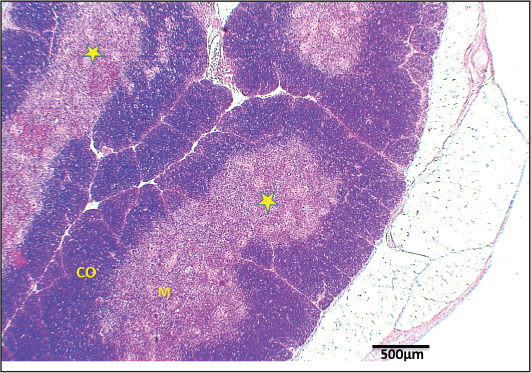
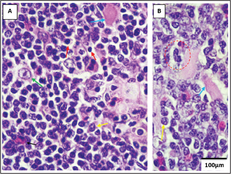
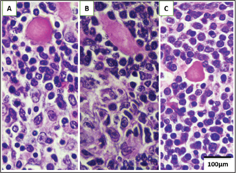
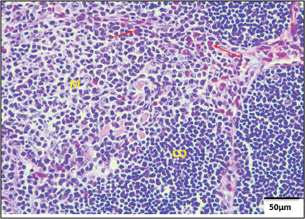
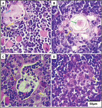
AbstractBackground: To ensure food security, it is crucial to examine the immune system of Ross broiler chickens, which are widely used locally. Aim: Though a major lymphoid organ, the thymus also exhibits secondary lymphoid traits. This study analyzed the post-hatching thymus and B-lymphocytes of this breed of chicken to establish baseline, age-related developmental, and immunological data for the thymus and B-lymphocyte ((Bu-1+)) population in unvaccinated Ross broiler chickens. And it provides crucial, normal histological, and morphometric parameters. Methods: H&E staining and a mouse anti-chicken monoclonal anti-Bu-1 antibody were used to analyze Ross grill chicken thymus from day 1 to day 58 post-hatching. The study examined B-lymphocytes, Hassall’s corpuscles, and their developmental alterations. The cortex and medulla thickness and thymic lobule size were also measured during the investigation. Results: A study found that the thymus was mostly made of lymphocytes, with additional cell types appearing as early as day 1 post-hatching. These cells proliferated with age. Myoid cells (MCs) and Hassall’s corpuscles (HCs) were found in the thymus cortex and medulla. MCs were classed as Kind 1, 2, and 3, while HCs were Types I, II, and III based on morphology. By day 7, MCs and HCs were more common, and by day 58, their numbers increased. Thymic lobules grew dramatically with age. By day 28, the cortex and medulla were significantly thicker than before. The medulla of the thymic lobules had Bu-1-positive B-lymphocytes from day 14 post-hatching. Conclusion: Thymic maturation in unvaccinated Ross broiler chickens is age-dependent with no mortality, indicating a functional immune system that may support future vaccination strategies. Keywords: Chicken, Histomorphometric, Immunohistochemistry, Thymus. IntroductionThe Ross 308 chicken, a prevalent hybrid breed, is now the foremost grill breed globally. In response to the escalating demand for animal protein, worldwide poultry meat output is projected to attain roughly 152.4 million tonnes in 2025, reflecting a year-on-year increase of 1.7% (FAO, meat and meat products, 2025). Chickens significantly impact the global economy and aid in poverty alleviation by supplying accessible supplies of meat and eggs. The immune system of chickens is essential for safeguarding against infectious illnesses and maximizing output in poultry husbandry (Buture and Milers, 2025). Furthermore, comprehending the avian immune system and its constituents is crucial for precise illness diagnosis and the formulation of efficient immunization strategies (Birhan, 2019). The thymus is a crucial organ in the avian immune system, responsible for the formation and maturation of T-lymphocytes (Ali, 2017). The thymus serves not only as a main lymphoid organ but has also been identified as a secondary lymphoid organ owing to the presence of B-lymphocytes and plasma cells (Treesh et al., 2014; Huralska et al., 2020). B-lymphocytes, located in diverse lymphoid organs in avians, can be distinguished using unique markers utilizing immunocytochemical methods (Al-Ogaili and Hameed 2021; Fejszák et al., 2022). Materials and MethodsAnimals and rearing conditionNinety healthy, unvaccinated male Ross breed chickens (RBCs) of genetic line 308 (Avex and Garanti Company) were procured from Jeen Hatchery in Marina Village, Duhok Province, Kurdistan Region of Iraq. The chicks were transported in sterile cages and housed in a sterile chamber under stringent hygienic conditions (dimensions: 3 m × 2.5 m × 2.4 m) at the poultry farm of the College of Veterinary Medicine, University of Duhok. They were granted unrestricted access to food and water. Management techniques and feeding regimens were consistently upheld throughout the study period (Sun and Pan, 2013). Study designThis study encompassed a histomorphometric and immunohistochemical analysis of the thymus in male RBCs, done from March 1, 2025, to April 27, 2025. The 90 chicks were categorized into nine age-specific groups based on post-hatching days: D1, D4, D7, D14, D21, D28, D35, D42, and D58 (Ayman et al., 2020). Each batch comprised 10 chicks. The D1 group was assessed immediately post-hatching, whereas the other groups were maintained under regulated circumstances and evaluated at their designated time intervals. Euthanization and collection of the samplesChicks from each group were euthanized by a simple method, CO2 (Ranjit et al., 2000; Wang et al., 2021). We put each pigeon on its back and made a cut in the middle of its chest and neck to get to and extract the thymus gland. The removed organs were carefully washed with regular saline. Using cross-sectional incisions, tissue samples about 0.5 cm long were taken from the front, middle, and back of the thymus. Some samples were kept in Bouin’s solution for 4–18 hours, while others were fixed in 10% neutral buffered formalin for 24–48 hours (Suvarna et al., 2013). Preparation of tissue specimensThe Duhok Research Centre, College of Veterinary Medicine, University of Duhok, Iraq, used normal paraffin embedding methods to prepare all of the tissue samples. After fixation, the specimens were rinsed with running tap water, dehydrated in a series of ethyl alcohol solutions, cleaned with xylene, and then embedded in paraffin wax at 58°C–60°C. Using a rotary microtome (Leica RM 2245, Germany), tissue slices that were 3–4 µm thick were made. Then, they were stained with Harris haematoxylin and eosin (H&E) for histological evaluation (Suvarna et al., 2013). Histomorphometrical measurementsHaematoxylin and eosin (H&E)-stained slides were employed for micromorphometric analysis (Madej et al., 2015; Sikandar et al., 2017). After making the slides, we examined different micromorphometric parameters of the thymus in specific areas at different times after hatching, by using ten slices were selected for each study group, and the parameters of both the cortex and medulla of thymus were calculated by counting ten slices, and their average was used in the statistical analysis, while for thymic lobule we used the common regular sections of lobule, then length and width of each of lobule were measured, and the two numbers were divided by two. The readings were then entered into the statistical analysis. The measurements were conducted with a light microscope (Olympus CX22, Japan) integrated with a digital camera (Omax 18.0 MP USB 3.0, code A35180U3; USA). Calibration was done either automatically or by hand using a stage micrometre and the Omax camera software. The software came with a lot of different measurement tools, such as ones that let you measure both regular and irregular areas by drawing dotted or solid lines. At 4X magnification, the parameters assessed were the thickness of the cortex and medulla of the thymic lobules and the diameter of the thymic lobules, both in micrometres. Data collection and statistical analysisAll data are displayed as mean ± standard error (SE) and were subjected to one-way analysis of variance (ANOVA) with a significance level established at p < 0.05. Tukey’s multiple comparison test was used to look at the differences between age groups after hatching. We used JMP Pro 14.3.0 software (JMP Pro Statistical Discovery LLC, Marlow, Buckinghamshire, England) to do the statistical analyses. Immunohistochemical preparation Immunohistochemical staining was conducted on formalin-fixed, paraffin-embedded tissue slices in accordance with the methodology established by Suvarna et al. (2013). Sections were deparaffinized in xylene during two successive 10-minute incubations, subsequently rehydrated using a graded ethanol series (two minutes each at 100%, 95%, 90%, 80%, and 70% v/v), followed by two washes in distilled water. Endogenous peroxidase activity was inhibited by incubating the sections in 3% hydrogen peroxide (H2O2) in methanol for 10 minutes. Antigen retrieval was performed via Heat-Induced Epitope Retrieval (HIER) in Ethylenediaminetetraacetic acid (EDTA) buffer at pH 9, utilizing 0.1-M citric acid monohydrate and sodium citrate dihydrate solutions for 35 minutes. Immunodetection was performed utilizing a mouse monoclonal anti-Bu-1 antibody (Catalogue no. MA5-28700; Invitrogen, Thermo Fisher Scientific, USA) diluted 1:100 in phosphate-buffered saline (PBS) and incubated on the sections for 55 minutes at ambient temperature. Slides were rinsed twice in T-TBS (pH 7.6) for three minutes each. The Zytochem Plus HRP polymer detection system (Zytomed Systems) was utilized for 30 minutes, followed by color development with diaminobenzidine (DAB) substrate (Spectrum, USA), with incubation durations varying from 3 to 15 minutes based on staining intensity (Jeurissen et al., 2000). Counterstaining was executed with Gill’s haematoxylin (Bio-Optica, Milano, Italy), and slides were affixed with either Kayser’s glycerin gelatin (Merck, Germany) or the synthetic aqueous mounting medium Aquatex (Merck, Germany). Every staining procedure used suitable positive and negative controls to confirm antibody specificity and staining integrity. Ethical approvalThis study follows the ethics guidelines of the College of Veterinary Medicine, Duhok University of Duhok, Iraq (ethics approval number: 1/3/2024, Reference No: CVM2024/0103UoD). ResultsThe thymus of male RBCs was meticulously analyzed on post-hatching day 42, deemed the standard slaughter day (Coban et al., 2014). The results from this time point were juxtaposed with those acquired from subsequent post-hatching days. Histological observationThe histological architecture of the thymus in male RBCs at day 42 comprised diffuse lymphatic tissue devoid of identifiable lymphatic nodules. Each thymic lobule had a distinct peripheral dark-staining cortex and a centrally positioned lighter-staining medulla. The medulla stretched throughout the center area of the thymic lobules (Fig. 1). The cortex and medulla of the thymic lobules comprised many lymphocytes of varying sizes, including lymphoblasts, epithelial reticular cells (ERCs), macrophages, and myoid cells (MCs) (Fig. 2A and 2B). The majority of these cells were distinguished by the attributes of their nucleus. Lymphocytes displayed densely pigmented, centrally positioned nuclei with little cytoplasm. Lymphoblasts exhibited big, prominent nuclei. Epithelial reticular cells (ERCs) possessed spherical or ovoid nuclei with mildly acidophilic cytoplasm. Macrophages exhibited indented or uneven nuclei. The medulla also had plasma cells, dendritic cells, and eosinophilic cells. Dendritic cells had pear-shaped or elongated nuclei, primarily characterized by peripheral heterochromatin and a condensed nucleolus. Myoid cells (MCs) were categorized into three kinds based on their morphology: Kind-1 MC, Kind-2 MC, and Kind-3 MC (Fig. 3A, B, and C). Kind-1 MCs were primarily spherical or oval; Kind-2 MCs were elongated or spindle-shaped; and Kind-3 MCs had triangular or irregular configurations. All MC types exhibited vibrant acidophilic cytoplasm with eccentrically located nuclei. The quantity of MCs was diminished in the cortex and elevated in the medulla. The corticomedullary junction (CMJ), situated between the cortex and medulla, was distinguished by a thick vascular network of blood capillaries (Fig. 4). A multitude of Hassall’s corpuscles (HCs) were identified in the thymus and categorized into three morphological types: Type-I, Type-II, and Type-III HCs. Type-I HCs were spherical formations consisting of hyalinized or cornified acidophilic reticular cells, encircled by flattened epithelial reticular cells (ERCs) (Fig. 5A and 5B). Type-II HCs displayed an irregular morphology characterized by a core, including hyalinized reticular cells, abundant lymphocytes, a limited number of macrophages, dendritic cells, and eosinophils, all encased by flattened ERCs (Fig. 5C). Type-III HCs were rounded and comprised randomly distributed lymphocytes and ERCs (Fig. 5D). Although all HC types were mostly situated in the medulla, they were sometimes sporadically observed in the cortex.
Fig. 1. Photomicrograph of the thymus in RMBCs at day 42 showing: thymic lobule (yellow stars), cortex (CO), and medulla (M). Stain: H & E 4X.
Fig. 2. Photomicrograph of the medulla of the thymus in RMBCs at day 42 showing: lymphoblasts (yellow arrows), lymphocytes (red arrows), epithelial reticular cells (green arrow), macrophages (red dotted circle), myoid cells (blue arrows), and eosinophils (black arrow). Stain: H & E (A & B) 100X.
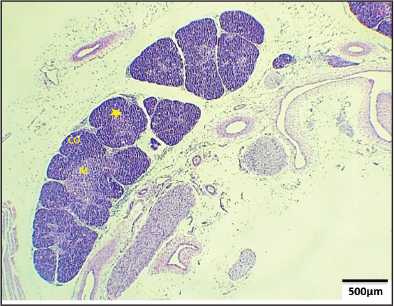
Fig. 3. Photomicrograph of the medulla of the thymus in RMBCs at day 42 showing: Kind-1 MC (A), Kind-2 MC (B), and Kind-3 MC (C). Stain: H & E 100X. Post-hatching maturation of the thymusFrom day 1 to day 4 postnatal, certain thymic lobules exhibited distinct separation between the cortex and medulla, but others did not, as illustrated in (Fig. 6). Blood capillaries were absent at the corticomedullary junction. The cells in both the brain and medulla resembled those in a 42-day-old thymus, with the exception that MCs were absent. Furthermore, Type I HC was observed from day 1 to day 4, albeit its structure was less distinct compared to that of the 42-day-old thymus.
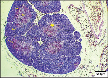
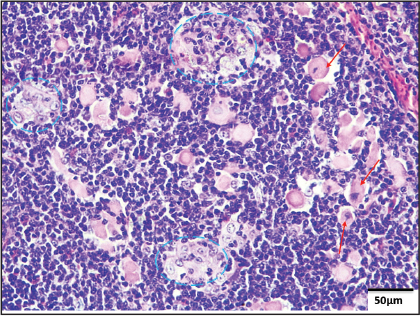
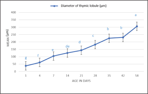
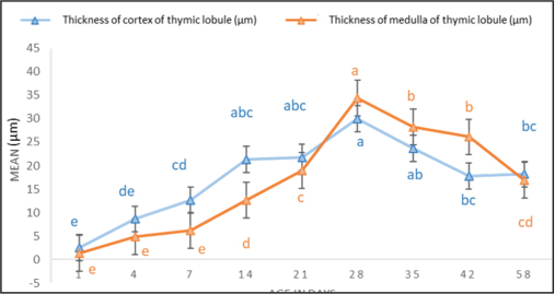
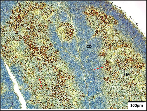
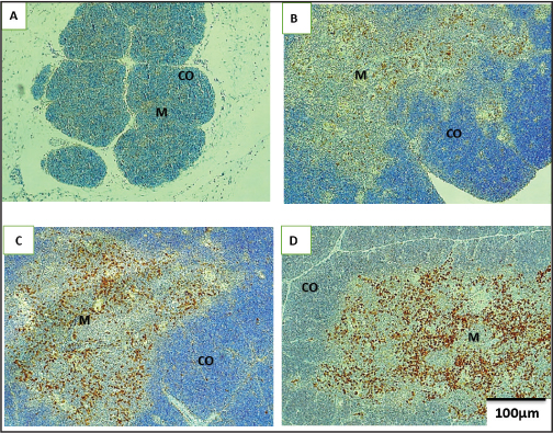
Fig. 4. Photomicrograph of the thymus in RMBCs at D42 old showing: cortex (CO), medulla (M), blood capillaries at corticomedullary junction (red arrows). Stain: H and E 40X. On day 7 post-hatching (D7), the delineation between the cortex and medulla became distinctly defined, corresponding with the emergence of many blood capillaries at the corticomedullary junction (CMJ) (Fig. 7). Myoid cells (MCs) were first identified at this stage, and all three types of Hassall’s corpuscles (HCs) exhibited increased structural differentiation. From D14 to 58 years old, all components of the thymic lobule were fully grown, akin to the thymus of a 42-day-old individual. Nevertheless, the quantity of MCs and HCs significantly escalated around 58 days of age, as illustrated in Figure 8. Micromorphometrical measurements of the thymusFigures 9 and 10 illustrate a substantial increase in the width of thymic lobules with age (p < 0.05). At 28 days of age, both the cortex and medulla exhibited a substantial increase in thickness compared to other time periods (p < 0.05), subsequently followed by a drop with advancing age. Immunohistochemical observationOn day 42 post-hatching (D42), Bu-1-positive B-lymphocytes (BLCs+) were mostly situated in the medulla of the thymic lobules and demonstrated robust immunoreactivity. The cells were examined either alone or in aggregates, as illustrated in Fig. 11. Post-hatching immunohistochemistry analysis indicated that the thymus had minimal Bu-1-positive B-lymphocyte (BLC+) expression on day 1 (D1) (Fig. 12A). On days 4 and 7 (D4 and D7), a limited number of BLC+ cells were detected in the medulla, exhibiting little immunoreactivity (Fig. 12B). On day 14 (D14), BLC+ cells were distinctly observable in the medulla with moderate expression (Fig. 12C). Between days 21 and 58 (D21–D58), BLC+ cells were mostly localized in the medulla, demonstrating robust expression akin to that observed on day 42 (Fig. 12D). DiscussionThis investigation revealed that the thymus of RBCs harbored a heterogeneous array of immune cells, including lymphocytes, ERCs, macrophages, plasma cells, and MCs, exhibiting morphological and cellular characteristics akin to those documented in other chicken breeds (Dahariya et al., 2020; Kanasiya et al., 2018). The medulla in RBCs contained a greater number of erythroid cells than lymphocytes. These ERCs were largely recognized for providing structural support to lymphocytes and other immune cells (Khan et al., 2014). Olah et al. (2014) similarly indicated that ERCs in chickens play a role in immunological control by guiding lymphocyte movement and positioning within thymic compartments. These cells exhibit glycoproteins that engage with adhesion molecules on lymphocytes. Macrophages, as elucidated by Kannan et al. (2015) in Nandanam chickens, are pivotal in antigen presentation and the release of immunological mediators, including cytokines. In the present investigation, plasma cells were also detected in the medulla of the thymic lobules within red blood cells. This discovery is consistent with earlier reports by Treesh et al. (2014) in Hubbard hens, Gülmez and Aslan (1999) in native geese, and Nnadozie et al. (2019) in turkeys. The existence of plasma cells at different ages substantiates the notion that the thymus, in addition to serving as a primary lymphoid organ, may also operate as a secondary lymphoid organ involved directly in immunological responses (Leena et al., 2008; Treesh et al., 2014).
Fig. 5. Photomicrograph of thymus in RMBCs at D42 old showing: Hassall’s corpuscles; Type- I HC (A and B); Type- II HC (C); Type- III HC (D), flattened epithelial reticular cells (yellow arrows), hyalinized reticular cells (blue arrow), cornified reticular cells (green arrow). Stain: H & E 100X.
Fig. 6. Photomicrograph of thymus in RMBCs at D1 old showing: thymic lobule (yellow star), cortex (CO), and medulla (M). Stain: H & E 4X.
Fig. 7. Photomicrograph of the thymus in RMBCs at D7 old showing: thymic lobule (yellow star), cortex (CO) and medulla (M), blood capillaries at corticomedullary junction (yellow arrows). Stain: H & E 4X.
Fig. 8. Photomicrograph of the thymus in RMBCs at D58 old showing: numerous myoid cells (red arrows) and Hassall’s corpuscles (blue dotted line). Stain: H & E 40X. This research discovered three categories of MCs in both the cortex and medulla of the thymus, corroborating the findings of Olah et al. (2014), Kanasiya et al. (2018), and Panwar et al. (2020). Nonetheless, other researchers, including Leena et al. (2008) and Tamilselvan et al. (2017), identified merely two varieties of MCs in avian thymus. This study characterized MCs according to their cytological characteristics observed via light microscopy, although they can also be identified using electron microscopy or immunohistochemistry (Panwar et al., 2020; Kannan et al., 2015). The precise biological function of these cells remains ambiguous, as indicated by Leena et al. (2008) and Kanasiya et al. (2018). Panwar et al. (2020) posited that MCs facilitate the contractile activity of the thymus, assisting in the discharge of lymphocytes as a component of the immune defense mechanism.
Fig. 9. Analysis of diameter of thymic lobule; mean ± standard error. Data with different letters within the same line (a–g) differ significantly (p ≤ 0 .05).
Fig. 10. Analysis of thickness of the cortex and medulla of the thymic lobule; mean ± standard error. Data with different letters within the same line (a–e) differ significantly (p ≤ 0 .05). Three separate types of HCs were found in RBCs: Type-I, Type-II, and Type-III HCs. The results aligned with those documented in broiler chickens (Wei et al., 2019). Conversely, Kanasiya et al. (2018) detected merely two categories of HCs in Kadaknath birds. In the present investigation, some disorganized Type-I HCs were noted at D1; however, all three types exhibited enhanced organization and a numerical rise from D7 to D58. These findings correspond with the discoveries made by Kanna et al. (2015) and Ayman et al. (2020) in different chicken breeds. Nonetheless, their results contrast with those of Treesh et al. (2014), who documented the emergence of HCs exclusively at D28. While the precise role of HCs is not fully understood, Watanabe et al. (2005) suggested that they may participate in the elimination of apoptotic lymphocytes and the maturation of new T-cells.
Fig. 11. Photomicrograph of thymus in RMBCs at D42 old showing: BLCs+ (red arrows), medulla (M), and cortex (CO). IHC (Bu-1 marker) 10X.
Fig. 12. Photomicrograph of the thymus in RMBCs at different age groups showing: BLCs+ in the medulla (M). A-very weak expression at D1 old, B- weak expression at D4 and D7 old, C- moderate expression at D14 old, D- strong expression at D21 old. BLCs- in the cortex (CO). IHC (Bu-1 marker) 10X. Histomorphometric analysis demonstrated significant age-related increases in the diameter of thymic lobules and in the thickness of the cortex and medulla, peaking at day 28, which may indicate enhanced lymphocyte proliferation and maturation, corroborating similar findings by Ayman et al. (2020), Khan et al. (2014), and Sohel et al. (2020) in other chicken breeds. The significant rise at D28 may indicate improved migration of mature lymphocytes from the cortex to the medulla, where they attain immunocompetence and the ability to identify pathogens. These developed lymphocytes may subsequently migrate to secondary lymphoid organs (Madej et al., 2015). The immunohistochemical investigation in this work demonstrated that Bu-1-positive B-lymphocytes (BLCs+) were mostly situated in the thymic medulla, with heightened expression from D1 to D58. This conclusion corroborates the work of Kozlu et al. (2019) in turkeys; however, they employed the CD79αcy marker rather than Bu-1. The progressive elevation of BLC+ expression with age indicates that the thymus may have a direct influence on immunological responses, as well as Bu-1-positive B-lymphocytes is regarded as superior to other B-cell markers and Linking Bu-1+ appearance to immune maturation because definitive marker expressed on both B-cell precursors and mature B-cells, allowing for tracking development from the early embryo (bursa of Fabricius) to maturity and it serves as a reliable marker to analyze the maturation of B-cell subsets and their transition during embryonic development (Houssaint et al.,1989), in other hand, the existence of these cells may also signify natural exposure to environmental antigens throughout development (Leena et al., 2008; Treesh et al., 2014). ConclusionThe post-hatch maturation of the thymus in unvaccinated RBCs transpired normally and was predominantly age-dependent. The findings correlate with the changes in thickness of the cortex and medulla of the thymic lobules, as well as diameters of the thymic lobules, which indicate the highest level of expressed functional activity of this organ during aging development. The data can yield new insights into developing preventive vaccination schemes. No mortality was observed during the trial period, indicating that the immune system of this hybrid breed was operational. This requires further comparative study with vaccinated birds to inform future vaccination programs. AcknowledgmentsThe authors would like to express their sincere gratitude to the staff of the College of Veterinary Medicine, University of Duhok, Iraq, for their valuable support and assistance throughout this study. We are especially thankful to the personnel of the Department of Anatomy and Histology for their technical support during sample collection, tissue processing, histological examinations, and immunohistochemical analyses. The authors also acknowledge the assistance of the poultry farm staff in the care and management of the experimental birds. Conflict of interestThere is no conflict of interest. FundingThe study was funded by the authors only. Authors’ contributionsYahaya A.Mohammed and Mahdi A. Abdullah contributed to the conception, study design, and methodology of the work. Shayma Z. Ameen performed sampling and laboratory techniques. Both authors interpreted the statistical analyses and read the histological, immunohistochemical, and microscopic slide results. Both drafted the main text. Yahaya A.Mohammed and Mahdi A. Abdullah supervised the study and revised the text. The authors read and approved the final version of the manuscript to be published. Data availabilityThe data supporting the findings of this study are available within the manuscript. ReferencesAli H. K. 2017. Histomorphological Study of Thymus in Local Chicken Gallus gallus domesticus. Diyala J. Pure Sci. 13(3),120–132; doi: 10.24237/djps.1303.294A Al-Ogaili A. S., Hameed S. S. 2021. Development of lymphocyte subpopulations in local breed chickens. Vet. World, 14(7), 1846–1852; doi: www.doi.org/10.14202/vetworld.2021.1846–1852 Ayman, U., Alam, M. and Das, S. 2020. Post-natal macro-and microscopic changes of the thymus of Sonali chicken in Bangladesh. J. Adv. Vet. Anim. Res. 7(2), 324–330; doi:10.5455/javar.2020.g425 Birhan M. 2019. Systemic review on avian immune systems. Live Sci. Biomed, 9 (5), 144–150 . Buture G. D., Milers R. D. The avian immune system. 2025. Available via https://edis.ifas.ufl.edu/publication/VM016 Coban, O., Lacin, E., Aksu, M.I., Kara, A. and Sabuncuoglu, N. 2014. The impact of slaughter age on performance, carcass traits, properties of cutup pieces of carcasses, and muscle development in broiler chickens. Eur. Poultry Science/Archiv. Für. Geflügelkunde. 78, 1–10; doi:10.1399/eps.2014.60 Dahariya N., Sathapathy S., Patra R., Mishra U. K., Sahu S. K., Samal L. and Joshi S. K. 2020. Age wise histological studies on the major lymphoid organs in various chicken genotypes. J. Anim. Res.,10(5), 811–819; doi:10.30954/2277-940X.05.2020.20 FAO. Meat and meat products. 2025. Available via https://openknowledge.fao.org > cd 5655en_meat Fejszák, N., Kocsis, K., Halasy, V., Szőcs, E., Soós, A., Roche, D.V.L., Härtle, S. and Nagy, N. 2022. Characterization and functional properties of a novel monoclonal antibody which identifies a B cell subpopulation in bursa of Fabricius. Poultry Sci. 101(22), 1–12. Gülmez, N. and Aslan. 1999. Histological and Histometrical Investigations on Bursa of Fabricius and Thymus of Native Geese. Turkish. J. Vet. Anim. Sci. 23(2), 163–172. Houssaint, E., Lassila, O. and Vainio, O. 1989. Bu-1 antigen expression as a marker for B cell precursors in chicken embryos. Eur. J. Immunol. 19(2), 239–243; doi:10.1002/eji.1830190204 Huralska, S., Kot, T., Koziy, V., Sokolyuk, V. and Khomenko, Z. 2020. Morphology and Immunohistochemistry of Thymus in Haysex Brown Cross Chickens. J. World’s. Poultry. Res. 10(3), 456–468; doi:10.36380/jwpr.2020.53 Jeurissen, S. 2000. Immunocytochemical techniques to investigate the pathogenesis of infectious micro-organisms and the concurrent immune response of the host. Develop. &. Comparative Immunol. 24(2-3), 141–151; doi:10.1016/s0145-305x(99)00069-5 Kanasiya, S., Karmore, S.K., Gupta, S.K., Barhaiya, R.K. and Rokde, K. 2018. Histomorphological characterization of the thymus gland in Kadaknath birds. Global. J. Bio-science. Biotechnol. 7, 396–399. Kannan, T.A., Ramesh, G., Ushakumari, S. and Basha, S.H. 2015a. Histological and Ultrastructural Studies of Thymic Myoid Cells in Nandanam Chicken. Int. J. Curr. Res. Rev. 7(14), 35–39. Kannan, T.A., Ramesh, G., Ushakumary, S., Dhinakarraj, G. and Vairamuthu, S. 2015b. Thymic Hassall’s corpuscles in Nandanam chicken-light and electronmicroscopic perspective (Gallus domesticus). J. Anim. Sci. Technol. 57, 1–4. Khan, M.Z.I., Masum, M., Khan, M.Z.I., Aziz, A.R., Nasrin, M. and Siddique, M.N.H. 2014. Histomorphology of the lymphoid tissues of broiler chickens in Kelantan, Malaysia (Histomorfologi Tisu Limfa Ayam Pedaging di Kelantan, Malaysia). Sains. Malaysiana. 43(8), 1175–1179. Kozlu, T., Sari, E.K., Bozkurt, Y.A. and Kurtdede, N. 2019. Immunohıstochemıcal staınıng of cd3, cd79acy and s-100 on bursa fabrıcıus, thymus and spleen of turkeys (Meleagrıs gallapavo). Indian J. Anim. Res. 53(11), 1450–1454; doi:10.18805/ijar.B-863. Leena, C., Prasad, R.V., Kakade, K. and Jamuna, K.V. 2008. Histology and age related involutary changes of the thymus of Giriraja bird (Gallus domesticus). J. Vet. Anim. Sci. 39, 40–43. Madej, J.P., Stefaniak, T. and Bednarczyk, M. 2015. Effect of in ovo-delivered prebiotics and synbiotics on lymphoid-organs’ morphology in chickens. Poultry Sci. 94(6), 1209–1219; doi:10.3382/ps/pev076 Nnadozie, O., Nlebedum, U.C., Agbakwuru, I. and Ikpegbu, E. 2019. Assessment of the morphological development of the thymus in Turkey (Meleagris gallopavo). J. Morphol. Anat. 8(1), 1431–1437. Olah, I., Nagy, N. and Vervelde, L. 2014. Structure of the avian lymphoid system. 2nd ed. Academic press. 2nd ed., London, UK: Academic Press, pp: 11–44; doi:10.1016/B978-0-12-818708-1.00027-0 Panwar, V.S., Gupta, S.K., Karmore, S.K., Jatav, G.P., Suman, A. and Yadav, M.K. 2020. Histological and Ultrastructural Studies on the Myoid Cells, Macrophages and Melanocytes in the Thymus of Kadaknath Fowl. Indian. J. Vet. Anatomy. 32(2), 35–37. Ranjit S. B., R. Jeff B., Caitlin H., Leonie J., Dianna B. 2000. Poultry Euthanasia: Single Bird Carbon Dioxide System,Extention university, 18 september. Sikandar A., Zaneb H., Younus M., Masood S., Aslam A., Shah M., Rehman H. 2017. Growth performance, immune status and organ morphometry in broilers fed Bacillus subtilis-supplemented diet. South African J. Anim. Sci., 47(3), 378–388; doi: 10.4314/sajas.v47i3.14 Sohel, S.H., Faruq, A.A., Shahjalal, M. and Rahman, M.L. 2020. Effect of farming condition on postnatal growth and development of lymphoid organs and tissues in deshi chicken (Gallus domesticus) of Bangladesh. Agricult. Sci. Digest-A. Res. J. 40(1), 69–76; doi:10.18805/ag.D-162 Sun Q. F., Pan H. H. 2013. RFID-Based Intelligent Management System of Poultry House. Appl. Mech. Materials, 433, 1511–1514; doi: 10.4028/www.scientific.net/AMM.433-435.1511. Suvarna, K.S., Layton, C. and Bancroft, J.D. 2013. Bancroft’s theory and practice of histological techniques. 7th ed., Philadelphia, PA: Elsevier Health Sci., 176, pp: 86–7. Tamilselvan, S., Balasundaram, K. and Jayachitra, S. 2017. Histomorphology of thymus gland in Guinea fowl (Numida meleagris). Int. J. Curr. Microbiol. Appl. Sci. 6(5), 1076–1083; doi:10.20546/ijcmas.2017.605.117 Treesh, S.A., Buker, A.O. and Khair, N.S. 2014. Histological, histochemical and immunohistochemical studies on thymus of chicken. Int. J. Histol. Cytol. 1(11), 103–111. Wang, X., Zhao, D., Milby, A.C., Archer, G.S., Peebles, E.D., Gurung, S. and Farnell, M.B. 2021. Evaluation of euthanasia methods on behavioral and physiological responses of newly hatched male layer chicks. Animals 11(6), 1–10; doi:10.3390/ani11061802 Watanabe N., Wang Y. H., Lee H. K., Ito T., Wang Y. H., Cao W., Liu Y. J. 2005. Hassall’s corpuscles instruct dendritic cells to induce CD4+ CD25+ regulatory T cells in human thymus. Nature, 436(7054), 1181–1185; doi:10.1038/nature03886 Wei, F.M., Li, Y.G., Ye, Y.L., Zhang, Y., Ma, Y.J. and Jiang, Q.Y. 2010. Localization and development of ghrelin-immunopositive cells in periphery organs of broiler chickens. Acta Vet. Zootech. Sin. 41(3), 341–346. | ||
| How to Cite this Article |
| Pubmed Style Ameen SZ, Mohammed YA, Abdullah MA. Establishing baseline parameters for thymic maturation in the absence of vaccine stimulation in Ross broiler chickens. Open Vet. J.. 2026; 16(8): 5352-5362. doi:10.5455/OVJ.2026.v16.i8.29 Web Style Ameen SZ, Mohammed YA, Abdullah MA. Establishing baseline parameters for thymic maturation in the absence of vaccine stimulation in Ross broiler chickens. https://www.openveterinaryjournal.com/?mno=309944 [Access: August 08, 2026]. doi:10.5455/OVJ.2026.v16.i8.29 AMA (American Medical Association) Style Ameen SZ, Mohammed YA, Abdullah MA. Establishing baseline parameters for thymic maturation in the absence of vaccine stimulation in Ross broiler chickens. Open Vet. J.. 2026; 16(8): 5352-5362. doi:10.5455/OVJ.2026.v16.i8.29 Vancouver/ICMJE Style Ameen SZ, Mohammed YA, Abdullah MA. Establishing baseline parameters for thymic maturation in the absence of vaccine stimulation in Ross broiler chickens. Open Vet. J.. (2026), [cited August 08, 2026]; 16(8): 5352-5362. doi:10.5455/OVJ.2026.v16.i8.29 Harvard Style Ameen, S. Z., Mohammed, . Y. A. & Abdullah, . M. A. (2026) Establishing baseline parameters for thymic maturation in the absence of vaccine stimulation in Ross broiler chickens. Open Vet. J., 16 (8), 5352-5362. doi:10.5455/OVJ.2026.v16.i8.29 Turabian Style Ameen, Shayma Z., Yahya Ahmed Mohammed, and Mahdi A. Abdullah. 2026. Establishing baseline parameters for thymic maturation in the absence of vaccine stimulation in Ross broiler chickens. Open Veterinary Journal, 16 (8), 5352-5362. doi:10.5455/OVJ.2026.v16.i8.29 Chicago Style Ameen, Shayma Z., Yahya Ahmed Mohammed, and Mahdi A. Abdullah. "Establishing baseline parameters for thymic maturation in the absence of vaccine stimulation in Ross broiler chickens." Open Veterinary Journal 16 (2026), 5352-5362. doi:10.5455/OVJ.2026.v16.i8.29 MLA (The Modern Language Association) Style Ameen, Shayma Z., Yahya Ahmed Mohammed, and Mahdi A. Abdullah. "Establishing baseline parameters for thymic maturation in the absence of vaccine stimulation in Ross broiler chickens." Open Veterinary Journal 16.8 (2026), 5352-5362. Print. doi:10.5455/OVJ.2026.v16.i8.29 APA (American Psychological Association) Style Ameen, S. Z., Mohammed, . Y. A. & Abdullah, . M. A. (2026) Establishing baseline parameters for thymic maturation in the absence of vaccine stimulation in Ross broiler chickens. Open Veterinary Journal, 16 (8), 5352-5362. doi:10.5455/OVJ.2026.v16.i8.29 |


