





| Research Article | ||
Open Vet. J.. 2026; 16(8): 5698-5708
Open Veterinary Journal, (2026), Vol. 16(8): 5698–5708 Research Article Histopathological study of the specific lobar distribution of lung lesions in a rat model of monocrotaline-induced pulmonary hypertensionMatilde Baltazares-Lipp1, Claudia Hernández-Jiménez1*, Rogelio Jasso-Victoria1, Virgilia Soto-Abraham2 , Axel Edmundo Guzmán-Cedillo1, Enrique Baltazares-Lipp3, Noé Hernández-García1, Miguel Gaxiola-Gaxiola4, Raúl Olmos-Zúñiga5 and Omar Martínez-Alarcón11Department of Surgical Research, Instituto Nacional de Enfermedades Respiratorias Ismael Cosío Villegas, Mexico City, Mexico 2Department of Pathology, Instituto Nacional de Cardiología Ignacio Chávez, Mexico City, Mexico 3Hemodynamics and Echocardiography Service, Instituto Nacional de Enfermedades Respiratorias Ismael Cosío Villegas, Mexico 4Department of Morphology, Instituto Nacional de Enfermedades Respiratorias Ismael Cosío Villegas, Mexico City, Mexico 5Experimental Lung Transplant Unit, Instituto Nacional de Enfermedades Respiratorias Ismael Cosío Villegas, Mexico City, Mexico *Corresponding Author: Claudia Hernández-Jiménez. Department of Surgical Research. Instituto Nacional de Enfermedades Respiratorias Ismael Cosío Villegas, Mexico City, Mexico. Email: claudia_herjim [at] yahoo.com Submitted: 04/02/2026 Revised: 25/06/2026 Accepted: 13/07/2026 Published: 20/08/2026 © 2025 Open Veterinary Journal
AbstractBackground: Pulmonary hypertension (PH) is a progressive disorder characterized by vasoconstriction, in situ thrombosis, and remodeling of small pulmonary arteries, leading to increased pulmonary vascular resistance and right ventricular failure. Despite extensive research using diverse animal models, the pathogenesis of this arteriopathy remains incompletely understood. The pyrrolizidine alkaloid monocrotaline (MCT) is a well-established agent for inducing PH in rats through endothelial injury and subsequent precapillary vascular remodeling. Aim: The objective of this study was to investigate the lobar distribution of pulmonary lesions in a well-characterized rat model of MCT pulmonary hypertension through hemodynamic, arterial blood gas analysis (ABG), and immunohistochemical assessments, in order to contribute to the pathophysiological characterization of this experimental model in veterinary and comparative medicine. Methods: Twenty rats were assigned to two groups: control (n=10) and PH (n=10; single subcutaneous dose of MCT, 80 mg/kg). After an eight-week induction period, animals underwent hemodynamic, ABG, and immunohistochemical evaluations. Results: Rats with PH exhibited significantly increased mean pulmonary arterial pressure (42.90 ± 9.04 mmHg vs. 21.2 ± 4.51 mmHg; p=0.0003, Student’s t-test) and decreased blood pH (p < 0.05, Student’s t-test). Immunohistochemistry demonstrated expression of CD31, von Willebrand factor, endothelin-1, and α-smooth muscle actin. Histopathological analysis confirmed vascular remodeling in 80% of PH animals (p < 0.05, χ2 test) and revealed a predominance of lesions in the cranial lobe (p < 0.05, χ2). Conclusion: These findings demonstrate marked lobar heterogeneity of pulmonary and vascular lesions in monocrotaline-induced PH and support the utility of this model for veterinary research, experimental animal medicine, and comparative pulmonary pathology. Keywords: Histopathology, Lung lobes, Pulmonary artery, Rat, Vascular remodeling. IntroductionPulmonary hypertension (PH) is a vascular disorder characterized by a mean pulmonary arterial pressure (mPAP) > 20 mmHg at rest and a pulmonary arterial wedge pressure (PAWP) < 15 mmHg. The pathogenesis of PH is complex and multifactorial, with endothelial dysfunction playing a central role in the development of structural changes in the pulmonary vasculature. This dysfunction promotes vasoconstriction, pulmonary vascular remodeling, and thrombosis, ultimately leading to increased pulmonary vascular resistance (Humbert et al., 2019, 2022; Rafikova et al., 2019; Simonneau et al., 2019). These alterations are mediated by the dysregulation of vasoactive molecules including endothelin-1 (ET-1), von Willebrand factor (VWF), CD31, and α-actin, as well as by changes in endothelial surface receptors involved in cell–cell and cell–matrix interactions (Tuder et al., 2020; Guignabert et al., 2024). PH is increasingly recognized in veterinary clinical practice, particularly in dogs (Reinero et al., 2020) and horses (Ferraro et al., 2023), where it is associated with congenital heart disease, chronic respiratory disorders, and parasitic infections, representing a significant cause of morbidity and mortality. Consequently, experimental animal models have become essential tools for improving the understanding of PH pathophysiology and for evaluating potential therapeutic strategies. Several well-characterized experimental PH models are currently available, including monocrotaline (MCT)-induced PH, chronic hypoxia models, multiple pathological insult models, and genetically modified models based on knockout or overexpression approaches. Among these, the PH-MCT model is one of the most widely used because it is reproducible, inexpensive, and does not require detailed technical skills (Hill et al., 2017; Sztuka et al., 2019). Although animal models do not fully reproduce the severity and complexity of human PH, the MCT model correlates well with the early stages of the disease, which are often underdiagnosed in clinical settings (Sztuka et al., 2019). Importantly, this model has contributed substantially to the understanding of pulmonary vascular remodeling and the underlying pathophysiological mechanisms of PH. Despite the progressive refinement of experimental PH models, important pathobiological differences remain between models, such as the PH-MCT and the chronic hypoxia-induced PH. Previous studies suggest that cellular signaling pathways involved in disease pathogenesis, as well as the resulting morphological changes in the pulmonary vasculature, may differ among models. These differences may ultimately lead to distinct patterns of pulmonary blood flow distribution (Schwenke et al., 2009). However, despite evidence suggesting regional differences in pulmonary blood flow distribution in experimental PH, it remains unclear whether pulmonary vascular and parenchymal lesions are heterogeneously distributed among lung lobes in the MCT model. This knowledge gap limits the pathological interpretation of lesion topography and may affect the translational relevance of this widely used experimental model. A more precise understanding of the spatial distribution of pulmonary lesions could improve the interpretation of molecular mechanisms involved in PH progression and therapeutic responses. Therefore, the aim of this study was to characterize and quantify the lobar distribution of pulmonary vascular histopathological lesions in a well-characterized rat model of PH-MCT. This was achieved through hemodynamic evaluation, arterial blood gas analysis, and immunohistochemical analyses. The findings of this study may improve the pathological interpretation of this experimental model in veterinary and comparative research, experimental animal medicine, and translational research. Materials and MethodsExperimental animalsTwenty 12-week-old male Wistar rats were used. This protocol was reviewed and approved by the Institutional Bioethics Committee of the National Institute of Respiratory Diseases Ismael Cosío Villegas (approval number IRB/B19-07). All animals were treated according to the Procedures and Specifications for the Care and Use of Laboratory Animals of the Official Mexican Standard NOM-062-ZOO-1999 (SAGARPA, 1999). Experimental procedures followed the principles described by Balls (1994) and Kilkenny (2010). The sample size was calculated using the WHO guidelines formula for comparison of two independent means. Assuming an α level of 0.05 and statistical power of 80%, and an expected large effect size (Cohen’s d ≈ 1.2), the required sample size was 10 animals per group ((Lwanga and Lemeshow, 1991). The animals were housed in individual cages (25 cm × 42 cm × 20 cm) under optimal laboratory conditions: controlled humidity (40%), temperature (22°C), and a 12-h light/dark cycle, with free access to water and standard chow ad libitum. Study groupsThe animals were assigned to the Control and PH-MCT groups using a simple randomization procedure. Control group I (Group I, n=10): received a single subcutaneous dose of 0.9% sodium chloride solution. PH-MCT group (Group II, n=10): received a single subcutaneous dose of 80 mg/kg of MCT (cat. #C2401, Sigma–Aldrich, Japan), as previously described (Stenmark et al., 2009; Lachant et al., 2018), which was dissolved in 1 N HCl and adjusted to pH 7.4 with 1 N NaOH. The selected dose of monocrotaline was based on previous studies demonstrating reliable induction of PH and pulmonary vascular remodeling. Experimental modelAnimals in both groups received a single subcutaneous dose of 2 mg/kg meloxicam (Melodex, Aranda, Mexico City, Mexico) for post-procedural analgesia. Following MCT administration, rats were allowed to develop PH for 8 weeks (Stenmark et al., 2009). This time point was selected to ensure established pulmonary vascular remodeling. EvaluationAt the end of the 8-week PH induction period, animals were anesthetized with intraperitoneal injections of xylazine (2.5 mg/kg) and ketamine (120 mg/kg) (Anesket, Pisa, Guadalajara, Mexico). With the rat in dorsal recumbency, a cervical incision and blunt dissection were performed to expose the trachea. The trachea was circumferentially dissected and cannulated, using a 16-G endotracheal cannula (Becton Dickinson, Belgium). The cannula was connected to a volume-controlled ventilator (Harvard Rodent Ventilator, Model 683, Massachusetts, USA) set to a respiratory rate of 80 breaths/min, tidal volume of 1 ml/100 g, and FiO2 of 21%. Systemic catheterizationThrough a cervical incision and under microsurgical magnification using a surgical microscope (OMP 19-FC, Carl Zeiss Surgical Microscope, Faserbeleuchtung, Germany), the left carotid artery was dissected. A proximal vascular microclamp and a second distal microclamp approximately 1 cm apart were placed. A small arteriotomy was then performed, and a 22-G catheter (Arrow International Inc., PA, USA) was advanced into the vessel and connected to a pressure transducer to record systolic blood pressure (SBP) and obtain arterial blood samples for blood gas analysis (Fig. 1A and B). Pulmonary catheterizationA median sternotomy was performed, and a Finochietto retractor was placed to expose the lungs and heart. The pericardium was incised, and a 7-0 polypropylene purse-string suture (Prolene, Ethicon, Somerville, NJ, USA) was placed on the main pulmonary artery. A 3-F arterial catheter was then inserted into the pulmonary artery and connected to a pressure transducer to record mean pulmonary arterial pressure (mPAP) (Fig. 1A, C, and D) and obtain pulmonary arterial blood samples. Both pressure transducers were connected to a multiparameter monitor (Carescape B650, General Electric, USA) for continuous monitoring of heart rate (HR) and body temperature. Hemodynamic evaluationThe parameters evaluated included HR, mPAP, systolic pulmonary arterial pressure, diastolic pulmonary arterial pressure, and SBP.
Fig. 1. Surgical instrumentation and hemodynamic monitoring in the rat model of pulmonary hypertension. (A) Intraoperative view of the surgical field during vascular catheter placement for hemodynamic assessment. The dashed boxes and arrows indicate magnified views of the surgical procedures shown in panels B and C. (B) Magnified view of carotid artery catheterization for systemic blood pressure monitoring. (C) Magnified view of pulmonary artery catheter placement. (D) Representative pulmonary artery waveform obtained during the procedure. Table 1. Hemodynamic and blood gases parameters.
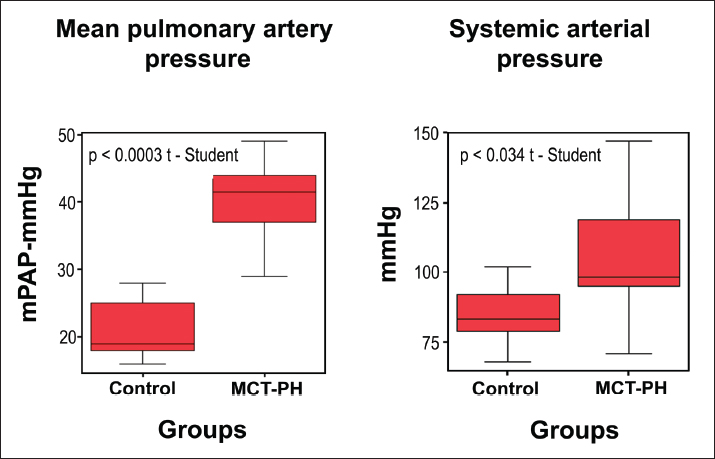
Blood gas analysisThe following parameters were measured: arterial partial pressure of oxygen (PaO2), arterial partial pressure of carbon dioxide (PaCO2), and pH. After the hemodynamic and blood gas evaluations, all animals were euthanized with an overdose of sodium pentobarbital (Anestesal, Pfizer SA de CV, Mexico) in accordance with the Official Mexican Standard NOM-062-ZOO-1999 (SAGARPA, 1999). Immediately thereafter, the vena cavae were ligated, and 10 ml of physiological saline solution was slowly injected through the pulmonary catheter to remove blood from the pulmonary vascular bed. Subsequently, 10 ml of 10% formalin was instilled through the endotracheal cannula, and the cardiopulmonary block was removed. Histological evaluationBiopsies were obtained from the pulmonary artery and each pulmonary lobe, fixed in 10% formalin, embedded in paraffin, and sectioned at 4 µm for hematoxylin and eosin (H&E) staining. Using light microscopy, the following arterial changes were evaluated: muscle hyperplasia, muscle hypertrophy, changes in muscle fiber orientation, and disruption of elastic fibers. In pulmonary tissue, fibrosis, interstitial inflammation, type II pneumocyte hyperplasia, regenerative pneumocytic changes, bronchiolar epithelial regeneration, interstitial edema, endothelial edema, and thrombotic microangiopathy were assessed. Histological findings were evaluated in a blinded manner and classified according to the PH grading system described by Heath and Edwards (1958). Histological evidence of PH was considered present when at least two parameters of the aforementioned classification were observed. Immunohistochemical evaluationImmunohistochemistry was performed in 3-µm-thick sections of lung tissue and pulmonary artery samples using the following antibodies: ET-1 monoclonal antibody (1:100; TR.ET.48.5, MA3-005, Invitrogen Thermo Fisher, Waltham, MA, USA), anti-α-SMA (alpha-smooth muscle actin) antibody (1:500; ab124964, Abcam, Cambridge, MA, USA), CD31 antibody (1:200; 250590, AbbioTec, CA, USA), and anti-VWF antibody (1:50; ab6994, Abcam, Cambridge, MA, USA). The MACH 1 Universal HRP-Polymer Detection system (M1U539G, L10 Biocare Medical, Pacheco, CA, USA) was used. 3-Amino-9-ethylcarbazole chromogen (AEC) (Substrate Pack Biogenex, 932-HK092-F, San Ramón, CA, USA) served as the chromogenic substrate, and sections were counterstained with hematoxylin (Biocare Medical, CA, USA). Lithium carbonate (Fisher Chemical, NY, USA) was applied for color development. Statistical analysisData analysis was performed using SPSS version 19.0 (SPSS Inc., Chicago, Illinois). Continuous variables were expressed as mean ± standard deviation (SD). Prior to statistical comparisons, normality of the data distribution was assessed using the Shapiro–Wilk test. Hemodynamic and blood gas variables showed normal distribution; therefore, comparisons between groups were performed using the independent-samples Student’s t-test. Histopathological variables were analyzed using the χ2 test. A p-value < 0.05 was considered statistically significant. Ethical approvalThis protocol was reviewed and approved by the Bioethics Committee of the National Institute of Respiratory Diseases Ismael Cosío Villegas (approval number IRB/B19-07). ResultsHemodynamic and arterial ABG analyses showed that HR did not differ significantly between groups (p > 0.05). The PH-MCT group exhibited significantly higher systolic and diastolic pulmonary artery pressures compared with the control group (p < 0.05; Table 1). Mean pulmonary artery pressure was 21.2 ± 4.51 mmHg in the control group and 42.90 ± 9.04 mmHg in the PH-MCT group (p < 0.05; Table 1). Systemic blood pressure was also significantly higher in the PH-MCT group (p < 0.05; Fig. 2). In ABG analysis, PaO₂ and PaCO₂ did not differ significantly between groups (p > 0.05). However, the PH-MCT group exhibited a significantly lower pH compared with the control group (p < 0.05; Table 1).
Fig. 2. Variation in vascular pressures. Comparative analysis of mean pulmonary arterial pressure and systolic blood pressure between control and PH-MCT groups. Values are expressed as mean ± SD. *p < 0.05, Student’s t-test. Table 2. Histological classification according to the Heath and Edwards grading system.
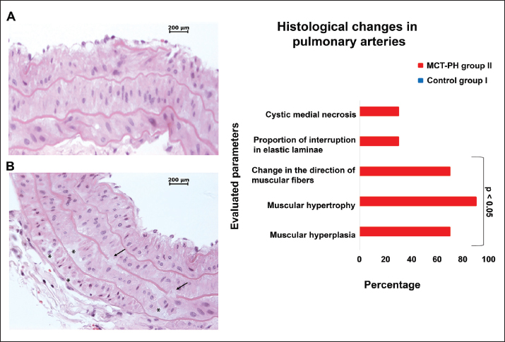
Pulmonary arterial histopathologyHistological analysis of control animals showed no vascular structural abnormalities (grade 0 according to the Heath and Edwards classification). Animals in the PH-MCT group presented different degrees of pulmonary arterial remodeling. Grade I lesions were observed in 50% of animals, grade II lesions in 20%, and grade III lesions in 10%, whereas two animals (20%) showed no evident histological alterations. Significant differences between groups were observed for muscle hyperplasia (χ2=7.912, p=0.005), muscle hypertrophy (χ2=12.929, p=0.0001), and changes in muscle fiber orientation (χ2=7.912, p=0.005). No significant differences were observed in elastic fiber disruption between groups (χ2=1.569, p=0.210). Mucinous deposits in the arterial wall were observed in some animals of the PH-MCT group, although differences between groups were not statistically significant (χ2=1.569, p=0.210; Table 2, Fig. 3). Lung histologyMicroscopically, lung tissue from control animals showed no evident histological alterations. In the PH-MCT group, type II pneumocyte hyperplasia was observed in 60% of animals. Significant differences between groups were identified in the cranial lobe (χ2=7.912, p=0.005), middle lobe (χc=5.952, p=0.015), and caudal lobe (χ2=5.952, p=0.015). Type II hyperplasia was significantly associated with the cranial lobe in the PH-MCT group (p=0.003), as well as in the middle lobe (p=0.011) and caudal lobes (p=0.011).
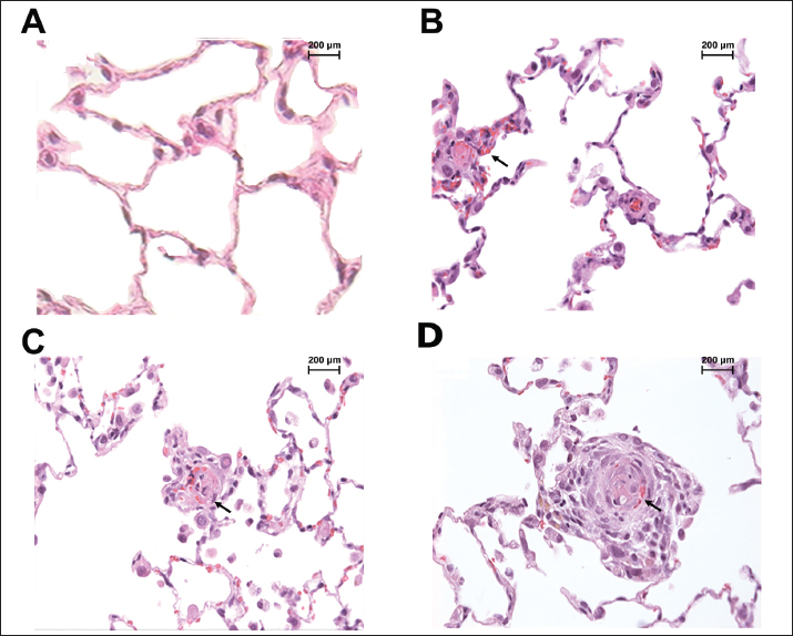
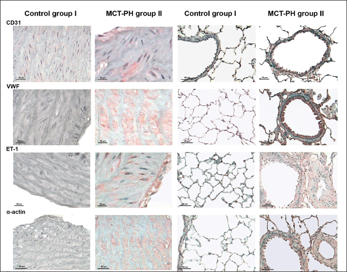
Fig. 3. Representative photomicrographs of pulmonary artery sections (H&E). (A) Control group showing preserved integrity of the elastic laminae between smooth muscle fibers. (B) PH-MCT group showing loss of elastic integrity, fragmentation of laminae (arrows), and accumulation of mucinous material consistent with cystic medial necrosis (asterisk). Morphological changes consistent with pneumocyte regeneration were observed in 50% of animals. Differences between groups were detected in the cranial lobe (χ2=5.952, p=0.015), but not in the middle (χ2=1.569, p=0.210) or caudal lobe (χ2=0.313, p=0.576). Regenerative pneumocytic changes were significantly more frequent in the cranial lobe or the PH-MCT- group (p=0.011). Regeneration of the bronchioalveolar epithelium was observed in 30% of animals, with significant differences in the cranial lobe (χ2=2.813, p=0.004) but not in the middle (χ2=0.000, p=1.000) or caudal lobe (χ2=0.556, p=0.456). Interstitial edema was observed in 30% of animals, without differences between groups in the cranial lobe (χ2=1.569, p=0.210), the middle lobe (χ2=2.813, p=0.094), or the caudal lobe (χ2=1.569, p=0.210). Endothelial edema did not differ between the control group and the group with PH in the cranial lobe (χ2=2.813, p=0.094), the middle lobe (χ2=1.569, p=0.210), or the caudal lobe (χ2=0.449, p=0.503). Interstitial fibrosis was present in 10% of animals, with no difference between groups in the cranial lobe (χ2=0.000, p=1.000), middle lobe (χ2=0.000, p=1.000), or caudal lobe (χ2=0.000, p=1.000). There were also no differences in interstitial inflammation in the cranial lobe (χ2=0.000, p=1.000), middle lobe (χ2=0.556, p=0.456), or caudal lobe (χ2=0.556, p=0.456). Thrombotic microangiopathy was observed in 20% of animals, which was not significantly different between groups in the cranial lobe (χ2=0.556, p=0.456), middle lobe (χ2=0.000, p=1.000), or caudal lobe (χ2=0.000, p=1.00; Table 2, Figs. 4 and 5). ImmunohistochemistryNo immunoreactivity for VWF, ET-1, or α-SMA was detected in pulmonary artery or lung tissue from control animals. Weak CD31 immunoreactivity was observed in lung tissue from control animals. In the PH-MCT group, endothelial immunoreactivity for CD31, VWF, and ET-1 was observed in the pulmonary artery and lung tissue. Diffuse cytoplasmic immunostaining for α-SMA was observed in smooth muscle cells in all cases (Fig. 6).
Fig. 4. Representative histopathological features of lung tissue (H&E). (A) Pulmonary parenchyma from PH-MCT group showing multiple lesions compared with normal lung architecture in controls. (B) Hyperplasia of type II pneumocytes with regenerative changes in PH-MCT group. (C) Acute thrombotic microangiopathy. (D) Medial hypertrophy of a medium-caliber pulmonary artery with concentric “onion-skin” appearance characteristic of pulmonary hypertension. DiscussionThe PH-MCT model revealed a heterogeneous topographic distribution of pulmonary lesions, predominantly affecting the cranial lung lobe. Hemodynamic assessment confirmed the development of PH, as demonstrated by the significant increase in mean pulmonary artery pressure in the PH-MCT group compared with the control group. These findings are consistent with previous studies describing hemodynamic and histopathological alterations associated with experimental PH (Sztuka et al., 2017; Konecny 2020; Guignabert et al., 2024). The PH-MCT rat model remains one of the most widely used experimental models because of its technical simplicity, reproducibility, and ability to reproduce key features of early pulmonary vascular disease (Hill et al., 2017). MCT-induced PH is primarily mediated by vascular endothelial injury, which disrupts the balance between vasodilator and vasoconstrictor mediators and promotes pulmonary vascular remodeling and increased vascular resistance (Lan et al., 2018; Xiao et al., 2019; Maron 2020). In agreement with previous reports, most animals in this study exhibited early-stage vascular lesions characterized by medial hypertrophy, muscularization, and early intimal proliferation, corresponding predominantly to grades I-II of the Heath and Edwards classification. These alterations are commonly associated with progressive vascular remodeling and increased pulmonary vascular resistance (Lan et al., 2018; Sztuka et al., 2017; Maron 2020; Guignabert et al., 2024). A smaller proportion of animals developed grade III lesions characterized by concentric intimal proliferation and partial vascular obliteration, consistent with advanced vascular remodeling (Guignabert et al., 2024).
Fig. 5. Distribution of pulmonary lesion types among lung lobes. Significant differences were observed between PH-MCT and control groups (p < 0.05, χ2 test). Although no significant differences in interstitial inflammation were identified in our study, inflammatory cells and cytokines have been implicated in early stages of pulmonary vascular remodeling in experimental PH (Li et al., 2024). Similarly, thrombotic microangiopathy lesions observed in the PH-MCT group support the presence of endothelial injury and microvascular dysfunction, which are recognized contributors to pulmonary vascular disease progression (Godbole et al., 2019). Macrophages and dendritic cells, both antigen-presenting cells, are thought to facilitate the inflammatory response in PH and thereby contribute to pulmonary vascular remodeling (Godbole et al., 2019; Liang et al., 2021). The MCT model induces alveolar epithelial and endothelial injury, promoting interstitial and alveolar edema. In response, type II pneumocytes proliferate as part of a reparative process aimed at restoring surfactant production and alveolar epithelial integrity (Castranova et al., 1988; Humbert et al., 2019). However, previous studies have also reported variable responses, including densities of type II pneumocytes, depending on MCT dose and exposure duration (Wilson and Segall, 1990). Chronic exposure of the alveolar–capillary barrier to elevated microvascular pressure in PH leads to adaptive structural modifications aimed at protecting this barrier from further vascular injury. These changes include thickening of the barrier and increased numbers of type II alveolar epithelial cells, thickened basement membranes, and expansion of the interstitial matrix (Zhou et al., 2018). Immunohistochemical findings further supported endothelial dysfunction and vascular remodeling in the PH-MCT group. Increased immunoreactivity for ET-1, VWF, CD31, and α-SMA reflected endothelial activation, smooth muscle cell proliferation, and vascular structural changes. These results complement the hemodynamic and histopathological results and are consistent with the established role of endothelial injury in the pathogenesis of MCT-induced PH. The principal finding of this study was the marked heterogeneity in lesion distribution among lung lobes, with a clear predominance in the cranial lobe. Previous studies have demonstrated differences in pulmonary blood flow distribution in experimental PH models, including the MCT model (Schwenke et al., 2009; Dickinson et al., 2013). Similarly, Zhu et al. (2020) demonstrated significant vascular alterations among pulmonary lobes in MCT-induced PH, including reductions in distal vascular branches and junctions. However, the relationship between regional blood flow patterns and lesion topography has not been fully explored. Unlike larger mammals, rats have limited gravitational gradients in ventilation and perfusion, and regional susceptibility may be determined by local bronchovascular anatomy, airflow exposure, and mechanical stress (Glenny et al., 2013). These factors may contribute to the preferential development of endothelial injury and vascular remodeling in the cranial region following monocrotaline administration.
Fig. 6. Representative immunohistochemical staining of lung tissue and pulmonary artery using 3-amino-9-ethylcarbazole (AEC) chromogen showing red immunolabeling. No immunoreactivity for von Willebrand factor (VWF), endothelin-1 (ET-1), or α-SMA was detected in the control group, whereas positive immunostaining for smooth muscle cells was observed in the PH-MCT group. Therefore, the heterogeneous distribution of pulmonary lesions observed in the PH-MCT model is likely multifactorial. Regional differences in pulmonary perfusion and vascular pressure gradients may lead to uneven exposure of the pulmonary endothelium to toxic monocrotaline metabolites, thereby promoting localized influences such as oxygen delivery, vascular tone, and local hemodynamic stress, affecting susceptibility to injury and contributing to regional differences in inflammatory responses, vascular remodeling, and parenchymal damage. Altered hypoxic pulmonary vasoconstriction and autonomic regulation may also participate in this process. Previous studies have suggested that chronic PH can modify sympathetic regulation of the pulmonary vasculature through changes in β-adrenergic receptor expression and activity, potentially affecting regional vascular tone and endothelial responses to injury. In the MCT model, reduced pulmonary β-adrenergic receptor density and attenuated sympathetic activity have been reported, which may further contribute to abnormal regional vasoreactivity and heterogeneous vascular remodeling (Schwenke et al., 2009). Collectively, these mechanisms may explain the predominance of lesions observed in the cranial lung lobe and support the concept that pulmonary lesion development in the PH-MCT model is not uniformly distributed throughout the lungs. Further studies are required to elucidate the underlying mechanisms. These observations highlight the importance of considering regional pulmonary variability in experimental PH studies. The heterogeneous distribution of lesions suggests that pulmonary sampling strategies may influence histopathological interpretation and lesion quantification, while recognition of regional susceptibility patterns may improve the assessment of molecular and cellular responses, disease progression, and therapeutic effects. It is important to acknowledge several limitations of this study. First, final body weight measurements were not recorded, preventing the assessment of weight changes throughout the experimental period. Second, right ventricular hypertrophy was not quantified using the Fulton index. While PH was confirmed by hemodynamic measurements and histopathological evaluation, the inclusion of the Fulton index would have complemented the characterization of right ventricular remodeling associated with disease progression. ConclusionThese findings reveal marked lobar heterogeneity in pulmonary pathology and highlight the translational relevance of the MCT-induced PH model in veterinary research and experimental animal medicine, as well as for comparative and translational research. Furthermore, they support its value as a preclinical platform for evaluating pharmacological and interventional therapies. AcknowledgmentsThe authors would like to express their sincere gratitude to David Rodríguez Santiago for providing the photographic documentation of the animal model, which contributed significantly to the visual presentation of this study. Conflict of interestEach author declares that they have no conflict of interest in relation to the submitted article. FundingThis study was supported by the Ismael Cosío Villegas National Institute of Respiratory Diseases. The funder did not participate in study design, data collection and analysis, decision to publish, or preparation of the manuscript Authors’ contributionsConception and Design: MBL, EBL, CHJ. Analysis and Interpretation: MBL, EBL, RJV, VSA, AEGC, NHG, MGG, ROZ. Data Collection: NHG, JVC, OMA. Writing the Manuscript: MBL, CHJ. Critical Revision: MBL, CHJ, EBL, RJV, VSA, AEGC, NHG, MGG, ROZ, OMA. Approval of the Manuscript: MBL, CHJ, EBL, RJV, VSA, AEGC, NHG, MGG, ROZ, OMA. Data availabilityAll data supporting the findings of this study are available within the manuscript. ReferencesBalls, M. 1994 Replacement of animal procedures: alternatives in research, education and testing. Lab. Anim. 28(3), 193–211. Castranova, V., Rabovsky, J., Tucker, J.H. and Miles, P.R. 1988 The alveolar type II epithelial cell: a multifunctional pneumocyte. Toxicol. Appl. Pharmacol. 93(3), 472–483. Dickinson, M.G., Bartelds, B., Borgdorff, M.A. and Berger, R.M. 2013 The role of disturbed blood flow in the development of pulmonary arterial hypertension: lessons from preclinical animal models. Am. J. Physiol. Lung Cell. Mol. Physiol. 305(1), L1–14; doi:10.1152/ajplung.00031.2013. Ferraro, A., Hartnack, S. and Schwarzwald, C.C. 2023 Diagnostic value of two-dimensional echocardiographic measurements of the pulmonary artery diameter and the pulmonary artery distensibility index to detect pulmonary hypertension in horses. J. Vet. Cardiol. 49, 52–66. Glenny, R.W., Bauer, C., Hofmanninger, J., Lamm, W.J., Krueger, M.A. and Beichel, R.R. 2013 Heterogeneity and matching of ventilation and perfusion within anatomical lung units in rats. Respir. Physiol. Neurobiol. 189(3), 594–606; doi:10.1016/j.resp.2013.07.027 Godbole, RH., Saggar, R. and Kamangar, N. 2019 Pulmonary tumor thrombotic microangiopathy: a systematic review. Pulm. Circ. 9(2), 2045894019851000. Guignabert, C., Aman, J., Bonnet, S., Dorfmüller, P., Olschewski, AJ., Pullamsetti, S., Rabinovitch, M., Schermuly, R.T., Humbert, M. and Stenmark, K.R. 2024 Pathology and pathobiology of pulmonary hypertension: current insights and future directions. Eur. Respir. J. 64(4), 2401095; doi: 10.1183/13993003.01095-2024 Heath, D. and Edwards, J.E. 1958 The pathology of hypertensive pulmonary vascular disease; a description of six grades of structural changes in the pulmonary arteries with special reference to congenital cardiac septal defects. Circulation 18(4 Part 1), 533–547. Hill, N.S., Gillespie, M.N. and McMurtry, I.F. 2017 Fifty years of monocrotaline-induced pulmonary hypertension: what has it meant to the field? Chest 152(6), 1106–1108. Humbert, M., Guignabert, C., Bonnet, S., Dorfmüller, P., Klinger, J.R., Nicolls, M.R., Olschewski, A.J., Pullamsetti, S.S., Schermuly, R.T., Stenmark, K.R. and Rabinovitch, M. 2019 Pathology and pathobiology of pulmonary hypertension: state of the art and research perspectives. Eur. Respir. J. 53(1), 1801887; doi:10.1183/13993003.01887-2018. Humbert, M., Kovacs, G., Hoeper, M.M., Badagliacca, R., Berger, R.M.F., Brida, M., Carlsen, J., Coats, A.J.S., Escribano-Subias, P., Ferrari, P., Ferreira, D.S., Ghofrani, H.A., Giannakoulas, G., Kiely, D.G., Mayer, E., Meszaros, G., Nagavci, B., Olsson, K.M., Pepke-Zaba, J., Quint, J.K., Rådegran, G., Simonneau, G., Sitbon, O., Tonia, T., Toshner, M., Vachiery, J.L., Vonk Noordegraaf, A., Delcroix, M. and Rosenkranz, S. 2022 ESC/ERS Scientific Document Group. 2022 ESC/ERS Guidelines for the diagnosis and treatment of pulmonary hypertension. Eur Heart J. 43(38), 3618–3731; doi: 10.1093/eurheartj/ehac237 Kilkenny, C., Browne, W.J., Cuthill, I.C., Emerson, M. and Altman, D.G. 2010 Improving bioscience research reporting: the ARRIVE guidelines for reporting animal research. PLoS Biol. 8(6), e1000412. Konecny, F. 2020 Hemodynamic characterization of pulmonary artery hypertension in a rat. Eur. J. Med. Health Sci. 2(3), 1–5; doi:10.24018/ejmed.2020.2.3.164. Lachant, D.J., Meoli, D.F., Haight, D., Lyons, J.A., Swarthout, R.F. and White, R.J. 2018 Low dose monocrotaline causes a selective pulmonary vascular lesion in male and female pneumonectomized rats. Exp Lung Res. 44(1), 51–61. Lan, N.S.H., Massam, B.D., Kulkarni, S.S. and Lang, C.C. 2018 Pulmonary arterial hypertension: pathophysiology and treatment. Diseases 6(2), 38. Li, Z., Ma, J., Wang, X., Zhu, L., Gan, Y. and Dai, B. 2024 The role of immune cells in the pathogenesis of connective tissue diseases-associated pulmonary arterial hypertension. Front. Immunol. 17(15); doi:10.3389/fimmu.2024.1464762. Liang, S., Desai, A.A., Black, S.M. and Cytokines, T.H., 2021 Chemokines, and Inflammation in Pulmonary Arterial Hypertension. Adv Exp Med Biol. 1303, 275–303; doi: 10.1007/978-3-030-63046-1_15. Lwanga, S.K. and Lemeshow, S. 1991 Sample size determination in health studies: a practical manual. Geneva, Switzerland: World Health Organization. Maron, B.A. 2020 Pulmonary arterial hypertension: cellular and molecular changes in the lung. Glob Cardiol. Sci. Pract. 2020(1), e202003. Rafikova, O., Al Ghouleh, I. and Rafikov, R. 2019 Focus on early events: pathogenesis of pulmonary arterial hypertension development. Antioxid. Redox. Signal. 31(13), 933–953. Reinero, C., Visser, L.C., Kellihan, H.B., Masseau, I., Rozanski, E., Clercx, C., Williams, K., Abbott, J., Borgarelli, M. and Scansen, B.A. 2020 ACVIM consensus statement guidelines for the diagnosis, classification, treatment, and monitoring of pulmonary hypertension in dogs. J. Vet. Intern. Med. 34(2), 549–573. Schwenke, D.O., Pearson, J.T., Shimochi, A., Kangawa, K., Tsuchimochi, H., Umetani, K., Shirai, M. and Cragg, P.A. 2009 Changes in pulmonary blood flow distribution in monocrotaline compared with hypoxia-induced models of pulmonary hypertension: assessed using synchrotron radiation. J. Hypertens. 27(7), 1410–1419. de Agricultura, Secretaría. and Ganadería, Desarrollo Rural. 1999 Pescay Alimentación (SAGARPA). Norma Oficial Mexicana NOM-062-ZOO-1999. Especificaciones técnicas para la producción, cuidado y uso de animales de laboratorio. Diario Oficial de la Federación. Simonneau, G., Montani, D., Celermajer, D.S., Denton, C.P., Gatzoulis, M.A., Krowka, M., Williams, P.G. and Souza, R.2019. Haemodynamic definitions and updated clinical classification of pulmonary hypertension. Eur. Respir. J. 53(1), 1801913. Stenmark, K.R., Meyrick, B., Galie, N., Mooi, W.J. and McMurtry, I.F. 2009 Animal models of pulmonary arterial hypertension: the hope for etiological discovery and pharmacological cure. Am. J. Physiol. Lung Cell Mol. Physiol. 297(6), L1013–L1032. Sztuka, K. and Jasińska-Stroschein, M. 2017 Animal models of pulmonary arterial hypertension: a systematic review and meta-analysis of data from 6126 animals. Pharmacol Res. 125(Pt B), 201–214. doi: 10.1016/j.phrs.2017.08.003. Tuder, R.M. and Stenmark, K.R. 2020 Perspective: pathobiological paradigms in pulmonary hypertension, time for reappraisal. Am. J. Physiol. Lung Cell Mol. Physiol. 318(6), L1131–L1137. Wilson, D.W. and Segall, H.J. 1990 Changes in type II cell populations in monocrotaline pneumotoxicity. Am. J. Pathol. 136(6), 1293–1299. Xiao, R., Zhu, L., Su, Y., Zhang, J., Lu, Y., Li, J., Wang, T., Fang, J., Jing, Z.C., Dupuis, J., Luo, S. and Hu, Q. 2019 Monocrotaline pyrrole induces pulmonary endothelial damage through binding to and release from erythrocytes in lung during venous blood reoxygenation. Am. J. Physiol. Lung Cell Mol. Physiol. 316(5), L798–L809. Zhou, C., Crockett, E.S., Batten, L., McMurtry, I.F. and Stevens, T. 2018 Pulmonary vascular dysfunction secondary to pulmonary arterial hypertension: insights gained through retrograde perfusion. Am. J. Physiol. Lung Cell Mol. Physiol. 314(5), L835–L845. Zhu, Z., Wang, Y., Long, A., Feng, T., Ocampo, M., Chen, S., Tang, H., Guo, Q., Minshall, R., Makino, A., Huang, W. and Chen, J. 2020 Pulmonary vessel casting in a rat model of monocrotaline-mediated pulmonary hypertension. Pulm. Circ. 10(3), 2045894020922129 | ||




| How to Cite this Article |
| Pubmed Style Baltazares-lipp M, Hernández-jiménez C, Jasso-victoria R, Soto-abraham V, Guzmán-cedillo AE, Baltazares-lipp E, Hernández-garcía N, Gaxiola-gaxiola M, Olmos-zúñiga R, Martínez-alarcón O. Histopathological study of the specific lobar distribution of lung lesions in a rat model of monocrotaline-induced pulmonary hypertension. Open Vet. J.. 2026; 16(8): 5698-5708. doi:10.5455/OVJ.2026.v16.i8.59 Web Style Baltazares-lipp M, Hernández-jiménez C, Jasso-victoria R, Soto-abraham V, Guzmán-cedillo AE, Baltazares-lipp E, Hernández-garcía N, Gaxiola-gaxiola M, Olmos-zúñiga R, Martínez-alarcón O. Histopathological study of the specific lobar distribution of lung lesions in a rat model of monocrotaline-induced pulmonary hypertension. https://www.openveterinaryjournal.com/?mno=309287 [Access: September 03, 2026]. doi:10.5455/OVJ.2026.v16.i8.59 AMA (American Medical Association) Style Baltazares-lipp M, Hernández-jiménez C, Jasso-victoria R, Soto-abraham V, Guzmán-cedillo AE, Baltazares-lipp E, Hernández-garcía N, Gaxiola-gaxiola M, Olmos-zúñiga R, Martínez-alarcón O. Histopathological study of the specific lobar distribution of lung lesions in a rat model of monocrotaline-induced pulmonary hypertension. Open Vet. J.. 2026; 16(8): 5698-5708. doi:10.5455/OVJ.2026.v16.i8.59 Vancouver/ICMJE Style Baltazares-lipp M, Hernández-jiménez C, Jasso-victoria R, Soto-abraham V, Guzmán-cedillo AE, Baltazares-lipp E, Hernández-garcía N, Gaxiola-gaxiola M, Olmos-zúñiga R, Martínez-alarcón O. Histopathological study of the specific lobar distribution of lung lesions in a rat model of monocrotaline-induced pulmonary hypertension. Open Vet. J.. (2026), [cited September 03, 2026]; 16(8): 5698-5708. doi:10.5455/OVJ.2026.v16.i8.59 Harvard Style Baltazares-lipp, M., Hernández-jiménez, . C., Jasso-victoria, . R., Soto-abraham, . V., Guzmán-cedillo, . A. E., Baltazares-lipp, . E., Hernández-garcía, . N., Gaxiola-gaxiola, . M., Olmos-zúñiga, . R. & Martínez-alarcón, . O. (2026) Histopathological study of the specific lobar distribution of lung lesions in a rat model of monocrotaline-induced pulmonary hypertension. Open Vet. J., 16 (8), 5698-5708. doi:10.5455/OVJ.2026.v16.i8.59 Turabian Style Baltazares-lipp, Matilde, Claudia Hernández-jiménez, Rogelio Jasso-victoria, Virgilia Soto-abraham, Axel Edmundo Guzmán-cedillo, Enrique Baltazares-lipp, Noé Hernández-garcía, Miguel Gaxiola-gaxiola, Raúl Olmos-zúñiga, and Omar Martínez-alarcón. 2026. Histopathological study of the specific lobar distribution of lung lesions in a rat model of monocrotaline-induced pulmonary hypertension. Open Veterinary Journal, 16 (8), 5698-5708. doi:10.5455/OVJ.2026.v16.i8.59 Chicago Style Baltazares-lipp, Matilde, Claudia Hernández-jiménez, Rogelio Jasso-victoria, Virgilia Soto-abraham, Axel Edmundo Guzmán-cedillo, Enrique Baltazares-lipp, Noé Hernández-garcía, Miguel Gaxiola-gaxiola, Raúl Olmos-zúñiga, and Omar Martínez-alarcón. "Histopathological study of the specific lobar distribution of lung lesions in a rat model of monocrotaline-induced pulmonary hypertension." Open Veterinary Journal 16 (2026), 5698-5708. doi:10.5455/OVJ.2026.v16.i8.59 MLA (The Modern Language Association) Style Baltazares-lipp, Matilde, Claudia Hernández-jiménez, Rogelio Jasso-victoria, Virgilia Soto-abraham, Axel Edmundo Guzmán-cedillo, Enrique Baltazares-lipp, Noé Hernández-garcía, Miguel Gaxiola-gaxiola, Raúl Olmos-zúñiga, and Omar Martínez-alarcón. "Histopathological study of the specific lobar distribution of lung lesions in a rat model of monocrotaline-induced pulmonary hypertension." Open Veterinary Journal 16.8 (2026), 5698-5708. Print. doi:10.5455/OVJ.2026.v16.i8.59 APA (American Psychological Association) Style Baltazares-lipp, M., Hernández-jiménez, . C., Jasso-victoria, . R., Soto-abraham, . V., Guzmán-cedillo, . A. E., Baltazares-lipp, . E., Hernández-garcía, . N., Gaxiola-gaxiola, . M., Olmos-zúñiga, . R. & Martínez-alarcón, . O. (2026) Histopathological study of the specific lobar distribution of lung lesions in a rat model of monocrotaline-induced pulmonary hypertension. Open Veterinary Journal, 16 (8), 5698-5708. doi:10.5455/OVJ.2026.v16.i8.59 |


