





| Research Article | ||
Open Vet. J.. 2026; 16(8): 5363-5375
Open Veterinary Journal, (2026), Vol. 16(8): 5363–5375 Research Article Relationship between quorum-sensing and virulence genes of Pseudomonas aeruginosa isolated from patients, and their association with antimicrobial resistanceAbdul-Nafea Ayad Hassan* and Ibtisam Qahtan Abdul-KareemDepartment of Veterinary Public Health, Zoonotic Disease Unit, College of Veterinary Medicine, University of Baghdad, Baghdad, Iraq *Corresponding Author: Abdul Nafea Ayad Hassan. Department of Veterinary Public Health, Zoonotic Disease Unit, College of Veterinary Medicine, University of Baghdad, Baghdad, Iraq. Email: Abdulnafie.ayad1304e [at] covm.uobaghdad.edu.iq Submitted: 04/02/2026 Revised: 01/06/2026 Accepted: 12/06/2026 Published: 08/08/2026 © 2025 Open Veterinary Journal
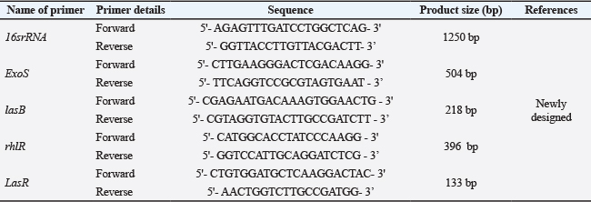
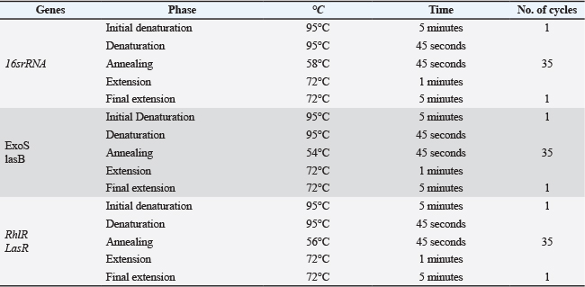
AbstractBackground: Zoonotic diseases are illnesses that humans can contract from animals and vice versa. Pseudomonas aeruginosa is an opportunistic organism that makes treating infections challenging, particularly in individuals with underlying medical conditions and those with impaired immune systems. The increasing number of multidrug-resistant forms of P. aeruginosa poses a serious threat to public health. Aim: This study examined the frequency of two virulence genes and two quorum-sensing genes. Methods: 100 samples (50 urine, 25 wound infections, and 25 burns) were collected from Medical City hospitals in Baghdad, Iraq, during the period (October 2023; February 2024). Determination of isolates was made according to colony features, Gram staining techniques, standard biochemical responses, and confirmed by the 16s rRNA gene. Some of the virulence factor genes (ExoS and lasB) and quorum-sensing genes (LasR and rhlR) were examined using specific primers with the aid of polymerase chain reaction (PCR). Then, the biofilm production test and antibiotic sensitivity test were conducted on these isolates using the microtiter plate method. Results: A total of 17 isolates (17%) were obtained from urine, burns, and wound infections. The isolates included 2 (40.00%) for the ExoS gene, 3(60.00%) for the lasB gene, and 2(60.00%) and 3(40.00%) for LasR and rhlR genes, respectively. The result showed that all the isolates 17(100%) produced biofilm, revealing that 60% of P. aeruginosa isolates were strong biofilm producers, while 30% and 10 % of the isolates were moderate and weak producers, respectively. The results also indicate that the isolates of P. aeruginosa resist meropenem, ceftazidime, and nalidixic 17(100%), while they were sensitive to gentamicin 8(74.0%), amikacin 7(41.1%), piperacillin 15(88.2%), ciprofloxacin, and levofloxacin 13(76.4%). Conclusion: Human isolates exhibit a higher prevalence of virulence genes, specifically ExoS (20%) and IasB (40%), relative to isolates from pet birds. Furthermore, human isolates demonstrate a greater prevalence of quorum-sensing genes, with LasR at 40% and rhlR at 60%, compared to pet bird isolates. Biofilm production among isolates obtained from human and pet bird sources was categorized as follows: 59.3% demonstrated strong production, 28.1% exhibited moderate production, and 12.5% displayed weak production. Keywords: Biofilm, Pseudomonas aeruginosa, Quorum-sensing genes, Virulence genes. IntroductionPseudomonas aeruginosa, Pseudomonas putida, Pseudomonas luteola, and Pseudomonas fluorescens are among the species that belong to the genus Pseudomonas, which is a member of the Pseudomonadaceae family (Razook et al., 2020). These species may exist in a variety of settings, some of which are external to the body as environmental isolates in soil and water, while most of them are harmful clinical isolates (Mohammed and Zgair, 2022). In addition, they can be found in some foodborne sources like dairy products (Qasim et al., 2023). They can grow in the best and significant mean under 25°C, 35°C, and 40°C (Al-Mamoory and Al-Mayaly, 2017). It is considered one of the most important hospital-acquired infections due to its ability to resist many antibiotics (Dawood and Alkazaz, 2024). According to Al-Qaissy and Al-Khafaji (2023), due to emerging resistance to antibiotics, the respiratory tract, placenta, urinary system, skin, and soft tissues are the most commonly infected areas by P. aeruginosa, with surgical wounds, burns, and pressure ulcers carrying the highest risk (Pachori et al., 2019). QS is a bacterial communication mechanism that controls gene expression in response to bacterial cell population density, which is indicated macroscopically by the production of pyocyanin (Hashim et al., 2024). The phenomenon impacts genes essential for virulence, horizontal gene transfer, and bacterial growth and function. The production of virulence components, such as protease, pyocyanin, exotoxin A—the extremely harmful virulence factor that attaches itself to certain cell receptors (Jawad and Rasheed, 2022)—alkaline protease, lectins, and rhamnosyltransferase, is significantly influenced by the QS system in P. aeruginosa (Hendiani et al., 2019), which may result in a substantial rise in the blood's mononuclear cells, primarily monocytes and lymphocytes (Razook et al., 2020). The Exoenzyme S (ExoS) protein has unique roles that control a variety of cellular processes, including phagocytosis, cell migration, and cytoskeleton organization (Barbieri and Sun 2004), impacting the host cell's cytoskeleton actin structure by decreasing cell–cell adhesion and promoting P. aeruginosa invasion through epithelial barriers (Yang et al., 2022). In addition, P. aeruginosa’s cell surface is covered with thin, flexible filaments called type IV pili (T4P), which are engaged in pathogenesis-related processes including cell adsorption, biofilm formation, and twitching motility (Hendrix et al., 2024). Pyoverdine and pyochelin, two kinds of siderophores produced by the opportunistic bacterium P. aeruginosa, are essential for scavenging iron from the environment and host cells. Siderophores of P. aeruginosa can carry out a number of tasks and act as virulence factors (Jeong et al., 2024). The majority of P. aeruginosa possess genes that encode different QS systems (rhlR and lasR); these genes are thought to be multidrug-resistant bacteria since they have resisted numerous antibiotic classes for more than three years (Ratajczak et al., 2021). A global issue is the spread of germs resistant to antibiotics. Given the significance of P. aeruginosa as a bacterium that is resistant to multiple drugs (Abdulameer and Abdulhassan, 2021). Centers for Disease Control and Prevention (CDC) have designated carbapenem-resistant as an urgent threat (Garcia et al. 2020). This study highlights the virulence and quorum-sensing genes in P. aeruginosa isolated from different samples of human patients. Materials and MethodsSample collection and processingHundred samples (50 urine, 25 wound infections, and 25 burns) were collected from Medical City Hospitals in Baghdad, Iraq, during the period (October 2023; February 2024). A highly aseptic condition was maintained during the sampling; the samples were labeled, placed in an ice box, and transported to the Microbiology Laboratory, Unit of Zoonotic Disease, College of Veterinary Medicine, University of Baghdad. Identification of P. aeruginosaUsing the sterile standard loop method, the isolates under examination were first identified on blood agar (Himedia, India) and MacConkey agar (Oxoid, UK). For a whole day, the media were incubated at 37°C according to Jibril et al. (2019). The bacterial diagnosis was validated by conventional morphological methods (beta-hemolytic, large, and gray-white on blood agar; and on MacConkey agar, large, mucoid, and lactose non-fermenting) (Mahmood and Aljobori, 2015), while on cetrimide agar as a selective medium, it appeared as a smooth and greenish-yellow colony. Then, Gram stain and biochemical tests were performed. Finally, bacterial isolates were confirmed using the 16s rRNA gene (Shakir et al., 2023). Molecular detectionA. Pseudomonas aeruginosa DNA extraction: DNA was extracted from P. aeruginosa using the FavorPrep Bacterial/Cultured Cells Genomic DNA Extraction Mini Kit, processed from FavorPrep® (Korea) according to the company’s instructions. B. DNA electrophoresis in agarose gel: The procedure was done following Santiago et al. (2021). C. DNA Primers: The primer sequences used in this study are detailed in Table 1. The primers for the lasB gene were newly designed for this work, while those for the lasR and rhlR genes were adopted from previous studies. The 16S rRNA and exoS primers were utilized as standard specific sequences for isolate confirmation and virulence detection, respectively. Studies by Ratajczak et al. (2021) and Ghanem et al. (2023). Program for Thermocycler for amplifying DNAThe enzyme polymerization method was carried out using a thermocycler PCR. This device was programmed for the genes under study using the interactions listed in Table 2. Antimicrobial susceptibility testingAccording to the guidelines of the Clinical and Laboratory Standards Institute, antibiotic susceptibility testing was conducted using the Kirby–Bauer disc diffusion method (CLSI, 2022), on the following antibiotics: ciprofloxacin (10 μg), levofloxacin (5 μg), meropenem (10 μg), piperacillin (100 μg), amikacin (30 μg), gentamicin (10 μg), ceftazidim (30 μg), and nalidixic (30 μg) Biofilm formation testingBiofilm formation was performed using the microtiter plate method. Quantification of biofilm formation by P. aeruginosa on abiotic surfaces was assessed as previously described (Tang et al., 2011). In brief, wells of sterile 96-well flat-bottomed polystyrene microplates were filled with 200 μl of an overnight Luria–Bertani (LB) broth supplemented with 1% glucose, with a bacterial concentration equivalent to McFarland standard no. 0.5, before the plates were covered and incubated aerobically at 37°C for 24 hours. Each bacterium was tested in triplicate. Control wells were prepared by adding bacteria-free LB. Table 1. The name, sequence, and product size of primers used in this study.
Table 2. PCR conditions for virulence ExoS, lasB, and quorum-sensing RhlR and LasR genes.
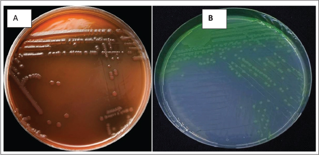
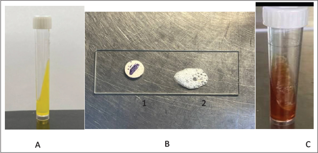
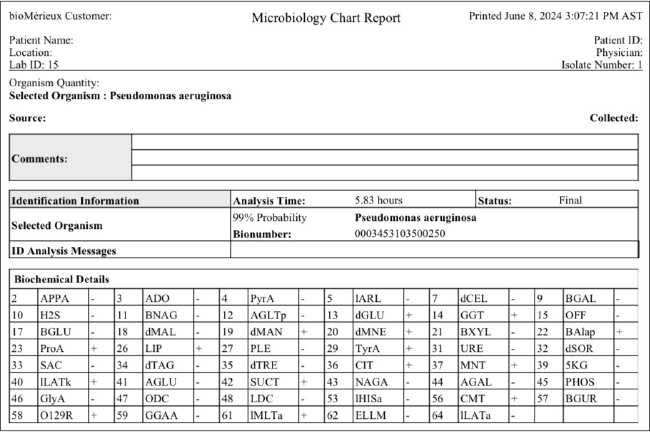
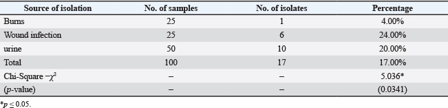
To visualize biofilms, the contents of the wells were aspirated, and the wells were washed three times with 200-μl sterile distilled water to remove loosely adherent cells. The remaining attached bacteria were fixed with 200-μl methanol for 15 minutes. After drying in air, the wells were stained with 200-μl 0.1% crystal violet solution for 15 minutes at room temperature. Excess stain was rinsed off by placing the plate under running tap water. Thereafter, the plates were dried at 37°C for approximately 30 minutes to ensure they were completely dry. Subsequently, the adherent cells were resolubilized with 200 μl of 33% glacial acetic acid for 15 minutes. Finally, the optical density of each well was obtained at 600 nm using a microplate reader. The cutoff value was calculated as the mean of the OD of control wells plus 3 SDs. Classification presented in Table 11, based on OD values obtained for individual isolates of P. aeruginosa, was used for the purpose of data simplification and calculation (Atshan et al., 2012). Statistical analysisThe Statistical Analysis System (SAS, 2018) program was used to detect the effect of different factors on study parameters. The chi-square test was used to compare percentages at 0.05 and 0.01 probability levels in this study. Ethical approvalThis research protocol was approved by our institutional ethics committee (University of Baghdad) via letter number PG-848/2025. ResultsAs shown in Figure 1, the isolates were identified on MacConkey agar and cetrimide agar. Biochemical testsAll isolates tested by biochemical tests for urease, oxidase, catalase, and triple sugar iron, as shown in Figure 2, 3, were confirmed by VITEK. From 100 samples that were obtained from burns, wound infections, and urine, 17 (17%) of the isolates were diagnosed with P. aeruginosa. From burns, the percentage was 1/25 (4.00 %), while from wound infections, it was 6/25 (24.00%), and from urine, it was 10/50 (20.00%). The percentage of male patients represented 10/48 (20.83%) of the total, while female patients made up 7/52 (13.46%), and the distribution according to age group showed the highest prevalence in the age group from 36 to >46, which was 7 (31.8%), as shown in Tables 3–5.
Fig. 1. Colonies characteristic on MacConkey agar and cetrimide agar, A: P. aeruginosa on MacConkey agar (non-lactose fermenter). B: P. aeruginosa on cetrimide agar (pyocyanin production).
Fig. 2. Biochemical tests. A: Urease; B: (1) Oxidase (2) Catalase. C: TSI.
Fig. 3. P. aeruginosa VITEK®2 compact system identification. Table 3. The number of P. aeruginosa isolates according to the source of isolation in humans.
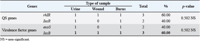
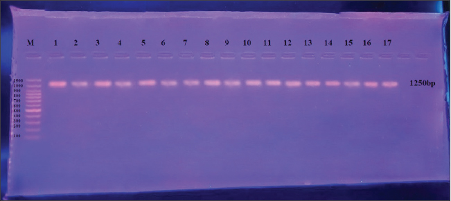
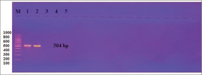
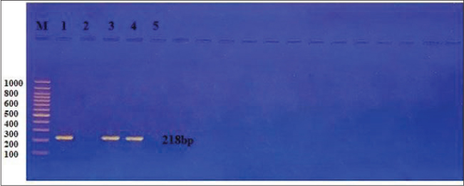
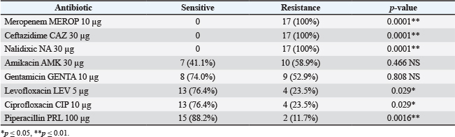
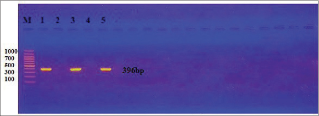
Molecular identification of P. aeruginosaFive isolates were chosen to be confirmed by the 16s rRNA gene and the PCR method to diagnose P. aeruginosa; the current study indicated that all the isolates were positive, as shown in Figure 4. Virulence genes of P. aeruginosaThe designated primers were employed to determine the presence of the ExoS and lasB genes. The PCR outcomes for the P. aeruginosa isolates showed that 25/(40.00%) for the ExoS gene and 35/ (60.00%) were positive for the lasB gene, as shown in the figures below (Tables 6 and 8; Fig. 5 and 6). Quorum-sensing of P. aeruginosaThe existence of the rhlR and LasR genes was verified using the specified primers. As shown in the figures below, the PCR results for the P. aeruginosa isolates revealed that 3/5 (60.00%) for the rhlR gene and 3/5 (40.00%) for the LasR gene (Tables 7 and 8; Fig. 7 and 8). Antibiotic susceptibility testThe findings explained that each clinical isolate of P. aeruginosa was sensitive to gentamicin 8 (47.1%), amikacin 7 (41.1%), piperacillin 15 (88.2%), ciprofloxacin, and levofloxacin 13 (76.4%), while resist for meropenem, ceftazidime, and nalidixic acid 17(100%). As shown in Table 9 and their relation with types of samples as in Table 10. Table 4. Distribution of samples according to age groups.
Table 5. Distribution of samples according to sex.
Table 6. Detection of virulence genes.
Table 7. Detection of quorum sensing.
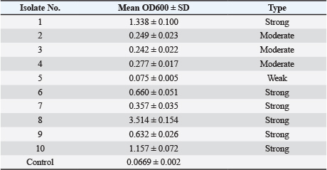
Biofilm formationTen isolates were chosen to be confirmed by the microtiter plate method. The results summarized in Table 11 revealed that 60% of P. aeruginosa isolates were strong biofilm producers, while 30% and 10% of the isolates were moderate and weak producers, respectively. DiscussionPseudomonas aeruginosa is ranked second among Gram-negative bacteria isolated in the hospital environment and is a leading cause of nosocomial infections responsible for a high morbidity and mortality rate. High prevalence of pseudomonal infections is common among critically ill patients upon admission to the intensive care unit and those with underlying clinical conditions. Our current study was obtained (1,6,10) from burns, wound infections, and urine, respectively, of P. aeruginosa isolates. This is in contrast to a study from Egypt, which reported a total of 57 P. aeruginosa strains (54 clinical and 3 environmental isolates) isolated from 287 clinical specimens and 40 urine specimens (17), and a total of 60 clinical and burn isolates of P. aeruginosa )Al-Yasseen et al., 2012(. The high number of P. aeruginosa isolates from the previous study could be due to differences in sample size and because many of the samples were from clinical sources (Silva et al., 2023). In other studies, among 1,049 samples showing growth of bacteria, P. aeruginosa was isolated from 68 (6.48%). This prevalence rate was somewhat higher and lower than other studies, 4.15% and 8.59%, respectively (Shidiki et al., 2019; Shrestha et al., 2021). Table 8. Distribution of quorum-sensing and virulence factor genes in P. aeruginosa isolated from different clinical specimens.
Fig. 4. Gel electrophoresis for PCR product of 16s rRNA gene shows 1,250 bp. Primer TM at (58 C), (1% agarose, at 85 volts, 1.5 hours). Visualized under UV light after staining with ethidium bromide. 5 volt/cm2 80, MA(75) for 1.5 hours. The prevalence of P. aeruginosa infection was higher in male patients than in female patients (52.5% and 47.5%, respectively), similar to a study in the northern part of Nigeria (Jombo et al., 2008; Al-Zaidi, 2016). The results of the current study are consistent with the results of different studies, including the study conducted by Kadhim and Ali (2014) in Baghdad city, which resulted in sixty isolates of P. aeruginosa isolated from different hospitals in Baghdad. The sources of these isolates were as follows: 22 (36.66%) isolates from burns, including 15% male and 21.6% female; 18 (30%) from wounds, including 26.6% male and 3.3% female; 9 (15%) from sputum, including 6.6% male and 8.3% female; 3 (5%) isolates from ear swabs, including 5 male and none female; 2 (3.3%) from urine, including 1.6% male and 1.6% female; and 6 (10%) isolates from blood. Also, the results showed that the highest rate of P. aeruginosa was 45% in older patients (ages 36–46 years), while the lowest rate, 20%, was found among the young age group <30 years. These results disagree with the studies of Shewatatek et al. (2014); their study reported a higher prevalence of these bacterial isolates in elderly sick people. On the other hand, our results agreed with the study in Nigeria, which reported the highest prevalence was (20.7%) recorded in 40 years old group and below (Okon et al., 2009; Al-Zaidi, 2016); this may be associated with the fact that the older group is more susceptible to infection due to weak immunity. The sequence of the highly conserved 16S rRNA gene region helps us predict correct taxonomy. Our present study was carried out to sequence the 16S rRNA gene based on PCR amplification for identification and genetic level confirmation of P. aeruginosa. The 16S rRNA gene is used for phylogenetic studies, as it is highly conserved between different species of bacteria and archaea (Weisburg et al., 1991; Coenye and Vandamme, 2003; Crone et al., 2020). In addition to highly conserved primer binding sites, 16S rRNA gene sequences contain hypervariable regions that can provide species-specific signature sequences useful for bacterial identification (Hussein et al., 2022). As a result, 16S rRNA gene sequencing has become prevalent in medical microbiology as a rapid and cheap alternative to phenotypic methods of bacterial identification. (Eremwanarue et al., 2021) Although it was originally used to identify bacteria, this sequencing was subsequently found to be capable of reclassifying bacteria into completely new species, or even genera. It has also been used to describe new species that have never been successfully cultured (Otu et al., 2023).
Fig. 5. Gel electrophoresis for PCR product of ExoS gene; the gene shows a band size of 504 bp. Primer TM at 58 C (1.5% agarose, at 85 volts, 1.5 hours.). Visualized under UV light after staining with ethidium bromide.
Fig. 6. Gel electrophoresis for PCR product of lasB gene, showing a band size of 218 bp. Primer TM at 58 C (1.5% agarose, at 85 volts, 1.5 hours.). Visualized under UV light after staining with ethidium bromide. Table 9. Antibiotic susceptibility test of P. aeruginosa isolates isolated from urine, burns, and wounds.
Fig. 7. Gel electrophoresis for PCR product of rhlR gene, showing a band size of 396 bp. Primer TM at 58 C (1.5% agarose, at 85 volts, 1.5 hours.). Visualized under UV light after staining with ethidium bromide.
Fig. 8. Gel electrophoresis for PCR product of LasR gene, showing a band size of 133 bp. Primer TM at 58 C (1.5% agarose, at 85 volts, 1.5 hours.). Visualized under UV light after staining with ethidium bromide. ExoS, lasB, rhlR, and LasR has long been considered a taxonomic “gold standard” in determining the phylogenies of bacterial species. Selective amplification of these Pseudomonas genes by PCR followed by restriction fragment length polymorphism analysis or denaturing gradient gel electrophoresis has been used to detect and differentiate Pseudomonas species from clinical and environmental samples (Yilmaz et al., 2023). In our study, the antimicrobial susceptibility test revealed that P. aeruginosa was resistant to most of the antibiotics tested. P. aeruginosa had 100% resistance to the antibiotics meropenem, ceftazidime, and nalidixic acid, which is the reason why this pathogen is considered MDR due to its resistance to more than ten percent of antimicrobial groups (EFSA and ECDC, 2021). However, piperacillin was the most effective drug against all P. aeruginosa strains. Ruh et al. (2016) studied the antibiotic resistance of P. aeruginosa between 2010 and 2014 in Northern Cyprus and reported that levofloxacin susceptibility was 79.8%. Based on our findings, Northern Cyprus's levofloxacin susceptibility rate dropped over the course of a year (2014–2015). According to the Southern Cyprus antimicrobial resistance survey from 2012 to 2015, P. aeruginosa isolates have greater rates of resistance (Martin-Loeches et al., 2015). According to scientific studies, multidrug-resistant strains in both hospitals and the community constitute a serious problem, especially infections induced by P. aeruginosa across the world (Gaouar-Borsali et al., 2012). Pseudomonas aeruginosa is a nosocomial bacterium that is present in disinfectants, respiratory equipment, sinks, taps, and mops in the hospital by forming a biofilm (Kareem, 2014a,b). Table 10. Prevalence of MDR isolates among the different types of infections.
Table 11. Biofilm formation of P. aeruginosa.
One of the most crucial aspects of microbiological pathogenicity is the organism's capacity to build a biofilm. The ability of an organism to form a biofilm is known as microbiological pathogenesis. Using practical and quantitative microtiter plates, bacterial isolates were tested to create a biofilm model. In this study, P. aeruginosa isolates formed biofilms in 100%, ranging from strong, moderate, and weak, with most isolates showing strong production for the biofilm (N=6/10). We agree our results with Karami et al. (2019); Campo-Pérez et al. (2023); Lima et al. (2018) who considered the bacteria to produce biofilm at a high percentage, and we agree with Bahador et al. (2019); Okafor et al. (2022) who reported that 60% produced biofilm strongly. On the other hand, our study disagrees with Obaid and Odudah, (2024) who discovered a low percentage of strong biofilm producers and a high percentage of moderate and weak biofilm producers. These differences in biofilm producers depend on several factors such as genetic diversity, environmental factors, high concentrations of chemicals, availability of oxygen, response to antibiotics, and the biological structure of biofilm. ConclusionHuman isolates exhibit a higher prevalence of virulence genes, specifically ExoS (20%) and IasB (40%), relative to isolates from pet birds. Furthermore, human isolates demonstrate a greater prevalence of quorum-sensing genes, with LasR at 40% and rhlR at 60%, compared to pet bird isolates. Biofilm production among isolates obtained from human and pet bird sources was categorized as follows: 59.3% demonstrated strong production, 28.1% exhibited moderate production, and 12.5% displayed weak production. AcknowledgmentsWe acknowledge the help and thank my coworkers, the kind staff helping obtain samples, the organization for guidance, the ethics and scientific team approving my project, and each other for contributing fairly. FundingNo funding was obtained for this research. Authors' contributionsThe first author contributed to the conception of the idea, isolation work, and laboratory work, as well as manuscript writing. The second author contributed to manuscript editing. Conflict of interestAll authors have no conflict of interest. Data availabilityAll data are available to the corresponding author upon request. ReferencesAbdulameer, H.H. and Abdulhassan, G.A. 2021. Occurrence of Point Mutations in gyrA and parC genes of ciprofloxacin-resistant Pseudomonas aeruginosa isolated from burn infections. Iraqi. J. Sci. 3457, 3457–3466; doi: 10.24996/ijs.2021.62.10.5 Aibuedefe, E.O., Udogadi, N.S. and Hakeem, S.O. 2021. Characterisation of the prevailing multidrug Pseudomonas aeruginosa strains from surgical wound using 16S rRNA sequencing technique. Malaysian. J. Med. Sci. MJMS. 28(4), 37. Al-Mamoory, M.H. and Al-Mayaly, I.K. 2017. Biodegradation of cypermethrin by two isolates of Pseudomonas aeruginosa. Iraqi J. Sci. 2309–2321. Al-Yasseen, A.K., Al-Yaqobbi, K.J. and Saleh, M. 2012. Distribution of resistance plasmid among clinical and environmental isolates of Pseudomonas aeruginosa. Kufa Med. Journal. 15(2), 11–15. Atshan, S.S., Nor Shamsudin, M., Sekawi, Z., Lung, L.T.T., Hamat, R.A., Karunanidhi, A. and Pei Pei, C. 2012. Prevalence of adhesion and regulation of biofilm‐related genes in different clones of Staphylococcus aureus. BioMed. Res. Int. 2012(1), 976972. Bahador, N., Shoja, S., Faridi, F., Dozandeh-Mobarrez, B., Izadpanah Qeshmi, F., Javadpour, S. and Mokhtary, S. 2019. Molecular detection of virulence factors and biofilm formation in Pseudomonas aeruginosa obtained from different clinical specimens in Bandar Abbas. Iranian J. Microbiol. 11(1), 25; doi:10.18502/ijm.v11i1.701 Barbieri, J.T. and Sun, J. 2004. Pseudomonas aeruginosa exos and exot. Rev. Physiol. Biochem. Pharmacol. 79–92. Campo-Pérez, V., Alcàcer-Almansa, J., Julián, E. and Torrents, E. 2023. A high-throughput microtiter plate screening assay to quantify and differentiate species in dual-species biofilms. Microorganisms 11(9), 2244. Centers for Disease Control Prevention (CDC). 2019. Antibiotic resistance threats in the United States. Atlanta, GA: US Department of Health and Human Services. Clinical and Laboratory Standards Institute. 2022. Performance Standards for Antimicrobial Susceptibility Testing. CLSI Supplement M100, 32th ed., PA. Coenye, T. and Vandamme, P. 2003. Intragenomic heterogeneity between multiple 16S ribosomal RNA operons in sequenced bacterial genomes. FEMS Microbiol. Lett. 228(1), 45–49; doi:10.1016/s0378-1097(03)00717-1 Crone, S., Vives., Flórez, M., Kvich, L., Saunders, A.M., Malone, M., Nicolaisen, M.H., Martínez., García, E., Rojas., Acosta, C., Gomez., Puerto, M.C., Calum, H., Whiteley, M., Kolter, R. and Bjarnsholt, T. 2019. The environmental occurrence of Pseudomonas aeruginosa. Apmis 128(3), 220–231; doi:10.1111/apm.13010 Crone, E.A., Achterberg, M., Dobbelaar, S., Euser, S., van den Bulk, B., van der Meulen, M. and van IJzendoorn, M.H. 2020. Neural and behavioral signatures of social evaluation and adaptation in childhood and adolescence: the Leiden consortium on individual development (L-CID). Developmental cognitive neuroscience, 45, 100805. Dawood, E.M. and Alkazaz, A.A. 2024. Evaluation of the expression of SIM and NDM genes in Pseudomonas aeruginosa isolated from clinical sources. Iraqi J. Sci. 5016–5025. Eremwanarue, O.A., Nwawuba, S.U. and Shittu, O.H. 2021. Characterisation of the prevailing multidrug Pseudomonas aeruginosa strains from surgical wound using 16S rRNA sequencing technique. MJMS 28(4), 37. European Centre for Disease Prevention and Control (ECDC). 2017. Antimicrobial resistance surveillance in Europe 2012. Annual report of the European Antimicrobial Resistance Surveillance Network (EARS‐Net). Stockholm, Sweden: ECDC. European Food Safety Authority, European Centre for Disease Prevention, Control. 2021. The European Union Summary Report on Antimicrobial Resistance in zoonotic and indicator bacteria from humans, animals and food in 2018/2019. EFSA. J. 19(4), 6490. European Food Safety Authority, & European Centre for Disease Prevention and Control. 2021. The European Union one health 2020 zoonoses report. EFSA J. 19(12), e06971. Gaouar-Borsali, N., Gaouar-Yadi, M., Babaahmed, Z. and Drissi, M. 2012. Antibiotic resistance study of some clinical strains of Pseudomonas aeruginosa characterization by conjugation and cleaning out of plasmid. Der Pharma Chemica 4(3), 1160–1163. Garcia, M., Lipskiy, N., Tyson, J., Watkins, R., Esser, E.S. and Kinley, T. 2020. Centers for disease control and prevention 2019 novel coronavirus disease (COVID-19) information management: addressing national health-care and public health needs for standardized data definitions and codified vocabulary for data exchange. J. Am. Med. Inform. Assoc. 27(9), 1476–1487. Ghanem, S.M., Abd El-baky, R.M., Abourehab, M.A., Fadl, G.F. and Gamil, N.G. 2023. Prevalence of quorum-sensing and virulence factor genes among Pseudomonas aeruginosa isolated from patients suffering from different infections and their association with antimicrobial resistance. Infectdrug. Resist. 2371, 2371–2385. Hashim, M.S., Naser, D.M. and Lafta, S.H. 2024. Hyperthermia efficiency of hydrothermal synthesized iron sulfide magnetic nanoparticles. Mustansiriyah J. Pure Appl. Sci. 2(2), 134–144. Hendiani, S., Pornour, M. and Kashef, N. 2019. Quorum-sensing-regulated virulence factors in Pseudomonas aeruginosa are affected by sub-lethal photodynamic inactivation. Photodiagnosis Photodynamic Therapy 26, 8–12. Hendrix, N., Hendrix, W., Maresch, B., Van Amersfoort, J., Oosterveld-Bonsma, T., Kolderman, S., Vestering, M., Zielinski, S., Rutten, K., Dammeier, J., Ong, L.S., Van Ginneken, B. and Rutten, M. 2024. Artificial intelligence for automated detection and measurements of carpal instability signs on conventional radiographs. European Radiol. 34(10), 6600–6613; doi: 10.1007/s00330-024-10744-1 Hussein, A.M., Muhialdin, A.J., Faraj, R.K., Majeed, N.R. and Hamad, W.N. 2022. 16s rRNA sequencing for Pseudomonas aeruginosa isolated from clinical samples. Mater. Today. Proc. 65, 2899–2903. Jawad, L.Q. and Rasheed, H.A.R.R. 2022. Isolation and Purification of anticancer protein Exotxin A from Pseudomonas aeruginosa. Iraqi J. Agricult. Sci. 53(1), 48–56. Jawad, R.A.Z. 2016. Antibiotic susceptibility patterns of Pseudomonas aeruginosa isolated from clinical and hospital environmental samples in Nasiriyah, Iraq. Afr. J. Microbiol. Res. 10(23), 844–849; doi:10.5897/ajmr2016.8042 Jeong, G., Khan, F., Tabassum, N. and Kim, Y. 2023. Natural and synthetic molecules with potential to enhance biofilm formation and virulence properties in Pseudomonas aeruginosa. Critical Rev. Microbiol. 50(5), 830–858; doi: 10.1080/1040841x.2023.2282459 Jibril, F.I., Hilmi, A.B.M. and Manivannan, L. 2019. Isolation and characterization of polyphenols in natural honey for the treatment of human diseases. Bull. Nat. Res. Centre. 43(1), 4. Jombo, G.T.A., Jonah, P. and Ayeni, J.A. 2008. Multidrug resistant Pseudomonas aeruginosa in contemporary medical practice: findings from urinary isolates at a Nigerian University Teaching Hospital. Nigerian. J. Physiol. Sci. 23(1–2), 105–109. Kadhim, D. and Ali, M.I.R. 2014. Prevalence study of quorum sensing groups among clinical isolates of Pseudomonas aeruginosa. Int. J. Curr. Microbiol. App. Sci. 3(11), 204–215. Karami, P., Mohajeri, P., Yousefi Mashouf, R., Karami, M., Yaghoobi, M.H., Dastan, D. and Alikhani, M.Y. 2019. Molecular characterization of clinical and environmental Pseudomonas aeruginosa isolated in a burn center. Saudi. J. Biol. Sci. 26(7), 1731–1736; doi:10.1016/j.sjbs.2018.07.009 Kareem, R.D. 2014a. Antibiotic susceptibility patterns of Pseudomonas aeruginosa strains isolated from various clinical specimens (Master's thesis, Fen Bilimleri Enstitüsü). Kareem, S.T. 2014b. Eighteenth-century fiction and the reinvention of wonder. Oxford University Press. Lafta, I.J. and Sadeq, Z. 2024. Pseudomonas aeruginosa is an effective indicator for screening of Quorum sensing inhibition by plant extracts. Iraqi J. Vet. Med. 48(1), 54–62. Lalucat, J., Gomila, M., Mulet, M., Zaruma, A. and García-Valdés, E. 2022. Past, present and future of the boundaries of the Pseudomonas genus: proposal of Stutzerimonas gen. nov. Systematic. Appl. Microbiol. 45(1), 126289. Lima, J.L.D.C., Alves, L.R., Jacomé, P.R.L.D.A., Bezerra Neto, J.P., Maciel, M.A.V. and Morais, M.M.C.D. 2018. Biofilm production by clinical isolates of Pseudomonas aeruginosa and structural changes in LasR protein of isolates non biofilm-producing. Braz. J. Infect. Dis. 22(2), 129–136; doi:10.1016/j.bjid.2018.03.003 Mahmood, A.N. and Aljobori, A.H. 2015. Isolation and identification of Pseudomonas aeruginosa from infected sheep and detection of phosolipase C (lecithinase). Iraqi. J. Vet. Med. 39, 28–32. Martin-Loeches, I., Torres, A., Rinaudo, M., Terraneo, S., de Rosa, F., Ramirez, P. and Ferrer, M. 2015. Resistance patterns and outcomes in intensive care unit (ICU)-acquired pneumonia. Validation of European Centre for Disease Prevention and Control (ECDC) and the Centers for Disease Control and Prevention (CDC) classification of multidrug resistant organisms. J. Infect. 70(3), 213–222. Mohammed, H.A. and Zgair, A.K. 2022. Detection of quorum sensing genes of Pseudomonas aeruginosa isolated from different areas in Iraq. Iraqi. J. Sci. 4665, 4665–4673. Obaid, W.A. and Oudah, I.S. 2024. A phenotypic and molecular study of biofilm production in Pseudomonas aeruginosa isolated from some selected Hospital wastewater samples in Baghdad, Iraq. J. Bioscience Appl. Res. 10(3), 302–317. Okafor, C.C., Nzekwe, C.A., Ajaero, C.C., Ibekwe, J.C. and Otunomo, F.A. 2022. Biomass utilization for energy production in Nigeria: a review. Cleaner Energy Systems 3, 100043; doi: 10.1016/j.cles.2022.100043 Okon, K., Agukwe, P., Oladosu, W., Balogun, S. and Uba, A. 2009. Antibiotic resistance pattern of Pseudomonas aeruginosa isolated from clinical specimens in a tertiary hospital in Northeastern Nigeria. J. Microbiol. 8(2), 5–7. Omaima S. Al-Qaissy. and Ahmed S. K. Al-Khafaji. 2023. Emergence of multidrug resistant bacteria among patients with respiratory tract infections. IRAQI. J. Agricult. Sci. 54(6), 1594–1602; doi:10.36103/ijas.v54i6.1860 Otu, J.U., Edim, S.N., Ugor, S.O. and Obiaje, J.U. 2023. 16S ribosomal gene sequencing, phylogeny and multidrug resistance of Pseudomonas aeruginosa isolated from clinical samples at a Tertiary Healthcare Facility in Nigeria. Eur. J. Med. Health. Res. 1(3), 87–97. Pachori, P., Gothalwal, R. and Gandhi, P. 2019. Emergence of antibiotic resistance Pseudomonas aeruginosa in intensive care unit; a critical review. Genes. &. Dis. 6(2), 109–119. Qasim, D., Lafta, I. and Iyiola, O. 2023. Antibacterial activity of Lactiplantibacillus plantarum from dairy products against some foodborne bacteria. Iraqi J. Vet. Med. 47(1), 44–51. Ratajczak, M., Kaminska, D., Dlugaszewska, J. and Gajecka, M. 2021. Antibiotic resistance, biofilm formation, and presence of genes encoding virulence factors in strains isolated from the pharmaceutical production environment. Pathogens, 10(2), 130; doi:10.3390/pathogens10020130 Razook, B.R.F., Al-Ani, A.N. and Mahmood, M.M. 2020. Hematological picture of rabbits immunized with Pseudomonas aeruginosa. Iraqi. J. Vet. Med. 44(E0), 64–68. Ruh, E., Gazi, U., Güvenir, M., Süer, K. and Çakır, N. 2016. Antibiotic resistance rates of Pseudomonas aeruginosa, Acinetobacter baumannii and Klebsiella pneumoniae isolated from a university-affiliated hospital in North Cyprus. Turk. Hij. Den. Biyol. Derg. 73(4), 333–344. SAS. 2018. Statistical Analysis System, User’s Guide. Statistical. Version 9.6th ed. SAS Institute Inc., Cary, NC. Shakir, Z.A., Al-Draghi, W.A.H. and Al-haboobi, H.M.R. 2023. Molecular identification, prevalence, and antibiotic resistance of Pseudomonas aeruginosa isolated from clinical and medical waste samples in Baghdad City, Iraq. HIV Nursing 23(2), 1216–1221. Shewatatek, G., Gizachew, T., Molalegne, B. and Terefe, G. 2014. Drug sensitivity of Pseudomonas aeruginosa from wound infections in Jimma University Specialized Hospital, Ethiopia. J. Med. Med. Sci. Res. 3(2), 13–18. Shidiki, A., Raj Pandit, B. and Vyas, A. 2019. Characterization and antibiotic profile of Pseudomonas aeruginosa isolated from patients visiting National Medical College and Teaching Hospital Nepal. Acta Sci. Pharma. Sci. 3, 2–6. Shrestha, M., Baral, R. and Shrestha, L.B. 2021. Metallo-β lactamase producing non-fermentative gram-negative bacilli from various clinical isolates in a tertiary care hospital: a descriptive cross-sectional study. J. Nepal. Med. Assoc. 59(241), 875. Silva, R.R.A., Marques, C.S., Arruda, T.R., Teixeira, S.C. and De Oliveira, T.V. 2023. Biodegradation of Polymers: Stages, measurement, standards and prospects. Macromol—A J. Macromolecular Res. 3(2), 371–399; doi:10.3390/macromol3020023 Tang, J.N., Kang, M.S., Chen, H.C., Shi, X.M., Zhou, R., Chen, J. and Du, Y. 2011. The staphylococcal nuclease prevents biofilm formation in Staphylococcus aureus and other biofilm-forming bacteria. Sci. China 54(9), 863–869. Weisburg, W.G., Barns, S.M., Pelletier, D.A. and Lane, D.J. 1991. 16S ribosomal DNA amplification for phylogenetic study. J. Bacteriol. 173(2), 697–703. Yang, J.J., Tsuei, K.S.C. and Shen, E.P. 2022. The role of Type III secretion system in the pathogenesis of Pseudomonas aeruginosa microbial keratitis. Tzu. Chi. Med. J. 34(1), 8–14. Yilmaz, N., Urganci, N.N. and Yildirim, Z. (2023). Isolation of Pseudomonas aeruginosa from Food and Determination of Their Antibiotic Resistance. | ||






| How to Cite this Article |
| Pubmed Style Hassan AA, Abdul-kareem IQ. Relationship between quorum-sensing and virulence genes of Pseudomonas aeruginosa isolated from patients, and their association with antimicrobial resistance. Open Vet. J.. 2026; 16(8): 5363-5375. doi:10.5455/OVJ.2026.v16.i8.30 Web Style Hassan AA, Abdul-kareem IQ. Relationship between quorum-sensing and virulence genes of Pseudomonas aeruginosa isolated from patients, and their association with antimicrobial resistance. https://www.openveterinaryjournal.com/?mno=309202 [Access: August 08, 2026]. doi:10.5455/OVJ.2026.v16.i8.30 AMA (American Medical Association) Style Hassan AA, Abdul-kareem IQ. Relationship between quorum-sensing and virulence genes of Pseudomonas aeruginosa isolated from patients, and their association with antimicrobial resistance. Open Vet. J.. 2026; 16(8): 5363-5375. doi:10.5455/OVJ.2026.v16.i8.30 Vancouver/ICMJE Style Hassan AA, Abdul-kareem IQ. Relationship between quorum-sensing and virulence genes of Pseudomonas aeruginosa isolated from patients, and their association with antimicrobial resistance. Open Vet. J.. (2026), [cited August 08, 2026]; 16(8): 5363-5375. doi:10.5455/OVJ.2026.v16.i8.30 Harvard Style Hassan, A. A. & Abdul-kareem, . I. Q. (2026) Relationship between quorum-sensing and virulence genes of Pseudomonas aeruginosa isolated from patients, and their association with antimicrobial resistance. Open Vet. J., 16 (8), 5363-5375. doi:10.5455/OVJ.2026.v16.i8.30 Turabian Style Hassan, Abdul-nafea Ayad, and Ibtisam Qahtan Abdul-kareem. 2026. Relationship between quorum-sensing and virulence genes of Pseudomonas aeruginosa isolated from patients, and their association with antimicrobial resistance. Open Veterinary Journal, 16 (8), 5363-5375. doi:10.5455/OVJ.2026.v16.i8.30 Chicago Style Hassan, Abdul-nafea Ayad, and Ibtisam Qahtan Abdul-kareem. "Relationship between quorum-sensing and virulence genes of Pseudomonas aeruginosa isolated from patients, and their association with antimicrobial resistance." Open Veterinary Journal 16 (2026), 5363-5375. doi:10.5455/OVJ.2026.v16.i8.30 MLA (The Modern Language Association) Style Hassan, Abdul-nafea Ayad, and Ibtisam Qahtan Abdul-kareem. "Relationship between quorum-sensing and virulence genes of Pseudomonas aeruginosa isolated from patients, and their association with antimicrobial resistance." Open Veterinary Journal 16.8 (2026), 5363-5375. Print. doi:10.5455/OVJ.2026.v16.i8.30 APA (American Psychological Association) Style Hassan, A. A. & Abdul-kareem, . I. Q. (2026) Relationship between quorum-sensing and virulence genes of Pseudomonas aeruginosa isolated from patients, and their association with antimicrobial resistance. Open Veterinary Journal, 16 (8), 5363-5375. doi:10.5455/OVJ.2026.v16.i8.30 |


