





| Research Article | ||
Open Vet. J.. 2026; 16(8): 5327-5339
Open Veterinary Journal, (2026), Vol. 16(8): 5327–5339 Research Article XXXX Structural characteristics and gastric endocrine cell distribution in the stomach of the Asian palm civet (Paradoxurus hermaphroditus)Teguh Budipitojo1*, Sekar Arum Krisna Putri2, Irma Padeta1, Arvendi Rachma Jadi1, Woro Danur Wendo1, Gretania Residiwati3, Handayu Untari4, Anni Nurliani5 and Muhammad Rizal61Department of Anatomy, Faculty of Veterinary Medicine, Universitas Gadjah Mada, Yogyakarta, Indonesia 2Doctoral Program in Veterinary Science, Faculty of Veterinary Medicine, Universitas Gadjah Mada, Yogyakarta, Indonesia 3Department of Veterinary Anatomy, Histology, and Embryology, Faculty of Veterinary Medicine, Universitas Brawijaya, Malang, Indonesia 4Laboratory of Veterinary Anatomy Pathology, Faculty of Veterinary Medicine, Universitas Brawijaya, Malang, Indonesia 5Department of Biology, Faculty of Mathematics and Natural Sciences, Universitas Lambung Mangkurat, Banjarbaru, Indonesia 6Department of Animal Science, Faculty of Agriculture, Universitas Lambung Mangkurat, Banjarbaru, Indonesia *Corresponding Author:Teguh Budipitojo. Department of Anatomy, Faculty of Veterinary Medicine, Universitas Gadjah Mada, Yogyakarta, Indonesia. Email: budipitojo [at] ugm.ac.id Submitted: 03/02/2026 Revised: 11/06/2026 Accepted: 23/06/2026 Published: 08/08/2026 © 2025 Open Veterinary Journal
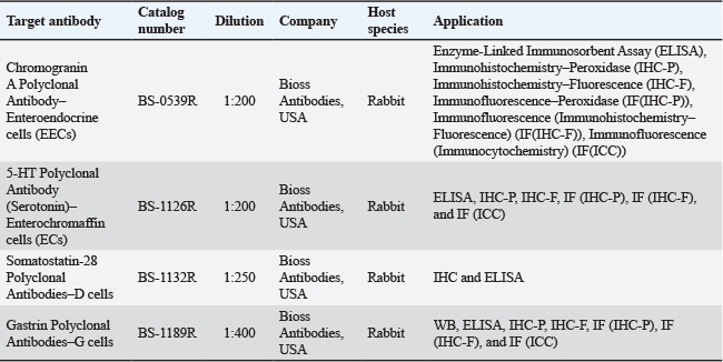
AbstractBackground: The gastrointestinal (GI) tract exhibits regional structural and endocrine specialization that is essential for digestive regulation. However, information on the gastric and intestinal endocrine organization of viverrid species remains limited. Aim: This study aims to investigate the anatomy, histology, and endocrine cell distribution in the stomach and duodenum of the Asian palm civet (Paradoxurus hermaphroditus). Methods: Stomach and duodenal segments were collected from three adult male P. hermaphroditus obtained from the special region of Yogyakarta, Indonesia. The animals were euthanized with ketamin–xylazine in accordance with ethical guidelines. Gross anatomical observations were conducted, followed by histological examination using haematoxylin–eosin staining. Immunohistochemistry was performed to identify enteroendocrine cells using antibodies against chromogranin A (CgA), gastrin, serotonin, and somatostatin (SST). Endocrine cell distribution was evaluated qualitatively in the part of the stomach (cardiac, fundic, and pyloric) and duodenal regions. Results: The stomach exhibited clear regional differentiation at both the gross and histological levels. CgA–immunoreactive cells were detected throughout all examined regions. Serotonin-immunoreactive cells were widely distributed in the stomach and were most prominent in the duodenum. SST-immunoreactive cells were observed in the fundic–body, pyloric region, and duodenal regions. Gastrin-immunoreactive cells were predominantly localized in the pyloric glands and represented the most abundant endocrine population in this region. Conclusion: The stomach and duodenum of P. hermaphroditus show a regionally organized endocrine system closely associated with gross and histological specialization. The pylorus region showed strong gastrin- and SST-immunoreactive cell distribution, resembling the typical endocrine regulatory pattern reported in other carnivorous mammals, while the duodenum demonstrated serotonin-dominant signaling associated with intestinal motility and secretion. The widespread distribution of CgA-positive cells throughout the GI tract further supports a conserved carnivoran-type GI endocrine organization in this viverrid species, despite its opportunistic omnivorous–frugivorous feeding behavior. Keywords: Asian palm civet (Paradoxurus hermaphroditus), duodenum, enteroendocrine cells, immunohistochemistry, stomach. IntroductionThe gastrointestinal (GI) tract is essential for digesting and absorbing nutrients in animals. In mammals, it shows clear regional specialization, with each part having its own structural features and physiological functions. These distinctions are evident in the anatomical and histological makeup of each segment, which is tailored to its specific role. Beyond its structural organization, this specialization is closely tied to endocrine regulation, ensuring that digestion and nutrient uptake are efficiently coordinated (Solcia et al., 2000; Guo et al., 2022; Yang et al., 2024; Nwako and McCauley, 2025). One of the key elements in the endocrine regulation of the digestive tract is the enteroendocrine cells (EECs), which are distributed throughout the epithelium of the stomach and intestines. These cells function as chemical sensors, detecting luminal and basal signals along the intestinal crypt–villus axis and translating them into hormonal responses through the secretion of hormones and bioactive amino acids. The secretory products of EECs play an important role in controlling gastric acid secretion, regulating GI motility, and influencing mucosal growth and differentiation. Moreover, hormones produced by EECs facilitate interorgan communication via endocrine, paracrine, and neurocrine pathways (Sanchez et al., 2023). Among the major endocrine markers, chromogranin A (CgA) is widely recognized as an enteroendocrine marker associated with large dense-core secretory vesicles and regulated monoamine secretion, including serotonin storage and release. Serotonin-producing enterochromaffin cells regulate GI motility, secretion, and luminal reflexes, whereas somatostatin (SST)-producing D cells exert inhibitory control over gastric acid secretion and the release of multiple GI hormones (Latorre et al., 2016). In addition to their local regulatory roles, EECs contribute significantly to GI peptide signaling and gut–brain communication. Serotonin released from enterochromaffin cells can activate enteric neural circuits and vagal afferent pathways, allowing luminal nutrient and chemical signals to be transmitted to the central nervous system. Through this gut–brain axis, EECs participate in the regulation of feeding behavior, gastric emptying, intestinal transit, and energy balance (Gunawardene et al., 2011; Grundeken and Aidy, 2025). Furthermore, increasing evidence suggests that EEC populations exhibit remarkable cellular plasticity, in which hormone expression profiles and cell densities can change in response to dietary composition, luminal stimulation, microbiota-derived metabolites, and physiological adaptation. Such plasticity enables the GI endocrine system to adapt dynamically to different feeding strategies and digestive demands (Fothergill et al., 2017). The stomach and duodenum form the starting point of the digestive tract, where chemical digestion begins and hormonal regulation takes shape. The stomach is the main site for releasing acid and digestive enzymes, initiating the breakdown of food. The duodenum, on the other hand, acts as a coordinating hub, integrating hormonal signals and managing the activity of the pancreas, biliary system, and small intestinal motility. Together, they ensure that digestion and nutrient absorption proceed smoothly and efficiently (Camilleri, 2019; Farmonovna, 2023; Niekerk et al., 2024). Both anatomical and histological features contribute to the variation and differentiation of EECs within the GI tract. Regional anatomical specialization of the stomach and intestine influences the morphology, localization, and functional characteristics of EEC populations, reflecting adaptations related to digestive physiology and nutrient sensing (Gunawardene et al., 2011). In carnivorous mammals, the stomach is generally divided into the cardiac, fundic–body, and pyloric regions, each characterized by unique histological features and endocrine cell composition (Salih and Hanash, 2024). The pyloric region serves as the main endocrine regulatory center, marked by a high density of gastrin- and SST -producing cells that regulate gastric acid secretion and gastric emptying (Schubert et al., 2020). Through the actions of these hormonal mediators, the stomach and duodenum function as an integrated communication system, ensuring a controlled and coordinated transition from gastric digestion to subsequent digestive processes in the small intestine (Cai et al., 2023). Members of the family Viverridae, including the Asian palm civet (Paradoxurus hermaphroditus), occupy an ecologically flexible niche and exhibit opportunistic feeding behavior (Kunteepuram et al., 2018; Khan et al., 2019; Akrim et al., 2023). Despite this dietary adaptability, little is known about the structural and endocrine organization of their GI tract. Existing studies on civets have primarily focused on ecology and conservation, with limited attention to digestive morphology and GI endocrinology (Dunn et al., 2022; Jennings and Veron, 2022). The present study therefore aims to describe the gross anatomical features, histological organization, and regional distribution of major endocrine cell types in the stomach and duodenum of male P. hermaphroditus. Understanding this structure provides baseline data for comparative GI endocrinology and contributes to knowledge of digestive adaptations in viverrid carnivorans. Materials and MethodsSample collectionThree adult male Asian palm civets (Paradoxurus hermaphroditus) were used in this study. Before tissue collection, the animals showed no gross abnormalities or visible pathological lesions in the GI tract during necropsy examination. This study was conducted at the Faculty of Veterinary Medicine, Universitas Gadjah Mada, Indonesia, and all procedures were approved by the institutional ethics committee and performed in accordance with accepted ethical standards for animal research. Following euthanasia, the abdominal cavity was opened, and the stomach, along with the proximal duodenum, was removed for further histological and immunohistochemical analyses. The stomach was examined macroscopically to identify its general shape, curvature, and regional differentiation. All animal samples were anesthetized and subsequently euthanized using a lethal dose based on the protocol described by Kusindarta et al. (2023). The organ was opened along the greater curvature, rinsed with saline, and photographed for documentation. The stomach and duodenum of each animal were collected and examined macroscopically, with detailed documentation captured using a Nikon D3500 digital camera (Nikon Corporation, Tokyo, Japan). Each stomach and duodenum sample was fixed in 10% neutral-buffered formalin for 24 hours, followed by dehydration through a series of graded ethanol concentrations and clearing with xylene. Tissues were then infiltrated and embedded in paraffin to ensure optimal preservation (Nurliani et al., 2015; Budipitojo et al., 2016; Suvarna et al., 2019; Aqiilah et al., 2024). Examination of histological featuresTissue samples from the stomach and proximal duodenum were fixed in 10% neutral buffered formalin, dehydrated through a graded ethanol series, cleared in xylene, and embedded in paraffin wax. Paraffin blocks were then sectioned at 4 µm thickness, deparaffinized, and stained with routine histological stains (haematoxylin–eosin) as described previously (Suvarna et al., 2019). Histological observations were performed using a light microscope (BX60, Olympus, Tokyo, Japan) and a stereobinocular microscope (SZ51, Olympus, Tokyo, Japan). Immunohistochemistry (IHC) stainingIn this study, as delineated in Table 1, primary antibodies were applied overnight at 4oC. Negative control sections were processed using the same staining protocol but without incubation with the primary antibodies to confirm the specificity of immunoreactive staining and exclude nonspecific antibody binding. Before primary antibody incubation, the sections were incubated with normal goat serum for 1 hour to reduce nonspecific staining. Subsequently, secondary antibodies were applied for 1 hour at room temperature, and immunoreactive sites were visualized using a diaminobenzidine solution. To enhance contrast, the sections were stained with Mayer’s hematoxylin. Following staining, the samples were rinsed in running water, dehydrated through graded alcohols, cleared with xylene, and mounted using Entellan (Budipitojo et al., 2016; Mahfud et al., 2020; Budipitojo et al., 2025). Data analysisHistological and IHC staining were meticulously examined using a light microscope (BX60, Olympus, Tokyo, Japan) and a stereobinocular microscope (SZ51, Olympus, Tokyo, Japan). Observations were documented using an OptiLab advance plus Binocular digital camera at various magnifications. Histological evaluation was conducted descriptively based on the regional organization of the gastric and duodenal wall, including epithelial morphology, gastric pits, glandular structure, and mucosal organization. The distribution of immunoreactive endocrine cells was assessed descriptively and semi-quantitatively. Semi-quantitative scoring of endocrine IR cells was performed subjectively and categorized into three levels: – (not detected), + (rare/scattered), ++ (moderate), +++ (numerous/prominent) at 400x magnification (Budipitojo et al., 2016; Budipitojo et al., 2025). Tissue sections exhibiting severe autolysis, marked degeneration, or substantial structural disruption were excluded from evaluation to ensure histological quality and interpretation reliability. Ethical approvalSample collection was conducted under the Ethical Clearance Number: 0016/EC-FKH/Int./2018 issued on June 25th, 2018. Table 1. The primary antibodies used in this study.
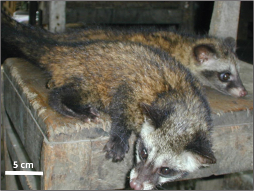
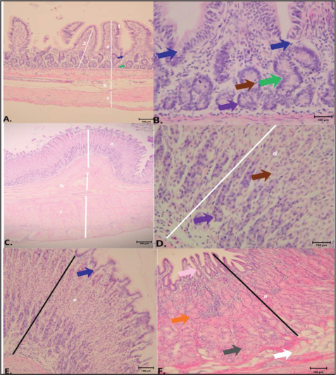
ResultsGross anatomyParadoxurus hermaphroditus exhibited the typical external morphological characteristics of the species, characterized by an elongated and slender body, coarse brownish-gray fur with darker pigmentation along the dorsal region, a pointed muzzle, and a long, well-developed, muscular tail (Fig. 1). The stomach of P. hermaphroditus (Fig. 2) appeared as a single-chambered, J-shaped organ with clear differentiation between the cardiac, fundic–body, and pyloric regions. Prominent mucosal folds were evident, particularly in the fundic–body region. The duodenum of Paradoxurus hermaphroditus appeared as a C-shaped duodenal loop located on the right side of the abdominal cavity and suspended from the dorsal abdominal wall by the duodenal mesentery together with the pancreas. Cranially, it was continuous with the pylorus, forming the first duodenal flexure, while caudally it curved leftward and transitioned into the jejunum. HistologyThe cardiac region follows a simple monogastric stomach plan and occupies a narrow area around the cardia, situated between the oesophageal squamous epithelium and the fundic–body mucosa. Histologically (Fig. 3a and b), the stomach wall consisted of the tunica mucosa, submucosa, and tunica muscularis. The tunica mucosa was composed of a surface simple columnar epithelium, gastric pits, lamina propria containing cardiac glands, and a distinct lamina muscularis mucosae. The cardiac glands were short, branched tubular structures located within the lamina propria, opening into relatively shallow gastric pits. These glands were composed predominantly of mucous-secreting cells characterized by lightly stained cytoplasm and basally located nuclei. In addition, a small number of chief cells and occasional parietal cells were present. Chief cells, with their basophilic cytoplasm, were generally located in the deeper portions of the glands, while parietal cells appeared as larger eosinophilic cells scattered among the glandular epithelium. Overall, the cardiac region was distinguished by shallow gastric pits and mucous glands, with only sparse populations of chief and parietal cells.
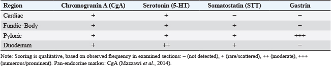
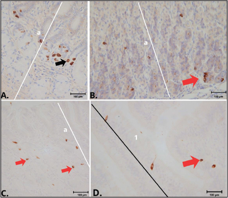
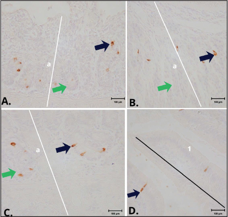
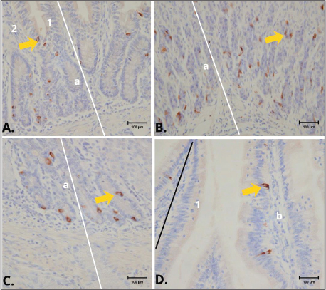
Fig. 1. Adult male Asian palm civet (Paradoxurus hermaphroditus) used in the present study. The animal exhibits the characteristic external morphology of the species, including an elongated body, coarse brownish-gray fur with darker dorsal regions, a pointed muzzle, and a long, muscular tail. The fundic–body region (Fig. 3c and d) showed densely packed tubular glands extending toward the muscularis mucosae. The pyloric region (Fig. 3e) was characterized by deep gastric pits and mucous pyloric glands. The duodenum (Fig. 3f) exhibited typical villi, crypts, and submucosal structures consistent with mammalian small intestine. ImmunohistochemistryThe qualitative distribution of immunoreactive endocrine cells in the stomach and duodenum of male P. hermaphroditus is summarized in Table 2. Gastrin-immunoreactive cells were predominantly localized in the pyloric glands and represented the most abundant endocrine population in this region (Fig. 4a). Somatostatin-immunoreactive cells were observed in the fundic–body, pyloric, and duodenal regions (Fig. 4b–d), mainly in glandular or crypt areas. Serotonin-immunoreactive cells were present throughout the stomach (Fig. 5a–c) and were particularly prominent in the duodenum (Fig. 5d). CgA-immunoreactive cells were detected in the cardiac–pyloric region and along the crypts and lower portions of the villi of the duodenum (Fig. 6a–d), appearing as scattered solitary endocrine cells.
Fig. 2. Gross anatomical appearance of the stomach and proximal duodenum of Paradoxurus hermaphroditus. The stomach appeared as a single-chambered, J-shaped organ with distinct cardiac, fundic–body, and pyloric regions, while the duodenum formed a C-shaped loop connected cranially to the pylorus and continuing caudally into the jejunum. DiscussionThis study offers a detailed look at both the gross anatomy and histology, along with the regional distribution of endocrine cells, in the stomach and proximal small intestine of the male Asian palm civet (Paradoxurus hermaphroditus). The stomach showed the typical glandular organization seen in carnivores, with clear distinctions between the cardiac, fundic–body, and pyloric regions (König and Liebich, 2020). Immunohistochemical analysis further highlighted a well-structured endocrine system, characterized by CgA-positive EECs and distinct hormonal profiles specific to each region (El-Salhy et al., 2014). Histology of the stomach of Paradoxurus hermaphroditusIn mammals, including the Asian palm civet (Paradoxurus hermaphroditus), the stomach wall is made up of four distinct layers: the mucosa, submucosa, muscularis externa, and serosa (Al-Shreefy and Al-Taai, 2024). However, the histological organization of the gastric mucosa varies among anatomical regions of the stomach, reflecting their specialized digestive and secretory functions. In carnivorous and omnivorous mammals, the cardiac region is typically characterized by relatively shallow gastric pits and mucous glands, whereas the fundic–body region contains densely packed oxyntic glands rich in parietal and chief cells. In contrast, the pyloric region exhibits deeper gastric pits with branched pyloric glands dominated by mucous and endocrine cells involved in the regulation of gastric secretion and motility (Natale et al., 2023; Naji, 2024). These regional anatomical and histological variations are closely linked to differences in enteroendocrine cell distribution and functional adaptation within the GI tract. The stomach of Paradoxurus hermaphroditus shows clear regional differentiation: a narrow cardiac region, a densely glandular fundic–body mucosa, and a well-developed pyloric region, all reflecting the close relationship between anatomical organization and functional specialization. In this study, the cardiac region was composed of short pits and mucous glands, whereas the fundic–body region contained densely packed tubular glands extending toward the muscularis mucosae. By contrast, the pyloric region was characterized by deep gastric pits and branched pyloric glands, supporting its regulatory role in gastric secretion and gastric emptying. In the cardiac region of the stomach, the gastric mucosa is characterized by short pits and branched tubular cardiac glands, which are composed mainly of mucous-secreting cells. Alongside these abundant mucous cells, both chief cells and parietal cells can also be found, though they are less prominent here than in the fundic–body region. Histologically, chief cells are distinguished by their basophilic cytoplasm and their position in the deeper portions of the glands, while parietal cells appear as larger, eosinophilic cells scattered within the glandular epithelium. The predominance of mucous cells highlights the protective role of this region at the gastroesophageal junction, where mucus secretion provides lubrication and shields the mucosa from mechanical stress and chemical injury (Natale et al., 2023; Al-Shreefy and Al-Taai, 2024).
Fig. 3. Histological appearance of the stomach regions of Paradoxurus hermaphroditus. The cardiac region shows a relatively thin gastric wall at low magnification (A), consisting of the tunica mucosa (a), tunica submucosa (b), and tunica muscularis (c). At higher magnification (B), shallow gastric pits (blue arrows) and short, branched cardiac glands (green arrows) composed predominantly of mucous-secreting cells are observed. Occasional parietal cells (brown arrows) and chief cells (purple arrows) are identified within the cardiac glands. The fundic–body region exhibits prominent mucosal folding at low magnification (C), while higher magnification (D) reveals long, straight fundic glands densely populated by parietal cells (brown arrows) and chief cells (purple arrows). The neck of the fundic glands is indicated by label (d), and the lamina muscularis mucosae is indicated by white arrows. The pyloric region is characterized by deep gastric pits (blue arrows) and short, coiled pyloric glands (e) composed mainly of mucous cells (E). The duodenum displays well-developed villi (pink arrows), crypts of Lieberkühn (orange arrows), and Brunner’s glands (grey arrows) within the submucosa (F). The simple columnar surface epithelium is indicated by label (1), and the lamina propria by label (2). Coloured arrows in the high-magnification panels identify the principal cellular and glandular components to facilitate histological interpretation. Hematoxylin and eosin staining; magnification 100× (A, C, E, and F) and 400× (B and D); scale bar: 100 µm. Table 2. Semi-quantitative number of endocrine cell immunoreactivity in the stomach and duodenum of male Paradoxurus hermaphroditus.
Fig. 4. Gastrin and somatostatin immunoreactivity (brown cytoplasmic staining) was observed across different stomach regions of Paradoxurus hermaphroditus. Numerous gastrin-immunoreactive cells (black arrows) were concentrated in the basal portions of the pyloric glands (A). In contrast, somatostatin-immunoreactive cells (red arrows) appeared more sparsely, distributed within the basal portions of the fundic–body glands (B), the pyloric glands (C), and the crypts and villi of the duodenum (D). Sections were processed using immunohistochemical stainingand examined at 400× magnification, with a scale bar of 100 µm. Label (a) indicates the tunica mucosa, label (1) marks the simple columnar surface epithelium, and the black line highlights the duodenal villi. In contrast, the fundic region displays a much thicker mucosa with densely packed, long, straight oxyntic glands. This area contains a far greater number of parietal and chief cells compared with the cardiac mucosa, underscoring its role as the primary site of hydrochloric acid and pepsinogen secretion. Parietal cells dominate within the fundic glands, reflecting their central function in acid production, while chief cells contribute to protein digestion through the release of pepsinogen. In addition, mucous neck cells and scattered EECs —including ECL cells, D cells, and ghrelin-producing cells—are present in the fundic mucosa of mammals, where they play key roles in regulating gastric activity (Natale et al., 2023; Al-Shreefy and Al-Taai, 2024). The pyloric mucosa is distinguished by deep gastric pits and branched pyloric glands, which are composed primarily of mucous cells. This region also contains numerous G cells, responsible for secreting gastrin, along with D cells. Together, these endocrine components make the pyloric mucosa a critical regulatory zone, coordinating both gastric acid secretion and the timing of gastric emptying (Fothergill et al., 2017; Kamal et al., 2017).
Fig. 5. Serotonin immunoreactivity (brown cytoplasmic staining) in the stomach regions of Paradoxurus hermaphroditus. Serotonin-immunoreactive cells (navy arrows) are distributed (A) among the epithelial cells of the gastric pits and glands of the cardiac region, (B) among glandular epithelial cells of the fundic–body region, (C) scattered among pyloric gland epithelial cells, and (D) within the crypt epithelium and lower villous epithelium of the duodenum. Immunohistochemical staining, examined at 400× magnification (scale bar=100 µm). Label (a) identifies the tunica mucosa, while green arrows highlight the branched cardiac glands. Label (1) denotes the simple columnar surface epithelium, and the black line marks the duodenal villi. Beneath the mucosa lies the submucosa, composed of loose to moderately dense connective tissue that contains collagen and elastic fibers, along with blood vessels, lymphatics, and the submucosal nerve plexus. In species such as pigs and guinea pigs, this layer is relatively thin and flexible, creating a compliant interface between the mucosa and the muscularis externa (Pejcinovska et al., 2025). The muscularis externa of the stomach is composed of three layers of smooth muscle: an inner oblique layer, a middle circular layer, and an outer longitudinal layer. Working together, these layers ensure efficient mixing and propulsion of gastric contents (Pangtey et al., 2017). Surrounding the stomach is the serosa, a thin sheet of loose connective tissue covered by simple squamous mesothelium, also known as the visceral peritoneum. In omnivorous mammals such as pigs and guinea pigs, this serosal covering envelops the external surface of the stomach (Kamal et al., 2017; Pejcinovska et al., 2025). Morphology as the structural basis for endocrine specializationThe fundic–body mucosa is packed with dense gastric glands, reflecting its role as a highly secretory region. In carnivorous mammals, this area drives strong acid and enzyme production, while the local endocrine network fine-tunes secretion to meet functional demands (König and Liebich, 2020; Hsu et al., 2025). The presence of CgA, serotonin, and SST-positive cells in the civet’s fundic–body region indicates a functional neuroendocrine microenvironment like that found in other carnivores. Within this system, endocrine signals help regulate acid release, blood flow in the mucosa, and overall glandular activity (Mazzawi et al., 2014; Engelstoft et al., 2015; Shulkes et al., 2006).
Fig. 6. Chromogranin A immunoreactivity (brown cytoplasmic staining) in the stomach regions of Paradoxurus hermaphroditus. Chromogranin A–immunoreactive cells (yellow arrows) are scattered within (A) the gastric pits and superficial cardiac glands of the cardiac region, (B) the gastric glands of the fundic–body region, (C) the pyloric glands of the pyloric region, and (D) the crypts of Lieberkühn and lower portions of the duodenal villi. Sections were processed using immunohistochemistry staining and examined at 400x magnification. Label (a) denotes the tunica mucosa, label (1) the simple columnar surface epithelium, and label (2) the lamina propria. The black line highlights the duodenal villi. Scale bar=100 µm. The pyloric region shows the typical structure of deep pits and mucous pyloric glands, serving as the main site for hormonal regulation of both gastric secretion and gastric emptying (Schubert et al., 2020). Reflecting this role, the pylorus displayed the widest range of endocrine activity in this study—CgA, gastrin, serotonin (5-HT), and SST—supporting its function as an endocrine “control center” that coordinates secretion in the fundic region and regulates the flow of contents into the duodenum (Schubert et al., 2020). CgA as a pan-enteroendocrine framework across regionsChromogranin A immunoreactivity was detected in all examined regions (cardiac, fundic–body, pyloric, and duodenum), supporting its use as a reliable marker for mapping the overall distribution of GI endocrine cells in P. hermaphroditus (Mazzawi et al., 2014). Across mammals, CgA labels diverse enteroendocrine cell populations and is widely used to define neuroendocrine compartments in comparative studies (Mazzawi et al., 2014; Romano et al., 2025). The civet’s diffuse, scattered CgA-positive pattern corresponds to the “solitary cell” distribution typical of EECs in gastric and intestinal epithelia (Engelstoft et al., 2015). Gastrin– SST balance in the pylorus and its functional implicationsOne of the key observations is the strong presence of gastrin-producing cells in the pyloric mucosa, alongside SST -producing cells. This setup reflects the typical balance seen in mammalian physiology: G cells release gastrin, which drives gastric acid secretion and promotes growth activity, both directly and indirectly through histamine pathways. In contrast, D cells release SST, which acts as a brake by inhibiting gastrin release and lowering acid output. Together, these opposing forces help keep stomach pH stable and prevent overstimulation (Schubert et al., 2020; Papantoniou et al., 2025). The presence of both gastrin and SST in the civet pylorus points to a tightly regulated feedback system, similar to what is seen in domestic carnivores and other carnivorans (Joseph et al., 2003). Functionally, this endocrine setup in the pylorus likely helps balance the strong secretory activity of the fundic glands with a controlled release of stomach contents into the duodenum (Engevik et al., 2019). Serotonin as a conserved motility–secretion integrator from stomach to duodenumSerotonin–producing cells were found throughout the stomach—cardiac, fundic–body, and pyloric regions—and were especially abundant in the duodenum. This pattern fits with the well-known role of enteroendocrine and enterochromaffin cells that release serotonin, helping regulate gut motility, secretion, and communication with the enteric nervous system, with their influence being particularly strong in the upper small intestine (Pyarokhil et al., 2017). In the 5-HT helps regulate gland activity and muscle function, including coordination at the pylorus. In the duodenum, it plays a central role in driving peristaltic reflexes and secretomotor pathways that respond to nutrients and incoming gastric chyme (Pyarokhil et al., 2017). The civet shows a pattern of widespread serotonin presence in the stomach and clear representation in the duodenum, reflecting the conserved mammalian organization. This supports the idea of serotonin acting as a gut-wide integrator, linking the gastric and intestinal phases of digestion (Pyarokhil et al., 2017). Duodenum: endocrine “receiving station” for gastric outputThe duodenum contained cells reactive to CgA, serotonin, and SST, underscoring its role as an endocrine hub for processing gastric output (Pyarokhil et al., 2017). Somatostatin here helps regulate secretion and hormone release by providing inhibitory control, maintaining balance at the gastroduodenal junction (Papantoniou et al., 2025). This endocrine profile complements the pyloric gastrin–SST system, suggesting that P. hermaphroditus has a coordinated stomach–duodenum endocrine axis that fine-tunes secretory activity and transit in response to luminal conditions (Camilleri, 2019). Comparative perspective within carnivora and viverridaeAlthough P. hermaphroditus is often described as an opportunistic omnivore with a substantial frugivorous component in its diet, the present findings demonstrate that its gastric endocrine organization closely resembles the conserved carnivoran pattern. The stomach structure, together with the strong gastrin immunoreactivity observed in the pyloric region, reflects a typical carnivoran endocrine blueprint associated with regulation of gastric acid secretion and digestive activity (Akrim et al., 2023; Clemente-Suárez et al., 2025). In mammals, dietary proteins and peptides are recognized as potent stimulators of gastrin release from pyloric G cells, thereby promoting acid secretion, pepsinogen release, and gastric motility required for protein digestion (Jahan-Mihan et al., 2011; Duan et al., 2026). However, the presence of a gastrin-dominant pyloric pattern does not necessarily imply continuous dependence on a chronically high-protein diet. Rather, it reflects a conserved anatomical and physiological regulatory capacity that can be dynamically modulated according to dietary composition and luminal conditions. Experimental studies have demonstrated that G-cell populations in the pyloric mucosa may remain relatively stable even during prolonged low-protein feeding, although their functional activity and circulating gastrin levels can be downregulated through feedback mechanisms involving luminal pH and SST release (Varagić et al., 1996). Thus, the gastrin-dominant pyloric organization observed in the Asian palm civet may represent an adaptive “ready-to-respond” endocrine framework that preserves the capacity for efficient protein digestion whenever animal-derived food resources become available. This interpretation is consistent with the feeding ecology of civets, which consume fruits opportunistically but also ingest insects, small vertebrates, and other protein-rich food items depending on seasonal availability. Furthermore, gastrin regulation is influenced not only by dietary protein but also by other luminal and neural factors, including gastric distension, lipid composition, vagal stimulation, and SST -mediated inhibitory pathways (Jahan-Mihan et al., 2011; Duan et al., 2026). Therefore, the endocrine architecture of the pyloric mucosa in P. hermaphroditus likely reflects functional flexibility rather than strict specialization for high-protein feeding alone. The persistence of this carnivoran-type endocrine organization despite a flexible omnivorous–frugivorous feeding strategy suggests that strong gastric secretory capacity and tight endocrine regulation remain advantageous for processing variable dietary components, particularly episodic protein and fat intake. Specifically, the endocrine pattern observed in P. hermaphroditus resembles the typical carnivoran organization characterized by: (1) strong gastrin immunoreactivity concentrated in the pyloric region, which functions as the principal regulatory site for gastric acid secretion; (2) diffuse CgA –positive EECs distributed throughout the gastric and intestinal mucosa; and (3) widespread serotonin-positive cells extending from the stomach into the duodenum, supporting coordinated regulation of motility and secretion. Similar endocrine distributions have been described in domestic carnivores such as dogs and cats, in which the pyloric mucosa contains abundant G cells, while serotonin-producing enterochromaffin cells are broadly distributed along the upper GI tract (Pyarokhil et al., 2017; Schubert et al., 2020). Comparable regional endocrine specialization has also been reported in other carnivoran mammals, suggesting that the civet retains a conserved GI endocrine framework despite its flexible omnivorous–frugivorous feeding ecology. Notably, a distinctive aspect of P. hermaphroditus lies in the coexistence of a conserved carnivoran endocrine architecture with a highly flexible and partially frugivorous feeding strategy. Field and ecological studies have shown that this species exhibits opportunistic feeding behavior, consuming fruits, insects, and small vertebrates depending on availability (Joshi et al., 1995; Nakashima et al., 2010). Despite this dietary flexibility, the structural organization of the GI endocrine system remains consistent with the conserved carnivoran pattern, which is generally associated with efficient processing of protein-rich diets (Stevens and Hume, 1995; Camilleri, 2019). Unlike obligate carnivores, whose endocrine activity is more consistently aligned with high protein intake, the civet appears to retain a structurally “carnivoran-type” pyloric organization while functionally modulating hormonal output in response to variable dietary inputs. This interpretation is supported by studies demonstrating that gastrin secretion and enteroendocrine activity are dynamically regulated by luminal nutrients and feedback mechanisms, rather than being fixed to dietary composition (Varagić et al., 1996; Jahan-Mihan et al., 2011; Duan et al., 2022). Furthermore, increasing evidence highlights the plasticity of EECs, which can adjust their functional output in response to environmental and dietary cues (Gribble and Reimann, 2016; Latorre et al., 2016). This combination suggests a degree of physiological plasticity that may represent an adaptive advantage within the Viverridae, allowing efficient processing of both animal-derived and plant-based resources. Therefore, P. hermaphroditus may be considered a flexible or intermediate model within carnivora, in which conserved endocrine morphology is retained while its functional expression is dynamically adjusted to ecological feeding diversity. Nevertheless, several limitations of this study should be acknowledged. The sample size was restricted to three adult male specimens, reflecting both limited availability and ethical considerations aligned with the principle of reduction in the 3Rs framework. As such, the findings are best viewed as descriptive baseline data rather than broad species-level generalizations. In addition, the distribution of endocrine cells was assessed using a semi-quantitative, descriptive approach. While this method is appropriate for regional comparisons, it is inherently less objective than fully quantitative, image-based analyses. The study also focused primarily on gross anatomy, histology, and IHC of the stomach and proximal duodenum. Complementary biochemical or molecular investigations—such as hormone quantification or gene expression profiling—were not conducted, as the limited tissue was prioritized for morphological and immunohistochemical evaluation. Future research incorporating larger and more diverse sample sets, quantitative morphometric techniques, and integrated molecular analyses will be essential to validate and expand upon these initial observations. ConclusionThe stomach and duodenum of P. hermaphroditus exhibit a regionally specialized endocrine system that closely parallels gross and histological organization. The pylorus functions as a major endocrine regulatory center through the gastrin–SST axis, while the duodenum is dominated by serotonin-mediated signaling. These findings indicate a conserved carnivoran-type GI endocrine pattern in this viverrid species and provide foundational data for future comparative and functional studies. AcknowledgmentsThe authors sincerely acknowledge the financial support provided by the Indonesian Endowment Fund for Education (LPDP) on behalf of the Indonesian Ministry of Higher Education, Science and Technology through the EQUITY Program (Contract Number: 4301/B3/DT.03.08/2025 and 10107/UN1.P/Dit.Keu/HK.08.00/2025). Conflict of interestThe authors declare that no financial or commercial relationships existed during the course of this research that could be understood as a potential conflict of interest. FundingThis research is funded by the Indonesian Endowment Fund for Education (LPDP) on behalf of the Indonesian Ministry of Higher Education, Science and Technology and managed under the EQUITY Program (Contract Number: 4301/B3/DT.03.08/2025 and 10107/UN1.P/Dit.Keu/HK.08.00/2025). Author’s contributionsTB contributed to the study design, supervised the research process, performed data analysis, and led the manuscript development. SAKP, IP, ARJ, and WDW conducted the experiments, collected and analyzed the data, and drafted the manuscript. GR, HU, AN, and MR critically reviewed and evaluated the manuscript draft. All authors read and approved the final version of the manuscript. Data vailabilityThe original data and contributions from this study are available in the article or supplementary materials. Furthermore, inquiries should be directed to the corresponding authors. ReferencesAkrim, F., Mahmood, T., Belant, J.L., Nadeem, M.S., Qasim, S., Dhendup, T., Fatima, H., Bukhari, S.A., Aslam, A., Younis, H., Rafique, A., Subhani, Z.A., Hashmi, S.A. and Munawar, N. 2023. Niche partitioning by sympatric civets in the Himalayan foothills of Pakistan. Peer J. 11, 1–15; doi: 10.7717/ peerj.14741 Aqiilah, K., Budipitojo, T., Wijayanto, H., Budiariati, V., Pangestiningsih, T.W., Saragih, A., Kustiati, U. and Wihadmadyatami, H. 2024. Morphological structure of the tongue of Gekko gecko in Yogyakarta, Indonesia. J. Trop. Biodiversity. Biotechnol. 9(4), 1–16; doi:10.22146/jtbb.90995 Budipitojo, T., Fibrianto, Y.H. and Mulyani, G.T. 2016. Gut endocrine cells in the stomach of sunda porcupines (Hystrix javanica). Asian J. Anim. Sci. 10(4–5), 268–272; doi10.3923/ajas.2016.268.272 Budipitojo, T., Larasati, A.D., Putri, S.A.K., Wendo, W.D. and Fibrianto, Y.H. 2025. Distribution of androgen receptor immunoreactive cells in the adrenal glands of Hystrix javanica. Open Vet. J. 15(9), 4090–4096; doi:10.5455/OVJ.2025.v15.i9.13 Cai, X., Bao, D., Hua, R., Cai, B., Wang, L., Dong, R. and Hua, L.A. 2023. Comparative Study on the Distribution Pattern of Endocrine Cells in the Gastrointestinal Tract of Two Small Alpine Mammals, Plateau Zokor (Eospalax baileyi) and Plateau Pika (Ochotona curzoniae). Animals 13, 640. Camilleri, M. 2019. Gastrointestinal hormones and regulation of gastric emptying. Curr. OpinEndocrinol. Diabetes Obes. 26(1), 3–10. Clemente-Suárez, V.J., Redondo-Flórez, L., BeltránVelasco, A.I., Yáñez-Sepúlveda, R., RubioZarapuz, A., Martín-Rodríguez, A., NavarroJimenez, E. and Tornero-Aguilera, J.F. 2025. Human digestive physiology and evolutionary diet: a metabolomic perspective on carnivorous and scavenger adaptations. Metabolites, 15(7), 1–58. Di Natale, M.R., Athavale, O.N., Wang, X., Du, P., Cheng, L.K., Liu, Z. and Furness, J.B. 2023. Functional and anatomical gastric regions and their relations to motility control. Neurogastroenterology. Motility. 35(9), 1–9; doi:10.1111/nmo.14560 Duan, S., Rico, K. and Merchant, J.L. 2026. Gastrin: from physiology to gastrointestinal malignancies. Function 2021(3), 1–13. Dunn, A., Amir, Z., Decoeur, H., Dehaudt, B., Nursamsi, I., Mendes, C., Moore, J.H., Negret, P.J., Sovie, A. and Scott, M. 2022. The ecology of the banded civet (Hemigalus derbyanus) in Southeast Asia with implications for mesopredator release , zoonotic diseases , and conservation. Ecosphere 2021, 1–13; doi:10.1002/ece3.8852 El-Salhy, M., Gilja, O.H. and Hausken, T. 2014. Chromogranin A cells in the stomachs of patients with sporadic irritable bowel syndrome. Mol. Med. Rep. 10, 1753–1757; doi:10.3892/mmr.2014.2472 Engelstoft, M.S., Lund, M.L., Grunddal, K.V., Egerod, K.L., Osborne-Lawrence, S., Poulsen, S.S., Zigman, J.M. and Schwartz, T.W. 2015. Research resource: a chromogranin a reporter for serotonin and histamine secreting enteroendocrine cells. Mol. Endocrinol. 29(11), 1658–1671. Engevik, A.C., Kaji, I. and Goldenring, J.R. 2019. The physiology of the gastric parietal cell. Physiol. Rev. 100, 573–602. Farmonovna, R.A. 2023. Digestion In The Duodum. Texas J. Med. Sci. 20, 67–69. Fothergill, L.J., Callaghan, B., Hunne, B., Bravo, D.M. and Furness, J.B. 2017. Costorage of enteroendocrine hormones evaluated at the cell and subcellular levels in male mice. Endocrinology 178(7), 2113–2123. Grundeken, E. and Aidy S El. 2025. Enteroendocrine cells: the gatekeepers of microbiome-gut-brain communication. Nature 11(179), 1–12. Gunawardene, A.R., Corfe, B.M. and Staton, C.A. 2011. Classification and functions of enteroendocrine cells of the lower gastrointestinal tract. Wiley, pp: 219–231; doi: 10.1111/j.1365-2613.2011.00767.x Guo, X., Lv, J. and Xi, R. 2022. The specification and function of enteroendocrine cells in Drosophila and mammals: a comparative review. Febs Press 289, 4773–4796. https://doi.org/10.1111/febs.16067 Hsu, M., Safadi, A.O. and Lui, F. 2025. Physiology, Stomach. StatPearls Publishing, p: 1–9. Jahan-Mihan, A., Luhovyy, B.L., El Khoury, D. and Anderson, G.H. 2011. Dietary proteins as determinants of metabolic and physiologic functions of the gastrointestinal tract. Nutrients 3(5), 574–603; doi:10.3390/nu3050574 Jennings, A.P. and Veron, G. 2022. Ecology and Conservation of Southeast Asian Civets (Viverridae) and Mongooses (Herpestidae). In Small carnivores: evolution, ecology, behaviour and conservation. Eds., San, E.D.L., Sato, J.J., Belant, J. and Somers, M.J. Wiley, pp: 393–427; doi:10.1002/9781118943274.ch19 Joseph, I.M.P., Zavros, Y., Merchant, J.L. and Kirschner, D. 2003. A model for integrative study of human gastric acid secretion. J. Appl. Physiol. 94, 1602–1618. Kamal, U., Sayed, A. and Gomaa, M. 2017. Studies on the stomach of the adult African catfish ( Clarias gariepinus ). J. Microsc. Ultrastructure. 5(3), 155–166; doi:10.1016/j.jmau.2016.08.002 Khan, K.A., Khan, J.A., Ahmad, K. and Mohan, N. 2019. Winter food habits of the common palm civet Paradoxurus hermaphroditus in Patna Bird Sanctuary, India. J. Threatened Taxa 11(12), 14490–14495; doi: 10.11609/jott.4165.11.12.14490-14495 Kunteepuram, V., Sreenivas, A., Tabasum, W., Challagandla, A.K. and Gaur, A. 2018. The complete mitochondrial genome of Asian palm civet (Paradoxurus hermaphroditus) with phylogenetic consideration. Mitochondrial DNA Part B. Resour. 3(1), 294–295; doi:10.1080/23802359.2018.1443853 Kusindarta, D.L., Saputra, F.C.E., Nurliani, A., Gracia, I.P.D.W. and Wihadmadyatami, H. 2023. The structure of the lingual papillary of the Asian palm civet (Paradoxurus hermaphroditus): The analysis of feeding habit adaption using gross-morphometric, scanning electron microscope, light microscope (histochemistry staining) [Preprint]. Authorea; doi: 10.22541/au.167482625.50810040/v1 Latorre, R., Sternini, C., De Giorgio, R. and Greenwood-van Meerveld, B. 2016. Enteroendocrine cells: a review of their role in brain – gut communication. Neurogastroenterology Motility 28(5), 620–630; doi:10.1111/nmo.12754 Mahfud, M., Ernawati, E., Mahmud, N.R.A., Budipitojo, T. and Wijayanto, H. 2020. An immunohistochemical study of endocrine cells in the digestive tract of Varanus salvator (Reptile: varanidae). Vet. World. 13(9), 1737–1742; doi:10.14202/vetworld.2020.1737-1742 Mazzawi, T., Gundersen, D., Hausken, T. and El-Salhy, M. 2014. Increased gastric chromogranin A cell density following changes to diets of patients with irritable bowel syndrome. Mol. Med. Rep. 10(5), 2322–2326; doi:10.3892/mmr.2014.2498 Naji, A.O. 2024. Histologic structure of stomach in carnivores. Wasit J. Pure Sci. 3(4), 197–200. Niekerk G Van., Kelchtermans, L., Broeckhoven, E., Coelmont, L., Alpizar, Y.A. and Dallmeier, K. 2024. Cytokine and Growth Factor Reviews Cholecystokinin and gastrin as immune modulating hormones: implications and applications. Cytokine. Growth. Factor. Rev. 80, 37–46; doi:10.1016/j.cytogfr.2024.11.003 Nurliani, A., Budipitojo, T. and Kusindarta, D.L. 2015. Morphological Characteristics of the Stomach of the Swamp Buffalo (Bubalus bubalis). Aceh. Int. J. Sci. Technol. 4(3), 78–82; doi:10.13170/aijst.4.3.3011 Nwako, J.G. and McCauley, H.A. 2025. Enteroendocrine cells regulate intestinal homeostasis and epithelial function. Mol. Cell. Endocrinol. 590, 112339; https://doi.org/10.1016/j.mce.2024.112339 Pangtey, B. 2017. Histogenesis of Muscularis Mucosa and Muscularis Externa of Stomach: a Human Foetal Study. J. Clin. Diagnostic. Res. 11(8), 24–26; doi:10.7860/JCDR/2017/26219.10323 Papantoniou, K., Aggeletopoulou, I., Pastras, P. and Triantos, C. 2025. The Role of Somatostatin in the Gastrointestinal Tract. Biology 14(5), 558. Pejcinovska, N., Dodovski, P., Karabolovski, N., Zdraveski, I., Dameski, P., Hristovska, T., Angelovska, M. and Ristevski, M. 2025. Morphological and histological features of the stomach in domestic pigs (Sus scrofa domestica). J. Hyg. Eng. Des. 51, 50–54. Pyarokhil, A.H., Sasaki, M., Tomikawa, S., Maetani, A., Yuhara, K., Kobayashi, Y. and Kitamura, N. 2017. Immunohistochemical study of endocrine cells in the stomach and small intestine of the lion (Panthera leo). Mammal. Study. 42(3), 179–184. Romano, E., Rinzivillo, M., Lamberti, G., Marasco, M., Esposito, G., Campana, D. and Panzuto, F. 2025. Rethinking chromogranin A: unveiling gastrointestinal factors beyond neuroendocrine neoplasms — a narrative review. Translational. Gastroenterol. Hepatol. 10(57), 1–12; doi:10.21037/tgh-24-113 Sanchez, J.G., Enriquez, J.R. and Wells, J.M. 2023. Enteroendocrine Cell Differentiation and Function in the Intestine. Curr. Opin. Endocrinol. Diabetes. Obes. 29(2), 1–14; doi:10.1097/MED.0000000000000709 Schubert, M.L. and Rehfeld, J.F. 2020. Gastric Peptides-Gastrin and Somatostatin. Compr. Physiol. 10, 197–228. Shulkes, A., Baldwin, G.S. and Giraud, A.S. 2006. Regulation of Gastric Acid Secretion. In Physiology of the Gastrointestinal Tract (Fourth Edition). Elsevier Inc, doi: 10.1016/B978-0-12-088394-3.50052-0 Solcia, E., Rindi, G., Buffa, R., Fiocca, R. and Capella, C. 2000. Gastric endocrine cells: types, function and growth. Regulatory. Peptides. 93, 31–35. Stevens, C.E. and Hume, I.D. 1995. Comparative Physiology of the Vertebrate Digestive System. Cambridge University Press. Varagić, J., Koko, V., Todorović, V., Božović, Z., Lacković, V. and Vuzevski, V.D. 1996. The effects of long-term low-protein intake on gastrin cells of the rat antral mucosa during adulthood. Histology Histopathology. Cellular Mol. Biol. 11(4), 837–843. Yang, Z., Yang, M., Rui, S., Hao, W., Wu, X., Guo, L., Armstrong, D.G., Yang, C. and Deng, W. 2024. Exosome-based cell therapy for diabetic foot ulcers: present and prospect. Heliyon 10(20), e39251; doi:10.1016/j.heliyon.2024.e39251 | ||

| How to Cite this Article |
| Pubmed Style Budipitojo T, Putri SAK, Padeta I, Jadi AR, Wendo WD, Residiwati G, Untari H, Nurliani A, Rizal M. Structural characteristics and gastric endocrine cell distribution in the stomach of the Asian palm civet (Paradoxurus hermaphroditus). Open Vet. J.. 2026; 16(8): 5327-5339. doi:10.5455/OVJ.2026.v16.i8.27 Web Style Budipitojo T, Putri SAK, Padeta I, Jadi AR, Wendo WD, Residiwati G, Untari H, Nurliani A, Rizal M. Structural characteristics and gastric endocrine cell distribution in the stomach of the Asian palm civet (Paradoxurus hermaphroditus). https://www.openveterinaryjournal.com/?mno=309077 [Access: August 08, 2026]. doi:10.5455/OVJ.2026.v16.i8.27 AMA (American Medical Association) Style Budipitojo T, Putri SAK, Padeta I, Jadi AR, Wendo WD, Residiwati G, Untari H, Nurliani A, Rizal M. Structural characteristics and gastric endocrine cell distribution in the stomach of the Asian palm civet (Paradoxurus hermaphroditus). Open Vet. J.. 2026; 16(8): 5327-5339. doi:10.5455/OVJ.2026.v16.i8.27 Vancouver/ICMJE Style Budipitojo T, Putri SAK, Padeta I, Jadi AR, Wendo WD, Residiwati G, Untari H, Nurliani A, Rizal M. Structural characteristics and gastric endocrine cell distribution in the stomach of the Asian palm civet (Paradoxurus hermaphroditus). Open Vet. J.. (2026), [cited August 08, 2026]; 16(8): 5327-5339. doi:10.5455/OVJ.2026.v16.i8.27 Harvard Style Budipitojo, T., Putri, . S. A. K., Padeta, . I., Jadi, . A. R., Wendo, . W. D., Residiwati, . G., Untari, . H., Nurliani, . A. & Rizal, . M. (2026) Structural characteristics and gastric endocrine cell distribution in the stomach of the Asian palm civet (Paradoxurus hermaphroditus). Open Vet. J., 16 (8), 5327-5339. doi:10.5455/OVJ.2026.v16.i8.27 Turabian Style Budipitojo, Teguh, Sekar Arum Krisna Putri, Irma Padeta, Arvendi Rachma Jadi, Woro Danur Wendo, Gretania Residiwati, Handayu Untari, Anni Nurliani, and Muhammad Rizal. 2026. Structural characteristics and gastric endocrine cell distribution in the stomach of the Asian palm civet (Paradoxurus hermaphroditus). Open Veterinary Journal, 16 (8), 5327-5339. doi:10.5455/OVJ.2026.v16.i8.27 Chicago Style Budipitojo, Teguh, Sekar Arum Krisna Putri, Irma Padeta, Arvendi Rachma Jadi, Woro Danur Wendo, Gretania Residiwati, Handayu Untari, Anni Nurliani, and Muhammad Rizal. "Structural characteristics and gastric endocrine cell distribution in the stomach of the Asian palm civet (Paradoxurus hermaphroditus)." Open Veterinary Journal 16 (2026), 5327-5339. doi:10.5455/OVJ.2026.v16.i8.27 MLA (The Modern Language Association) Style Budipitojo, Teguh, Sekar Arum Krisna Putri, Irma Padeta, Arvendi Rachma Jadi, Woro Danur Wendo, Gretania Residiwati, Handayu Untari, Anni Nurliani, and Muhammad Rizal. "Structural characteristics and gastric endocrine cell distribution in the stomach of the Asian palm civet (Paradoxurus hermaphroditus)." Open Veterinary Journal 16.8 (2026), 5327-5339. Print. doi:10.5455/OVJ.2026.v16.i8.27 APA (American Psychological Association) Style Budipitojo, T., Putri, . S. A. K., Padeta, . I., Jadi, . A. R., Wendo, . W. D., Residiwati, . G., Untari, . H., Nurliani, . A. & Rizal, . M. (2026) Structural characteristics and gastric endocrine cell distribution in the stomach of the Asian palm civet (Paradoxurus hermaphroditus). Open Veterinary Journal, 16 (8), 5327-5339. doi:10.5455/OVJ.2026.v16.i8.27 |


